Hyperpolarized Nuclei for NMR Imaging and Spectroscopy - Lunds ...
Hyperpolarized Nuclei for NMR Imaging and Spectroscopy - Lunds ...
Hyperpolarized Nuclei for NMR Imaging and Spectroscopy - Lunds ...

Create successful ePaper yourself
Turn your PDF publications into a flip-book with our unique Google optimized e-Paper software.
22<br />
250<br />
RBC<br />
A (brain gray matter?)<br />
200<br />
205<br />
150<br />
200<br />
B (brain white matter?)<br />
195<br />
ppm<br />
100<br />
ppm<br />
190<br />
C (?)<br />
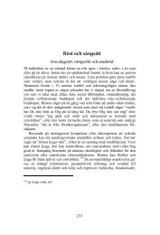
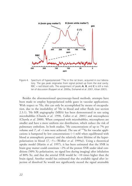
Figure 4. Spectrum of hyperpolarized 129 Xe in the rat brain, acquired in our laboratory.<br />
The gas peak originates from signal picked up from the oral cavity.<br />
RBC = red blood cells. The assignment of peaks A, B, <strong>and</strong> C is still a matter<br />
of discussion (Ruppert et al. 2000a, Duhamel et al. 2001, Kilian 2001).<br />
Besides the a<strong>for</strong>ementioned spectroscopy-based methods, attempts have<br />
been made to employ hyperpolarized noble gases in vascular applications.<br />
With respect to 3 He, this can only be accomplished by means of encapsulation,<br />
due to the insolubility of 3 He in blood <strong>and</strong> other fluids (see section<br />
2.3.1). 3 He MR angiography (MRA) has been demonstrated in rats using<br />
microbubbles (Chawla et al. 1998, Callot et al. 2001) <strong>and</strong> microspheres<br />
(Chawla et al. 2000). When compared with microbubbles, microspheres are<br />
smaller <strong>and</strong> have a more uni<strong>for</strong>m size distribution, which reduce the risk of<br />
pulmonary embolism. In both studies, 3 He concentrations of up to 7% per<br />
volume <strong>and</strong> T 1 of ∼1 min were achieved. The use of 129 Xe <strong>for</strong> vascular applications<br />
is hampered by low concentrations (∼1 mM when equilibrated with<br />
blood at atmospheric pressure) <strong>and</strong> the relatively short lifetime of the hyperpolarization<br />
in blood (T 1 ∼5 s (Wolber et al. 1999a)). Using a theoretical<br />
uptake model (Martin et al. 1997), it has been estimated that the SNR in<br />
brain gray matter could constitute ∼2% of the proton SNR under ideal conditions<br />
(50% Xe polarization, no signal loss during imaging) after inhalation<br />
of 80% Xe, <strong>and</strong> that the arterial SNR would be ∼10 times higher than the<br />
brain signal. Another model has estimated that the available signal after injection<br />
of dissolved Xe would not significantly exceed the signal attainable<br />
50<br />
185<br />
gas<br />
0