Viable Titer and Pure Culture
Viable Titer and Pure Culture
Viable Titer and Pure Culture
- TAGS
- viable
- titer
- culture
- www.fiu.edu
Create successful ePaper yourself
Turn your PDF publications into a flip-book with our unique Google optimized e-Paper software.
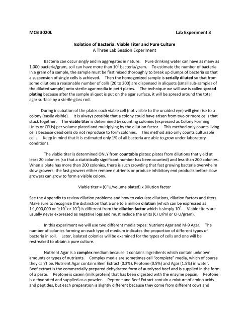
MCB 3020L Lab Experiment 3<br />
Isolation of Bacteria: <strong>Viable</strong> <strong>Titer</strong> <strong>and</strong> <strong>Pure</strong> <strong>Culture</strong><br />
A Three Lab Session Experiment<br />
Bacteria can occur singly <strong>and</strong> in aggregates in nature. <strong>Pure</strong> drinking water can have as many as<br />
1,000 bacteria/gram, soil can have more than 10 6 bacteria/gram. To estimate the number of bacteria<br />
in a gram of a sample, the sample must be first mixed thoroughly to break up clumps of bacteria so that<br />
a suspension of single cells is achieved. Then the homogenized sample is serially diluted so that from<br />
some dilutions a reasonable number of cells (20 to 200) are dispensed in aliquots (small sub-samples of<br />
the diluted sample) onto sterile agar media in petri plates. The technique we will use is called spread<br />
plating because after the sample aliquot is put on the agar surface, it will be spread around the total<br />
agar surface by a sterile glass rod.<br />
During incubation of the plates each viable cell (not visible to the unaided eye) will give rise to a<br />
colony (easily visible). It is always possible that a colony could have arisen from two or more cells that<br />
stuck together. The viable titer is determined by counting colonies (expressed as Colony Forming<br />
Units or CFUs) per volume plated <strong>and</strong> multiplying by the dilution factor. This method only counts living<br />
cells because dead cells do not reproduce to form colonies. This method also only counts culturable<br />
cells. Keep in mind that it is estimated only 1% of all bacteria are able to grow under laboratory<br />
conditions.<br />
The viable titer is determined ONLY from countable plates: plates from dilutions that yield at<br />
least 20 colonies (so that a statistically significant number has been counted) <strong>and</strong> less than 200 colonies.<br />
When a plate has more than 200 colonies, there is such crowding that fast growing bacteria overwhelm<br />
slow growers: the fast growers either remove nutrients or produce inhibitory end products before slow<br />
growers can grow to form a visible colony.<br />
<strong>Viable</strong> titer = (CFU/volume plated) x Dilution factor<br />
See the Appendix to review dilution problems <strong>and</strong> how to calculate dilutions, dilution factors <strong>and</strong> titers.<br />
Make sure to recognize the distinction that a one to a million dilution (which can be expressed as<br />
1:1,000,000 or 1:10 6 or 10 -6 ) is different from the dilution factor which is simply 10 6 . <strong>Viable</strong> titers are<br />
usually never expressed as negative logs <strong>and</strong> must include the units (CFU/ml or CFU/gram).<br />
In this experiment we will use two different media types: Nutrient Agar <strong>and</strong> M-9 Agar. The<br />
number of colonies forming on each type of medium indicates the proportion of different types of<br />
bacteria in soil. Later, isolated colonies will be examined for the types of cells <strong>and</strong> one will be<br />
restreaked to obtain a pure culture.<br />
Nutrient Agar is a complex medium because it contains ingredients which contain unknown<br />
amounts or types of nutrients. Complex media are sometimes call “complete” media, which of course<br />
they can’t be. Nutrient Agar contains Beef Extract (0.3%), Peptone (0.5%) <strong>and</strong> Agar (1.5%) in water.<br />
Beef extract is the commercially prepared dehydrated form of autolyzed beef <strong>and</strong> is supplied in the form<br />
of a paste. Peptone is casein (milk protein) that has been digested with the enzyme pepsin. Peptone<br />
is dehydrated <strong>and</strong> supplied as a powder. Peptone <strong>and</strong> Beef Extract contain a mixture of amino acids<br />
<strong>and</strong> peptides, but each preparation is slightly different because they come from different cows <strong>and</strong>
milkings. Beef Extract also contains water soluble digest products of all other macromolecules (nucleic<br />
acids, fats, polysaccharides) as well as vitamins <strong>and</strong> trace minerals. Although we know <strong>and</strong> can define<br />
Beef Extract in these terms, each batch can not be chemically defined. There are many media<br />
ingredients which are complex: yeast extract, tryptone, <strong>and</strong> others. The advantage of complex media<br />
is that they support the growth of a wide range of microbes.<br />
Agar is purified from red algae in which it is an accessory polysaccharide (poly-galacturonic acid)<br />
in their cell walls. Agar is added only as a solidification agent. Agar for most purposes has no<br />
nutrient value. Agar dissolves >95 o C <strong>and</strong> solidifies at 45 o C.<br />
M-9 Agar is a chemically defined medium. All the ingredients are chemically defined: the<br />
chemical identity of each ingredient as well as the amount added to the medium is known. M-9 Agar<br />
usually contains glucose as the sole carbon <strong>and</strong> energy source <strong>and</strong> is used by most bacteria growing on<br />
M-9 as the energy source. Ammonium chloride is the nitrogen source <strong>and</strong> magnesium sulfate is the<br />
sulfur <strong>and</strong> magnesium source. The only bacteria that can grow on this medium will be those that can<br />
transport the chemicals into the cells <strong>and</strong> metabolize it to produce energy <strong>and</strong> all the metabolic<br />
intermediates <strong>and</strong> monomers for biosynthesis.<br />
A pure culture is defined as the progeny from one cell. Actually we will be making an axenic<br />
culture from a clone (colony). Assuming that one cell could have given rise to the colony, we call these<br />
pure cultures even though we have no technical proof of that. Proof of pure culture involves showing<br />
that all the colonies on the restreak are identical, <strong>and</strong> Gram staining these to demonstrate all the cells in<br />
the resulting colonies are identical <strong>and</strong> the same as those on the original plate.<br />
Materials per Pair<br />
Lab Session One<br />
1. Four 9ml sterile dilution blanks (16x150 mm capped tube) <strong>and</strong> one 99 ml sterile dilution blank (in a<br />
square bottle)<br />
2. Sterile 1,000 µl pipette tips.<br />
3. 5 Nutrient Agar <strong>and</strong> 5 M-9 Agar plates<br />
4. Sterile 200 µl pipette tips.<br />
5. Balances <strong>and</strong> weighing boats.<br />
6. Glass spreader <strong>and</strong> alcohol in a beaker.<br />
Lab Session Two<br />
1. Gram staining reagents, slides.<br />
2. 1 Nutrient Agar <strong>and</strong> 1 M-9 Agar plates.<br />
Lab Session Three<br />
1. 1 Nutrient Agar Slant<br />
Procedure<br />
Lab Session One - <strong>Viable</strong> <strong>Titer</strong><br />
1. Mark the sterile dilution blanks in the following manner: the 100 ml dilution blank is 10 -2 <strong>and</strong><br />
the tubes sequentially are 10 -3 , 10 -4 , 10 -5 , 10 -6 .<br />
2. Weigh one gram of your sample out in a weigh-boat. Add that gram to the 10 -2 dilution blank<br />
<strong>and</strong> shake vigorously for at least 2 full minutes. Make sure the cap is securely tightened during<br />
the shaking.<br />
3. Allow the 10 -2 dilution to sit for a short period. Then aseptically transfer 1 ml from this dilution<br />
to the 10 -3 tube. The instructor will demonstrate how to make an aseptic transfer. Mix
thoroughly.<br />
4. Using a fresh, sterile pipette for each succeeding step, transfer 1ml from the 10 -3 dilution to the<br />
10 -4 dilution blank, then from the 10 -4 to the 10 -5 , then from the 10 -5 to the 10 -6 . Each time, the<br />
sample transferred must be thoroughly mixed with the dilution fluid before being transferred to<br />
the next tube. Use the Vortex-mixers. The dilution <strong>and</strong> plating outline is like the one in<br />
Figure 6.11 in the text (page 146, 11th Edition of Brock).<br />
5. Mark one plate for each tube dilution on the bottom with the dilution it will receive. From<br />
each dilution beginning with 10 -2 dilution pipette 50 μL of dilution fluid onto each of the<br />
corresponding five sterile petri plates. Be sure to use aseptic technique. The lab instructor<br />
will demonstrate.<br />
6. Take a glass spreader soaking in alcohol, shake of excess alcohol, <strong>and</strong> pass the spreader quickly<br />
though a Bunsen burner flame. The spreader is sterilized by the alcohol, the flame only<br />
removes (rapidly) any excess alcohol.<br />
7. Then using the cool, sterile spreader, spread the aliquot on the plate all over the surface of the<br />
agar. Continue to spread all other plates, each with a sterile spreader.<br />
8. Invert the plates <strong>and</strong> stack into pipette canisters <strong>and</strong> place in the incubator or at room<br />
temperature until next period (2 days).<br />
Lab Session Two<br />
<strong>Viable</strong> <strong>Titer</strong><br />
1. Retrieve your plates. Count the number of colonies on each plate beginning with those of the<br />
highest dilution. Be sure to count all colonies. When the number of colonies exceeds 200,<br />
those plates are considered "Too Numerous To Count" <strong>and</strong> can be recorded in your results as<br />
TNTC.<br />
2. Do the CFU counts make sense? Are there about 10X less between diltuions?<br />
3. Calculate the viable titer for each plating medium. Record your results on the blackboard with<br />
those of the rest of the class.<br />
Population Survey<br />
1. Find five different colonies that are well isolated from other colonies. Circle each on the back<br />
of the plate <strong>and</strong> assign a number to each. You will need to refer back to each colony later.<br />
2. Describe each colony morphology. Refer to the Appendix to describe the size, shape, margin,<br />
elevation, consistency, color, transparency <strong>and</strong> other terms to give an accurate description of<br />
the colonies.<br />
3. From each marked colony, preform the Gram Stain using a freshly sterilized loop for each<br />
colony. It is important to keep the Gram Stain observation of each colony with the colony<br />
description: they are properties of the same organism.<br />
4. Are all the colonies composed of only one cell type? What types of cells predominate in your<br />
population survey?<br />
Begin to Obtain a <strong>Pure</strong> <strong>Culture</strong><br />
1. Choose one colony that appears to be composed of only one cell type.<br />
2. Flame an inoculating loop, when cool gently touch it to the surface of the colony you will<br />
restreak onto a Nutrient Agar plate.<br />
3. Streak these cells as a primary streak on the surface of a Nutrient Agar Plate. This streak<br />
should not occupy more than 1/4 of the plate (see the diagram).<br />
4. Flame the loop, allow it to cool <strong>and</strong> streak across the primary streak once or twice <strong>and</strong> then
continue streaking the secondary streak without going back into the primary streak.<br />
5. Flame the loop, allow it to cool <strong>and</strong> streak across the secondary streak once or twice <strong>and</strong> then<br />
continue streaking the tertiary streak without going back into the secondary or primary streak.<br />
The plate should be streaked as:<br />
6. Incubate the plates at 30 o C or room temperature until next lab period.<br />
Lab Session Three<br />
1. Examine the plates streaked for isolation of a pure culture. Are all the colonies identical? If<br />
they are, Gram stain a representative colony. Is it the same bacterium found in the original<br />
colony?<br />
2. If your plate contains more than one colony type, Gram stain each <strong>and</strong> determine which has the<br />
identical cell morphology <strong>and</strong> Gram reaction as the original colony. At this point, you still do<br />
not have a pure culture. You will need to restreak another plate with the correct colony, <strong>and</strong><br />
then next period repeat step one above.<br />
3. When you have achieved a pure culture, then streak that colony that you Gram stained onto a<br />
Nutrient Agar slant. Incubate for 1 day <strong>and</strong> place in the refrigerator. This is your pure culture<br />
isolate from your sample <strong>and</strong> you will be using this strain in future experiments.<br />
Questions<br />
1. State a good reason for cooling the melted agar to 45 o C before pouring into the petri plates.<br />
2. Are all the bacteria in your sample counted by this procedure? Is this an overestimate or<br />
underestimate of the actual number of viable bacteria? Why?<br />
3. What is a pure culture? How do you prove a culture is pure?<br />
4. Why is it important to obtain pure cultures in research?


