

10 - World Journal of Gastroenterology
10 - World Journal of Gastroenterology
10 - World Journal of Gastroenterology

Create successful ePaper yourself
Turn your PDF publications into a flip-book with our unique Google optimized e-Paper software.
Chen Y et al . IL-8 and pancreatic cancer<br />
diseases [1,2] . Recently, it has been shown that IL-8 plays a<br />
critical role in cancer invasion, angiogenesis and metastasis<br />
[3-6] and is considered as an important component <strong>of</strong><br />
tumor microenvironment [7,8] . The significance <strong>of</strong> tumor<br />
microenvironment, which can be described as the “soil”<br />
<strong>of</strong> cancer cells, has been emphasized, especially the cancer-stroma<br />
interaction, which has become critical determinants<br />
<strong>of</strong> cancer behavior [9] . Stromal cells can produce<br />
IL-8 to influence the ability <strong>of</strong> invasion or metastasis <strong>of</strong><br />
cancer cells, and the cancer cells themselves can also secret<br />
IL-8 in an autocrine or paracrine manner, such as in<br />
breast cancer [<strong>10</strong>] , gastric cancer [4] , colon cancer [11] , cervical<br />
cancer [12] , pancreatic cancer [8,13] and leukemia [14,15] .<br />
Pancreatic cancer is one <strong>of</strong> the most aggressive human<br />
malignancies and <strong>of</strong>ten with a dismal prognosis.<br />
The overall 5-year survival rate is only 5% or even less.<br />
Patients with pancreatic cancer have already been at<br />
advanced stage in their first visit to hospital. Around<br />
15%-20% <strong>of</strong> the patients have the chance for tumor resection<br />
and the rest can only receive adjuvant therapies [16] .<br />
Although new drugs and techniques have been developed<br />
in treating pancreatic cancer, their therapeutic effects<br />
varied. Even the first-line anticancer drug, gemcitabine,<br />
still has not achieved satisfactory results in improving patients’<br />
outcome. The overall survival <strong>of</strong> pancreatic cancer<br />
patients is quite low, it is therefore, very important to predict<br />
the prognosis <strong>of</strong> the patients so as to enable more<br />
active treatment to prolong their lives.<br />
Many studies [8] have revealed that pancreatic cancer<br />
highly produces IL-8, which can promote angiogenesis<br />
and invasion <strong>of</strong> tumors. Serum IL-8 levels were elevated<br />
in pancreatic cancer patients [17] , suggesting the feasibility<br />
<strong>of</strong> IL-8 to be a fine marker in predicting the outcome <strong>of</strong><br />
the patients. The main aim <strong>of</strong> the present study is to investigate<br />
the prognostic value <strong>of</strong> IL-8 in pancreatic cancer<br />
patients. We examined the expression and secretion<br />
levels <strong>of</strong> IL-8 in both tumor tissue and human blood.<br />
Furthermore, we implanted the cancer tissues from patients<br />
with various serum IL-8 levels subcutaneously to<br />
the nude mice and observed the growth <strong>of</strong> each xenograft.<br />
Based on these in vitro and in vivo analyses, we aim<br />
to more precisely define the role <strong>of</strong> IL-8 in predicting the<br />
prognosis <strong>of</strong> pancreatic cancer patients.<br />
MATERIALS AND METHODS<br />
Sample collection<br />
Eighty-one pancreatic ductal adenocarcinoma (PDC) specimens<br />
with matched para-cancerous pancreas were collected<br />
during resection in the surgically treated patients.<br />
Five cases <strong>of</strong> chronic pancreatitis (CP) served as controls.<br />
All patients with pancreatic cancer had been followed<br />
up for survival and outcome until October 2008.<br />
Except for four patients who were alive till the end <strong>of</strong><br />
follow-up, the rest all died.<br />
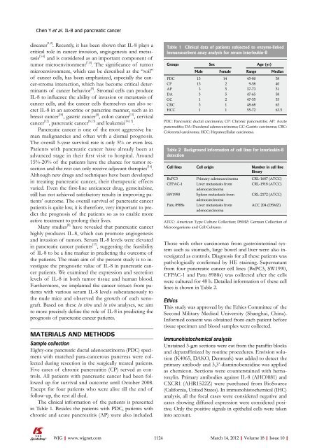
The clinical information <strong>of</strong> the patients is presented<br />
in Table 1. Besides the patients with PDC, patients with<br />
chronic and acute pancreatitis (AP) were also included.<br />
WJG|www.wjgnet.com<br />
Table 1 Clinical data <strong>of</strong> patients subjected to enzyme-linked<br />
immunosorbent assay analysis for serum interleukin-8<br />
Groups Sex Age (yr)<br />
Male Female Range Median<br />
PDC 13 14 45-80 58<br />
CP 13 2 9-58 40<br />
AP 3 5 37-73 51<br />
DA 3 5 47-65 58<br />
GC 1 2 47-55 53<br />
CRC 3 1 48-68 63<br />
HCC 1 1 55-72 63.5<br />
PDC: Pancreatic ductal carcinoma; CP: Chronic pancreatitis; AP: Acute<br />
pancreatitis; DA: Duodenal adenocarcinoma; GC: Gastric carcinoma; CRC:<br />
Colorectal carcinoma; HCC: Hepatocellular carcinoma.<br />
Table 2 Background information <strong>of</strong> cell lines for interleukin-8<br />
detection<br />
Cell lines Cell origin Number in cell line<br />
library<br />
BxPC3 Primary adenocarcinoma CRL-1687 (ATCC)<br />
CFPAC-1 Liver metastasis from<br />
adenocarcinoma<br />
CRL-1918 (ATCC)<br />
SW1990 Spleen metastasis from<br />
adenocarcinoma<br />
CRL-2172 (ATCC)<br />
Patu 8988s Liver metastasis from<br />
adenocarcinoma<br />
ACC 204 (DSMZ)<br />
ATCC: American Type Culture Collection; DSMZ: German Collection <strong>of</strong><br />
Microorganisms and Cell Cultures.<br />
Those with other carcinomas from gastrointestinal system<br />
such as stomach, large bowel and liver were also investigated<br />
as controls. Diagnosis for all these patients was<br />
pathologically confirmed by HE staining. Supernatant<br />
from four pancreatic cancer cell lines (BxPC3, SW1990,<br />
CFPAC-1 and Patu 8988s) was collected after the cells<br />
were cultured for 48 h. Detailed information <strong>of</strong> these cell<br />
lines is shown in Table 2.<br />
Ethics<br />
This study was approved by the Ethics Committee <strong>of</strong> the<br />
Second Military Medical University (Shanghai, China).<br />
Informed consent was obtained from each patient before<br />
tissue specimen and blood samples were collected.<br />
Immunohistochemical analysis<br />
Unstained 3-μm sections were cut from the paraffin blocks<br />
and deparaffinized by routine procedures. Envision solution<br />
(K4065, DAKO, Denmark) was added to detect the<br />
primary antibody and 3,3’-diaminobenzidine was applied<br />
as chemicon. Sections were counterstained with hematoxylin.<br />
Primary antibodies against IL-8 (AHC0881) and<br />
CXCR1 (AHR1522Z) were purchased from BioSource<br />
(California, United States). In immunohistochemical (IHC)<br />
analysis, all the focal cases were considered negative and<br />
cases showing diffused expression were considered positive.<br />
Only the positive signals in epithelial cells were taken<br />
into account.<br />
1124 March 14, 2012|Volume 18|Issue <strong>10</strong>|


