

10 - World Journal of Gastroenterology
10 - World Journal of Gastroenterology
10 - World Journal of Gastroenterology

Create successful ePaper yourself
Turn your PDF publications into a flip-book with our unique Google optimized e-Paper software.
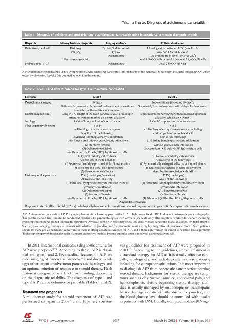
Table 1 Diagnosis <strong>of</strong> definitive and probable type 1 autoimmune pancreatitis using international consensus diagnostic criteria<br />
Diagnosis Primary basis for diagnosis Imaging evidence Collateral evidence<br />
Definitive type 1 AIP Histology Typical/indeterminate Histologically confirmed LPSP (level 1 H)<br />
Imaging Typical Any non-D level 1/level2<br />
indeterminate Two or more from level 1 (+ level 2 D 1 )<br />
Response to steroid Level 1 S/OOI + Rt or level 1 D + level 2 S/OOI/H + Rt<br />
Probable type 1 AIP Indeterminate Level 2 S/OOI/H + Rt<br />
AIP: Autoimmune pancreatitis; LPSP: Lymphoplasmacytic sclerosing pancreatitis; H: Histology <strong>of</strong> the pancreas; S: Serology; D: Ductal imaging; OOI: Other<br />
organ involvement. 1 Level 2 D is counted as level 1 in this setting.<br />
Table 2 Level 1 and level 2 criteria for type 1 autoimmune pancreatitis<br />
Criterion Level 1 Level 2<br />
Parenchymal imaging Typical: Indeterminate (including atypia 2 ):<br />
Diffuse enlargement with delayed enhancement (sometimes<br />
associated with rim-like enhancement)<br />
Segmental/focal enlargement with delayed enhancement<br />
Ductal imaging (ERP) Long (> 1/3 length <strong>of</strong> the main pancreatic duct or multiple<br />
strictures without marked up stream dilatation<br />
In 2011, international consensus diagnostic criteria for<br />
AIP were proposed [23] . According to these, AIP is classified<br />
into type 1 and 2. Five cardinal features <strong>of</strong> AIP are<br />
used: imaging <strong>of</strong> pancreatic parenchyma and ducts; serology;<br />
other organ involvement; pancreatic histology; and<br />
an optional criterion <strong>of</strong> response to steroid therapy. Each<br />
feature is categorized as a level 1 or 2 finding, depending<br />
on the diagnostic reliability. The diagnosis <strong>of</strong> type 1 and<br />
type 2 AIP can be definitive or probable (Tables 1 and 2).<br />
Treatment and prognosis<br />
A multicenter study for steroid treatment <strong>of</strong> AIP was<br />
performed in Japan in 2009 [24] , and Japanese consen-<br />
WJG|www.wjgnet.com<br />
Takuma K et al . Diagnosis <strong>of</strong> autoimmune pancreatitis<br />
Segmental/focal narrowing without marked upstream<br />
dilatation (duct size, < 5 mm )<br />
Serology IgG4, > 2x upper limit <strong>of</strong> normal value IgG4, 1-2x upper limit <strong>of</strong> normal value<br />
other organ involvement a or b a or b<br />
a: Histology <strong>of</strong> extrapancreatic organs a: Histology <strong>of</strong> extrapancreatic organs including<br />
Any three <strong>of</strong> the following: endoscopic biopsies <strong>of</strong> bile duct 3 :<br />
(1) Marked lymphoplasmacytic infiltration Both <strong>of</strong> the following:<br />
with fibrosis and without granulocytic infiltration (1) Marked lymphoplasmacytic infiltration<br />
(2) Storiform fibrosis without granulocytic infiltration<br />
(3) Obliterative phlebitis (2) Abundant (> <strong>10</strong> cells/HPF) IgG-positive cells<br />
(4) Abundant (> <strong>10</strong> cells/HPF) IgG4-positive cells<br />
b: Typical radiological evidence b: Physical or radiological evidence<br />
At least one <strong>of</strong> the following: At least one <strong>of</strong> the following:<br />
(1) Segmental/multiple proximal (hilar/intrahepatic) (1) Symmetrically enlarged salivary/lachrymal glands<br />
or proximal and distal bile duct stricture (2) Radiological evidence <strong>of</strong> renal involvement<br />
(2) Retroperitoneal fibrosis described in association with AIP<br />
Histology <strong>of</strong> the pancreas LPSP (core biopsy/resection) LPSP (core biopsy)<br />
At least 3 <strong>of</strong> the following: Any 2 <strong>of</strong> the following:<br />
(1) Periductal lymphoplasmacytic infiltrate without (1) Periductal lymphoplasmacytic infiltrate without<br />
grnulocytic infiltration grnulocytic infiltration<br />
(2) Obliterative phlebitis (2) Obliterative phlebitis<br />
(3) Storiform fibrosis (3) Storiform fibrosis<br />
(4) Abundant (> <strong>10</strong> cells/HPF) IgG4-positive cells (4) Abundant (> <strong>10</strong> cells/HPF) IgG4-positive cells<br />
Diagnostic steroid trial<br />
Response to steroid (Rt) 1 Rapid (≤ 2 wk) radiologicallydemonstrable resolution or marked improvement in pancreatic/extrapancreatic manifestations<br />
AIP: Autoimmune pancreatitis; LPSP: Lymphoplasmacytic sclerosing pancreatitis; HPF: High power field; ERP: Endoscopic retrograde pancreatography.<br />
1 Diagnostic steroid trial should be conducted carefully by pancreatologists with caveats (see text) only after negative workup for cancer including<br />
endoscopic ultrasound-guided fine needle aspiration; 2 Atypical: Some AIP cases may show low-density mass pancreatic ductal dilatation, or distal atrophy.<br />
Such atypical imaging findings in patients with obstructive jaundice and/or pancreatic mass are highly suggestive <strong>of</strong> pancreatic cancer. Such patients<br />
should be managed as pancreatic cancer unless there is strong collateral evidence for AIP, and a thorough workup for cancer is negative (see algorithm);<br />
3 Endoscopic biopsy <strong>of</strong> duodenal papilla is a useful adjunctive method because ampulla <strong>of</strong>ten is involved pathologically in AIP.<br />
sus guidelines for treatment <strong>of</strong> AIP were proposed in<br />
20<strong>10</strong> [25] . According to the guidelines, steroid treatment is<br />
a standard therapy for AIP, as it is usually effective clinically,<br />
serologically, and radiologically in these patients,<br />
including for extrapancreatic lesions. It is most important<br />
to distinguish AIP from pancreatic cancer before starting<br />
steroid therapy. Indications for steroid therapy are symptoms<br />
such as obstructive jaundice, abdominal pain, and<br />
hydronephrosis. Before beginning steroid therapy, jaundice<br />
is usually managed by endoscopic or transhepatic<br />
biliary drainage in patients with obstructive jaundice, and<br />
the blood glucose level should be controlled with insulin<br />
in patients with DM. Initially, oral prednisolone (0.6 mg/<br />
<strong>10</strong>17 March 14, 2012|Volume 18|Issue <strong>10</strong>|


