

10 - World Journal of Gastroenterology
10 - World Journal of Gastroenterology
10 - World Journal of Gastroenterology

Create successful ePaper yourself
Turn your PDF publications into a flip-book with our unique Google optimized e-Paper software.
Survival distribution function<br />
1.00<br />
0.75<br />
0.50<br />
0.25<br />
0.00<br />
Statistical analysis<br />
All the data were presented as mean ± SD. The survival<br />
distribution function was evaluated by the Kaplan-Meier<br />
survival curve and the others were determined using<br />
Students T-test. These analysis were performed with the<br />
statistical s<strong>of</strong>tware SAS 9.1.3 (SAS Institute Inc., North<br />
Carolina State University, United States), and UPLC-MS<br />
data were analyzed by PLS-DA with the SIMCA-P+ 12<br />
S<strong>of</strong>tware (Umetrics, Sweden). A P-value <strong>of</strong> less than 0.05<br />
was considered statistically significant.<br />
RESULTS<br />
0 50 <strong>10</strong>0 150 200 250 300 350 400<br />
t /d<br />
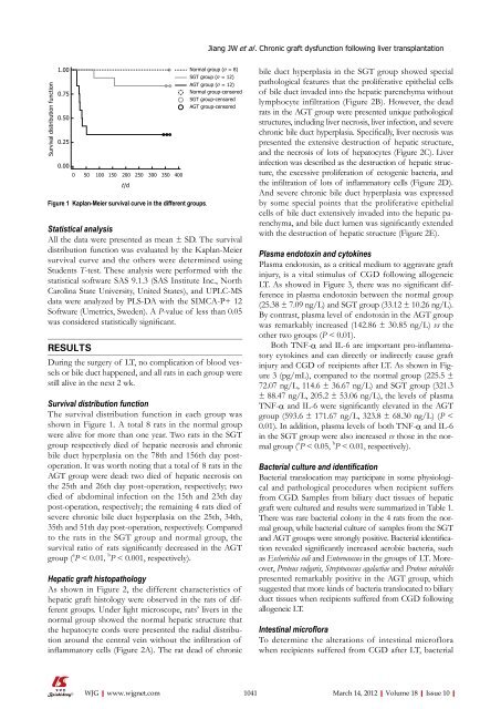
Figure 1 Kaplan-Meier survival curve in the different groups.<br />
During the surgery <strong>of</strong> LT, no complication <strong>of</strong> blood vessels<br />
or bile duct happened, and all rats in each group were<br />
still alive in the next 2 wk.<br />
Survival distribution function<br />
The survival distribution function in each group was<br />
shown in Figure 1. A total 8 rats in the normal group<br />
were alive for more than one year. Two rats in the SGT<br />
group respectively died <strong>of</strong> hepatic necrosis and chronic<br />
bile duct hyperplasia on the 78th and 156th day postoperation.<br />
It was worth noting that a total <strong>of</strong> 8 rats in the<br />
AGT group were dead: two died <strong>of</strong> hepatic necrosis on<br />
the 25th and 26th day post-operation, respectively; two<br />
died <strong>of</strong> abdominal infection on the 15th and 23th day<br />
post-operation, respectively; the remaining 4 rats died <strong>of</strong><br />
severe chronic bile duct hyperplasia on the 25th, 34th,<br />
35th and 51th day post-operation, respectively. Compared<br />
to the rats in the SGT group and normal group, the<br />
survival ratio <strong>of</strong> rats significantly decreased in the AGT<br />
group ( a P < 0.01, b P < 0.001, respectively).<br />
Hepatic graft histopathology<br />
As shown in Figure 2, the different characteristics <strong>of</strong><br />
hepatic graft histology were observed in the rats <strong>of</strong> different<br />
groups. Under light microscope, rats’ livers in the<br />
normal group showed the normal hepatic structure that<br />
the hepatocyte cords were presented the radial distribution<br />
around the central vein without the infiltration <strong>of</strong><br />
inflammatory cells (Figure 2A). The rat dead <strong>of</strong> chronic<br />
WJG|www.wjgnet.com<br />
Jiang JW et al . Chronic graft dysfunction following liver transplantation<br />
Normal group (n = 8)<br />
SGT group (n = 12)<br />
AGT group (n = 12)<br />
Normal group-censored<br />
SGT group-censored<br />
AGT group-censored<br />
bile duct hyperplasia in the SGT group showed special<br />
pathological features that the proliferative epithelial cells<br />
<strong>of</strong> bile duct invaded into the hepatic parenchyma without<br />
lymphocyte infiltration (Figure 2B). However, the dead<br />
rats in the AGT group were presented unique pathological<br />
structures, including liver necrosis, liver infection, and severe<br />
chronic bile duct hyperplasia. Specifically, liver necrosis was<br />
presented the extensive destruction <strong>of</strong> hepatic structure,<br />
and the necrosis <strong>of</strong> lots <strong>of</strong> hepatocytes (Figure 2C). Liver<br />
infection was described as the destruction <strong>of</strong> hepatic structure,<br />
the excessive proliferation <strong>of</strong> ectogenic bacteria, and<br />
the infiltration <strong>of</strong> lots <strong>of</strong> inflammatory cells (Figure 2D).<br />
And severe chronic bile duct hyperplasia was expressed<br />
by some special points that the proliferative epithelial<br />
cells <strong>of</strong> bile duct extensively invaded into the hepatic parenchyma,<br />
and bile duct lumen was significantly extended<br />
with the destruction <strong>of</strong> hepatic structure (Figure 2E).<br />
Plasma endotoxin and cytokines<br />
Plasma endotoxin, as a critical medium to aggravate graft<br />
injury, is a vital stimulus <strong>of</strong> CGD following allogeneic<br />
LT. As showed in Figure 3, there was no significant difference<br />
in plasma endotoxin between the normal group<br />
(25.38 ± 7.09 ng/L) and SGT group (33.12 ± <strong>10</strong>.26 ng/L).<br />
By contrast, plasma level <strong>of</strong> endotoxin in the AGT group<br />
was remarkably increased (142.86 ± 30.85 ng/L) vs the<br />
other two groups (P < 0.01).<br />
Both TNF-α and IL-6 are important pro-inflammatory<br />
cytokines and can directly or indirectly cause graft<br />
injury and CGD <strong>of</strong> recipients after LT. As shown in Figure<br />
3 (pg/mL), compared to the normal group (225.5 ±<br />
72.07 ng/L, 114.6 ± 36.67 ng/L) and SGT group (321.3<br />
± 88.47 ng/L, 205.2 ± 53.06 ng/L), the levels <strong>of</strong> plasma<br />
TNF-α and IL-6 were significantly elevated in the AGT<br />
group (593.6 ± 171.67 ng/L, 323.8 ± 68.30 ng/L) (P <<br />
0.01). In addition, plasma levels <strong>of</strong> both TNF-α and IL-6<br />
in the SGT group were also increased vs those in the normal<br />
group ( a P < 0.05, b P < 0.01, respectively).<br />
Bacterial culture and identification<br />
Bacterial translocation may participate in some physiological<br />
and pathological procedures when recipient suffers<br />
from CGD. Samples from biliary duct tissues <strong>of</strong> hepatic<br />
graft were cultured and results were summarized in Table 1.<br />
There was rare bacterial colony in the 4 rats from the normal<br />
group, while bacterial culture <strong>of</strong> samples from the SGT<br />
and AGT groups were strongly positive. Bacterial identification<br />
revealed significantly increased aerobic bacteria, such<br />
as Escherichia coli and Enterococcus in the groups <strong>of</strong> LT. Moreover,<br />
Proteus vulgaris, Streptococcus agalactiae and Proteus mirabilis<br />
presented remarkably positive in the AGT group, which<br />
suggested that more kinds <strong>of</strong> bacteria translocated to biliary<br />
duct tissues when recipients suffered from CGD following<br />
allogeneic LT.<br />
Intestinal micr<strong>of</strong>lora<br />
To determine the alterations <strong>of</strong> intestinal micr<strong>of</strong>lora<br />
when recipients suffered from CGD after LT, bacterial<br />
<strong>10</strong>41 March 14, 2012|Volume 18|Issue <strong>10</strong>|


