MDR1/P-glycoprotein and MRP-1 mRNA and Protein Expression in ...
MDR1/P-glycoprotein and MRP-1 mRNA and Protein Expression in ...
MDR1/P-glycoprotein and MRP-1 mRNA and Protein Expression in ...

You also want an ePaper? Increase the reach of your titles
YUMPU automatically turns print PDFs into web optimized ePapers that Google loves.
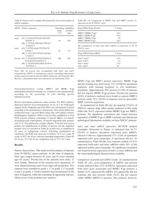
Table II. Primers used to amplify cDNA formed by reverse transcription of<br />
<strong>mRNA</strong> templates.<br />
cDNA Primer sequences Anneal<strong>in</strong>g Amplified<br />
temp. product<br />
(ÆC) (bp)<br />
mrp1 (F): 5’ GTACATTAACATGATC<br />
TGGTC 3’ 54 203<br />
(R): 5’ CGTTCATCAGCTTGAT<br />
CCGAT 3’<br />
mdr1 (F): 5’ GTTCAAACTTCTGCTC<br />
CTGA 3’ 55 157<br />
(R): 5’ CCCATCATTGCAATAG<br />
CAGG 3’<br />
‚-act<strong>in</strong> (F): 5’ GAAATCGTGCGTGACA AR 383<br />
TTAAGGAGAAGCT 3’<br />
(R): 5’ TCAGGAGGAGCAATGA<br />
TCTTGA 3’<br />
Note AR: As ‚-act<strong>in</strong> was co-amplified with mrp1 <strong>and</strong> mdr1<br />
(respectively) cDNAs as endogenous control, anneal<strong>in</strong>g temperature<br />
(temp.) used was that of relevant cDNA of <strong>in</strong>terest. (F): Forward; (R):<br />
Reverse. (All primers used were selected <strong>in</strong> our laboratory (19)).<br />
Immunohistochemical scor<strong>in</strong>g. <strong>MRP</strong>-1 <strong>and</strong> <strong>MDR1</strong> P-gp<br />
immunohistochemical sta<strong>in</strong><strong>in</strong>g was evaluated semi-quantitatively,<br />
accord<strong>in</strong>g to the percentage of cells show<strong>in</strong>g specific<br />
immunoreactivity.<br />
Reverse transcriptase-polymerase cha<strong>in</strong> reaction. For RNA analyses,<br />
dissected tumours were homogenised, on ice, <strong>in</strong> 2 ml TriReagent<br />
(Sigma; Poole, Engl<strong>and</strong>) <strong>and</strong> total RNA was subsequently isolated<br />
accord<strong>in</strong>g to the manufacturer's <strong>in</strong>structions. First-str<strong>and</strong> cDNA was<br />
synthesised from 1 Ìg total RNA us<strong>in</strong>g oligo (dT) primer (Oswel;<br />
Southampton, Engl<strong>and</strong>). cDNA (5 Ìl) was then amplified <strong>in</strong> a 50 Ìl<br />
PCR reaction solution conta<strong>in</strong><strong>in</strong>g 1.5 mmol/l MgCl 2 , 0.2 mmol/l<br />
deoxynucleotide triphosphates, 20 Ìmol/l oligonucleotide primers<br />
<strong>and</strong> 2.5 U Taq polymerase enzyme (Sigma). Forward <strong>and</strong> reverse<br />
primers for amplification of cDNAs of <strong>in</strong>terest <strong>and</strong> the amplified<br />
product sizes are detailed <strong>in</strong> Table II. ‚-act<strong>in</strong> was co-amplified <strong>in</strong><br />
all cases as endogenous control. Follow<strong>in</strong>g optimisation of<br />
conditions, the PCR cycle used was as follows: 94ÆC for 2 m<strong>in</strong>; 30<br />
cycles of 94ÆC for 30 sec, relevant anneal<strong>in</strong>g temperature (Table II)<br />
for 30 sec, 72ÆC for 30 sec; completion step of 72ÆC for 5 m<strong>in</strong>.<br />
Results<br />
Patient characteristics. This study <strong>in</strong>volved analysis of tumours<br />
from 38 NSCLC cancer patients. At the time of diagnosis,<br />
their ages ranged between 41 years <strong>and</strong> 81 years (median<br />
age=65 years). Twenty-one of the patients were male; 17<br />
were female. N<strong>in</strong>eteen of the tumours were squamous, 15<br />
were adenocarc<strong>in</strong>omas <strong>and</strong> 4 were large cell carc<strong>in</strong>omas. Two<br />
tumours were classified as grade 1, 17 as grade 2, 17 as grade<br />
3 <strong>and</strong> 2 as grade 4. Twelve tumours had metastasised at the<br />
time of diagnosis, while the rema<strong>in</strong><strong>in</strong>g 26 apparently had not.<br />
Thirty-seven of the patients were smokers.<br />
Roy et al: Multiple Drug Resistance <strong>in</strong> Lung Cancer<br />
Table III. (A) Comparison of <strong>MDR1</strong> P-gp <strong>and</strong> <strong>MRP</strong>-1 prote<strong>in</strong> coexpression<br />
<strong>in</strong> all 38 NSCLC cases.<br />
Drug Efflux Pump <strong>Prote<strong>in</strong></strong> % Cases<br />
<strong>MRP</strong>1 + /<strong>MDR1</strong> P-gp + 18.4<br />
<strong>MRP</strong>1 – /<strong>MDR1</strong> P-gp + 52.6<br />
<strong>MRP</strong>1 + /<strong>MDR1</strong> P-gp – 2.6<br />
<strong>MRP</strong>1 – /<strong>MDR1</strong> P-gp – 26.4<br />
(B) Comparison of mdr1 <strong>and</strong> mrp1 <strong>mRNA</strong> co-expression <strong>in</strong> all 30<br />
NSCLC cases.<br />
Drug Efflux Pump <strong>mRNA</strong> % Cases<br />
mrp1 + /mdr1 + 13.3<br />
mrp1 – /mdr1 + 3.3<br />
mrp1 + /mdr1 – 73.4<br />
mrp1 – /mdr1 – 10<br />
<strong>MDR1</strong> P-gp <strong>and</strong> <strong>MRP</strong>-1 prote<strong>in</strong> expression. <strong>MDR1</strong> P-gp<br />
specific sta<strong>in</strong><strong>in</strong>g was observed <strong>in</strong> 71% of NSCLC specimens<br />
analysed, with sta<strong>in</strong><strong>in</strong>g localised to cell membrane/<br />
cytoplasm. Approximately 29% percent (11/38) of tumours<br />
did not express <strong>MDR1</strong> P-gp prote<strong>in</strong>. Twenty-one percent<br />
(8/38) of tumours analysed were found to express <strong>MRP</strong>-1<br />
prote<strong>in</strong>, while 79% (30/38) of tumours had no detectable<br />
<strong>MRP</strong>-1 prote<strong>in</strong> expression.<br />
As summarised <strong>in</strong> Table III (A), the majority (73.6%) of<br />
NSCLCs express drug efflux pumps analysed <strong>in</strong> this study<br />
while only 26.4% expressed neither <strong>MDR1</strong> P-gp nor <strong>MRP</strong>-1<br />
prote<strong>in</strong>s. No significant association was found between<br />
expression of <strong>MDR1</strong> P-gp or <strong>MRP</strong>-1 prote<strong>in</strong>s <strong>and</strong> cl<strong>in</strong>ical <strong>and</strong><br />
pathological <strong>in</strong>formation available on these NSCLC patients.<br />
mdr1 <strong>and</strong> mrp1 <strong>mRNA</strong> expression. RT-PCR analysis<br />
(examples illustrated <strong>in</strong> Figure 1) <strong>in</strong>dicated that 86.7%<br />
(26/30) of tumour specimens expressed mrp1 <strong>mRNA</strong>,<br />
whereas 4 did not. Approximately 17% (5/30) of specimens<br />
expressed mdr1 gene transcripts, 25 specimens did not. As<br />
summarised <strong>in</strong> Table III (B), four tumours studied (13.3%)<br />
expressed both mdr1 <strong>and</strong> mrp1 <strong>mRNA</strong>s while 10% (3/30)<br />
expressed neither gene transcripts. No significant correlation<br />
was found between expression of mdr1 or mrp1 <strong>mRNA</strong>s <strong>and</strong><br />
the cl<strong>in</strong>icopathological characteristics available.<br />
Comparison of prote<strong>in</strong> <strong>and</strong> <strong>mRNA</strong> results. As summarised <strong>in</strong><br />
Table IV (A), cross-comparison of <strong>mRNA</strong> <strong>and</strong> prote<strong>in</strong><br />
results <strong>in</strong>dicated that 4.3% of NSCLC specimens expressed<br />
both mdr1 gene transcripts <strong>and</strong> <strong>MDR1</strong> P-gp prote<strong>in</strong>; a<br />
further 4.3% expressed the <strong>mRNA</strong>, but apparently did not<br />
translate this <strong>in</strong>to prote<strong>in</strong>; while 30.4% did not express<br />
either <strong>MDR1</strong> <strong>mRNA</strong> or prote<strong>in</strong>. This analysis, however,<br />
1327