B-The Chemical & Physical Structure of Merino Wool.cdr - csiro
B-The Chemical & Physical Structure of Merino Wool.cdr - csiro
B-The Chemical & Physical Structure of Merino Wool.cdr - csiro
- TAGS
- merino
- csiro
- www.csiro.au

You also want an ePaper? Increase the reach of your titles
YUMPU automatically turns print PDFs into web optimized ePapers that Google loves.
<strong>The</strong> <strong>Chemical</strong> & <strong>Physical</strong> <strong>Structure</strong> <strong>of</strong> <strong>Merino</strong> <strong>Wool</strong><br />
<strong>The</strong>se contaminants are removed during processing.<br />
Clean wool, together with other animal fibres,<br />
belongs to a group <strong>of</strong> proteins known as keratins.<br />
Unlike cotton and the majority <strong>of</strong> synthetic fibres,<br />
wool does not have a homogeneous structure. <strong>Wool</strong><br />
fibres have highly complex physical and chemical<br />
compositions that have evolved over millions <strong>of</strong><br />
years to protect sheep from extremes <strong>of</strong> heat and<br />
cold.<br />
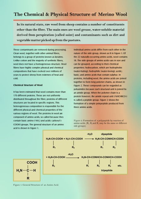
In its natural state, raw wool from sheep contains a number <strong>of</strong> constituents<br />
other than the fibre. <strong>The</strong> main ones are wool grease, water-soluble material<br />
derived from perspiration (called suint) and contaminants such as dirt and<br />
vegetable matter picked up from the pastures.<br />
<strong>Chemical</strong> <strong>Structure</strong> <strong>of</strong> <strong>Wool</strong><br />
It has been estimated that wool contains more than<br />
170 different proteins. <strong>The</strong>se are not uniformly<br />
distributed throughout the fibre; proteins <strong>of</strong> different<br />
structures are located in specific regions. This<br />
heterogeneous composition is responsible for the<br />
different physical and chemical properties <strong>of</strong> the<br />
various regions <strong>of</strong> wool. <strong>The</strong> proteins in wool are<br />
composed <strong>of</strong> amino acids; so called because they<br />
contain basic amino (-NH ) and acidic carboxyl (-<br />
2<br />
COOH) groups. <strong>The</strong> general structure <strong>of</strong> an amino<br />
acid is shown in Figure 1.<br />
COOH<br />
HN C H<br />
2<br />
R<br />
Figure 1: General <strong>Structure</strong> <strong>of</strong> an Amino Acid.<br />
H2N-CH-COOH + H2N-CH-COOH<br />
Individual amino acids differ from each other in the<br />
nature <strong>of</strong> the side group, shown as R in Figure 1. Of<br />
the 22 naturally-occurring amino acids, wool contains<br />
18. <strong>The</strong> side groups <strong>of</strong> amino acids vary in size and<br />
can be grouped, according to their chemical<br />
properties: hydrocarbon, which are hydrophobic<br />
(water-hating); hydrophilic (water-loving); acidic;<br />
basic; and amino acids that contain sulphur. In<br />
proteins, including wool, the amino acids are joined<br />
together to form long polymer chains, as shown in<br />
Figure 2. <strong>The</strong>se compounds can be regarded as<br />
polyamides because each structural unit is joined by<br />
an amide group. When the polymer chain is a<br />
protein however, the amide repeat unit (-NHCHRCO-)<br />
is called a peptide group. Figure 2 shows the<br />
formation <strong>of</strong> a simple polypeptide produced from<br />
three amino acids.<br />
Figure 2: Formation <strong>of</strong> a polypeptide by reaction <strong>of</strong><br />
amino acids. (R 1, R2 and R 3 may be the same or different<br />
side groups).<br />
-H2O dipeptide<br />
H2N-CH-CONHCH-COOH<br />
R1 R2 R1 R2<br />
-H2O<br />
+ H2N-CH-COOH H2N-CH-CONHCH-CONHCH-COOH<br />
R3<br />
R1 R2 R3 etc.<br />
tripeptide
HN<br />
intermolecular<br />
disulfide crosslink<br />
HN<br />
Figure 3: Bonds in <strong>Wool</strong>.<br />
In wool, individual polypeptide chains are joined<br />
together to form proteins by a variety <strong>of</strong> covalent<br />
(chemical bonds), called crosslinks, and non-covalent<br />
physical interactions (Figure 3).<br />
S<br />
S<br />
O<br />
CH 2<br />
HN<br />
CH 2<br />
<strong>The</strong> most important crosslinks are the sulphur-<br />
containing disulphide bonds, which are formed<br />
during fibre growth by a process called<br />
“keratinisation”. <strong>The</strong>se make keratin fibres insoluble<br />
in water and more stable to chemical and physical<br />
attack than other types <strong>of</strong> proteins. Disulphide bonds<br />
are involved in the chemical reactions that occur in<br />
the 'setting' <strong>of</strong> fabrics during finishing. In this<br />
process, disulphide crosslinks are rearranged to give<br />
wool fabrics smooth-drying properties so that ironing<br />
is not required after laundering. Another type <strong>of</strong><br />
crosslink is the isopeptide bond, formed between<br />
amino acids containing acidic or basic groups. In<br />
addition to the chemical crosslinks, some other types<br />
<strong>of</strong> interactions also help to stabilize the fibre under<br />
both wet and dry conditions. <strong>The</strong>se arise from<br />
interactions between the side groups <strong>of</strong> the amino<br />
acids that constitute wool proteins. Thus,<br />
hydrophobic interactions occur between<br />
hydrocarbon side groups; and ionic interactions<br />
H<br />
N<br />
occur between groups that can exchange protons.<br />
<strong>The</strong>se ionic interactions or 'salt linkages' between<br />
acidic (carboxyl) and basic (amino) side chains are<br />
the most important <strong>of</strong> the non-covalent interactions.<br />
<strong>The</strong> most important <strong>of</strong> the non-covalent interactions<br />
are the ionic, or 'salt linkages' between acidic<br />
(carboxyl) and basic (amino) side groups. <strong>The</strong><br />
O<br />
R<br />
R<br />
O<br />
H<br />
N<br />
CH<br />
O R<br />
N<br />
H<br />
H 3C CH 3<br />
hydrophobic interaction<br />
O<br />
N<br />
H<br />
CH 2<br />
H<br />
N<br />
O R<br />
H<br />
N<br />
O CH 2<br />
O<br />
H 2C<br />
H 2C<br />
N<br />
H<br />
H 2C<br />
CH 2<br />
O<br />
NH 3<br />
COO<br />
O<br />
Figure 4: Amphoteric behaviour <strong>of</strong> wool.<br />
carboxyl and amino groups in wool are also<br />
important because they give wool its amphoteric or<br />
pH buffering properties. This is its ability to absorb<br />
and desorb both acids and alkalis, as shown in<br />
Figure 4. <strong>The</strong> ionic groups also control the dyeing<br />
behaviour <strong>of</strong> the fibre, as a result <strong>of</strong> their<br />
interactions with negatively charged dye molecules.<br />
<strong>The</strong> <strong>Physical</strong> <strong>Structure</strong> <strong>of</strong> <strong>Wool</strong><br />
In addition to its chemical complexity, wool also has<br />
a very complex physical structure, as shown<br />
schematically in Figure 5. A wool fibre can be<br />
considered as a biological composite consisting <strong>of</strong><br />
regions that are both chemically and physically<br />
different.<br />
N<br />
H<br />
H<br />
N<br />
O<br />
H<br />
N<br />
Australian merino wool fibres range in diameter<br />
typically from 17 to 25 m. <strong>The</strong>y are composed <strong>of</strong> two<br />
types <strong>of</strong> cell: the internal cells <strong>of</strong> the cortex and<br />
external cuticle cells that form a sheath around the<br />
fibre, shown in Figure 5.<br />
R O R<br />
O<br />
ionic<br />
interaction<br />
O<br />
H 2C<br />
C<br />
H 2C<br />
N<br />
H<br />
CH 2<br />
NH<br />
CH 2<br />
CH 2<br />
N<br />
H<br />
H<br />
N<br />
R O R<br />
H +<br />
H3N + - WOOL – COOH H3N + - WOOL – COO -<br />
isopeptide<br />
crosslink<br />
OH -<br />
O<br />
O<br />
H<br />
N<br />
O<br />
H 2C<br />
N<br />
H<br />
H<br />
CH<br />
O<br />
C<br />
C<br />
H2N – WOOL – COO -<br />
acidic isoelectric (neutral) basic
c<br />
CSIRO Textile & Fibre Technology<br />
Graphics by H.Z. Roe, 1992<br />
based on a drawing by<br />
R.D.B. Fraser, 1972.<br />
right<br />
handed<br />
a - helix<br />
low-S<br />
proteins<br />
left<br />
handed<br />
coiled-coil<br />
rope<br />
high-S<br />
proteins<br />
high-tyr<br />
proteins<br />
matrix<br />
intermediate<br />
filament<br />
(micr<strong>of</strong>ibril)<br />
1 2 7<br />
Cuticle cells (or scales), which overlap like tiles on a<br />
ro<strong>of</strong>, make wool unique amongst textile fibres. <strong>The</strong><br />
complex physical structure <strong>of</strong> cuticle cells is shown<br />
in Figure 6. An important function <strong>of</strong> cuticle cells is<br />
to anchor wool fibres in the skin <strong>of</strong> sheep. <strong>The</strong><br />
exposed edge <strong>of</strong> each cuticle cell points from the<br />
fibre root towards the tip. This gives rise to a larger<br />
surface frictional value when a fibre is drawn in the<br />
against-scale direction than in the with-scale<br />
direction. <strong>The</strong> frictional difference helps to expel dirt<br />
and other contaminants from the fleece, but it is also<br />
responsible for wool's property <strong>of</strong> felting when<br />
agitated in water. This characteristic, which is not<br />
shared by any other textile fibre, enables fabrics with<br />
very dense structures to be produced, such as<br />
blankets, felts and overcoat materials. When felting is<br />
regarded as undesirable (for example in knitted<br />
garments that will be machine-washed), processes<br />
are available to remove the frictional difference and<br />
make wool shrinkresistant. <strong>The</strong> fibre surface is also<br />
largely responsible for the natural s<strong>of</strong>tness <strong>of</strong> wool<br />
and its property as one <strong>of</strong> the smoothest textile<br />
fibres.<br />
Even after the natural wool grease has been<br />
removed by scouring with a detergent, wool fibres<br />
are relatively difficult to wet compared with other<br />
textile materials. This natural water repellency makes<br />
wool fabrics 'shower-pro<strong>of</strong> ' and able to resist water-<br />
cell<br />
membrane<br />
complex<br />
cortex<br />
root end<br />
para-cortical<br />
ortho-cortical<br />
cell<br />
cell<br />
macr<strong>of</strong>ibril meso-cortical cell<br />
200<br />
Figure 5: CSIRO schematic diagram <strong>of</strong> wool fibre.<br />
nuclear<br />
remnant<br />
2 000<br />
epicuticle<br />
exocuticle<br />
a endocuticle<br />
cuticle<br />
20 000 nm<br />
based stains. This property is the result <strong>of</strong> a waxy,<br />
hydrocarbon coating that is chemically bound to the<br />
surface <strong>of</strong> each scale. <strong>The</strong> coating survives processes<br />
such as dyeing and can only be removed by a severe<br />
chemical treatment.<br />
Figure 6: SEM <strong>of</strong> wool fibre.
<strong>The</strong> cortex <strong>of</strong> wool comprises approximately 90%<br />
<strong>of</strong> the fibre. It consists <strong>of</strong> overlapping spindle-<br />
shaped cells cortical cells, shown schematically in<br />
Figure 7. Both the cuticle and cortical cells have<br />
highly complex substructures, as shown in<br />
Figure 5.<br />
Cortical cells are held together by the cell<br />
membrane complex (CMC), which also separates<br />
cortical cells from those <strong>of</strong> the cuticle. <strong>The</strong> CMC is<br />
a continuous region, containing relatively lightly-<br />
crosslinked proteins and waxy lipids, that extends<br />
throughout the whole fibre. Although it comprise<br />
only around 5% <strong>of</strong> the total fibre mass, it plays an<br />
important role in the overall properties <strong>of</strong> wool. It<br />
is a region <strong>of</strong> relatively low mechanical strength in<br />
the fibre composite. When wool worsted fabrics<br />
are abraded during prolonged wear, breakdown<br />
tends to occur mainly by fracture along the<br />
boundaries between cortical cells, resulting in<br />
fibrillation. Figure 8 shows separation <strong>of</strong> individual<br />
cortical cells in a fibre taken from a severely<br />
abraded fabric.<br />
Cuticle Cells<br />
Figure 7: Schematic <strong>of</strong> a wool fibre showing cuticle<br />
and cortical cells.<br />
Because the CMC is only slightly crosslinked, it is<br />
also more susceptible to chemical attack. than<br />
other regions <strong>of</strong> the fibre; for example if strongly<br />
alkaline conditions or very high temperatures are<br />
Cell Membrane Complex<br />
Cortical Cells<br />
used during fabric manufacturing processes. Being<br />
the only continuous phase in the fibre, it also<br />
provides a channel by which dyes and chemicals can<br />
diffuse in and out <strong>of</strong> wool.<br />
Fine wool fibres contain two main types <strong>of</strong> cortical<br />
cell (ortho- and para-). In the case <strong>of</strong> merino wool,<br />
these are arranged bilaterally. Coarser types <strong>of</strong> wool<br />
(diameters >25 m) tend to have less distinct<br />
segmentation <strong>of</strong> the two types <strong>of</strong> cortical cells. <strong>The</strong><br />
bilateral segmentation <strong>of</strong> merino wool is associated<br />
with the highly desirable natural crimp <strong>of</strong> the fibres.<br />
An interesting feature is that the orthocortex is<br />
always orientated towards the outside radius <strong>of</strong> the<br />
Figure 8: SEM showing fibre fibrillation along cortical<br />
cell boundaries following prolonged abrasion.
Orthocortex<br />
Orthocortex<br />
Paracortex<br />
Paracortex<br />
Side view End view<br />
Figure 9: Diagram showing relationship between<br />
ortho/para segmentation and crimp in a merino fibre.<br />
crimp. This occurs as a result <strong>of</strong> the two segments<br />
rotating around the fibre in phase with the crimp, as<br />
shown in Figure 4.<br />
<strong>The</strong> structure <strong>of</strong> the proteins in wool differs between<br />
the various regions <strong>of</strong> the fibre. Some <strong>of</strong> the proteins<br />
in the micr<strong>of</strong>ibrils are helical, like a spring, which<br />
gives wool its flexibility, elasticity, resilience and<br />
good wrinkle recovery properties. Other proteins,<br />
particularly in the matrix that surrounds the<br />
micr<strong>of</strong>ibrils, have a more amorphous structure and<br />
are responsible for wool's advantage over other<br />
fibres <strong>of</strong> absorbing a relatively large amount <strong>of</strong> water<br />
without feeling wet (up to around 30% <strong>of</strong> the mass<br />
<strong>of</strong> the dry fibre). <strong>The</strong> matrix proteins are also<br />
responsible for wool's property <strong>of</strong> absorbing and<br />
retaining large amounts <strong>of</strong> dyestuffs.<br />
<strong>Wool</strong>, a fibre that has evolved over thousands <strong>of</strong><br />
years to insulate and protect sheep, is the most<br />
complex and versatile <strong>of</strong> all textile fibres. It can be<br />
used to make products as diverse as cloth for billiard<br />
tables to the finest woven and knitted fabrics. <strong>The</strong><br />
insulating and moisture absorbing properties <strong>of</strong> the<br />
fibre make fine wool products extremely comfortable<br />
to wear. <strong>The</strong> chemical composition <strong>of</strong> wool enables<br />
it to be easily dyed to shades ranging from pastels to<br />
full, rich colours. It is indeed justified to call wool:<br />
“Natures Wonder Fibre”.<br />
Further Reading:<br />
Rippon, J. A. (1992) <strong>The</strong> <strong>Structure</strong> <strong>of</strong> <strong>Wool</strong>; Chapter 1,<br />
In: <strong>Wool</strong> Dyeing, Lewis, D.M.<br />
(Ed.), Bradford (UK): Society <strong>of</strong> Dyers and Colourists<br />
Leeder, J. D. (1984) <strong>Wool</strong> - nature's wonder fibre, Ocean<br />
Grove, Vic.: Australasian Textiles Publishers,<br />
Morton, W.E. and Hearle, J.W.S., [1993] <strong>Physical</strong><br />
Properties <strong>of</strong> Textile Fibres, 3rd Ed., Manchester, UK.:<br />
<strong>The</strong> Textile Institute.<br />
Rippon, J. A. et al, (2003) <strong>Wool</strong>, in Encyclopedia <strong>of</strong><br />
Polymer Science and Technology, New York : Interscience<br />
Publishers.