Acne and Sebaceous Gland Function - Städtisches Klinikum Dessau
Acne and Sebaceous Gland Function - Städtisches Klinikum Dessau
Acne and Sebaceous Gland Function - Städtisches Klinikum Dessau

Create successful ePaper yourself
Turn your PDF publications into a flip-book with our unique Google optimized e-Paper software.
<strong>Acne</strong> <strong>and</strong> <strong>Sebaceous</strong> Gl<strong>and</strong> <strong>Function</strong><br />
CHRISTOS C. ZOUBOULIS, MD<br />
Abstract: The embryologic development of the human sebaceous gl<strong>and</strong> is closely related to the differentiation of the hair follicle<br />
<strong>and</strong> the epidermis. The number of sebaceous gl<strong>and</strong>s remains approximately the same throughout life, whereas their size tends to<br />
increase with age. The development <strong>and</strong> function of the sebaceous gl<strong>and</strong> in the fetal <strong>and</strong> neonatal periods appear to be regulated<br />
by maternal <strong>and</strong>rogens <strong>and</strong> by endogenous steroid synthesis, as well as by other morphogens. The most apparent function of the<br />
gl<strong>and</strong>s is to excrete sebum. A strong increase in sebum excretion occurs a few hours after birth; this peaks during the first week<br />
<strong>and</strong> slowly subsides thereafter. A new rise takes place at about age 9 years with adrenarche <strong>and</strong> continues up to age 17 years, when<br />
the adult level is reached. The sebaceous gl<strong>and</strong> is an important formation site of active <strong>and</strong>rogens. Androgens are well known for<br />
their effects on sebum excretion, whereas terminal sebocyte differentiation is assisted by peroxisome proliferator-activated receptor<br />
lig<strong>and</strong>s. Estrogens, glucocorticoids, <strong>and</strong> prolactin also influence sebaceous gl<strong>and</strong> function. In addition, stress-sensing cutaneous<br />
signals lead to the production <strong>and</strong> release of corticotrophin-releasing hormone from dermal nerves <strong>and</strong> sebocytes with subsequent<br />
dose-dependent regulation of sebaceous nonpolar lipids. Among other lipid fractions, sebaceous gl<strong>and</strong>s have been shown to<br />
synthesize considerable amounts of free fatty acids without exogenous influence. <strong>Sebaceous</strong> lipids are responsible for the<br />
three-dimensional skin surface lipid organization. Contributing to the integrity of the skin barrier. They also exhibit strong innate<br />
antimicrobial activity, transport antioxidants to the skin surface, <strong>and</strong> express proinflammatory <strong>and</strong> anti-inflammatory properties.<br />
<strong>Acne</strong> in childhood has been suggested to be strongly associated with the development of severe acne during adolescence. Increased<br />
sebum excretion is a major factor in the pathophysiology of acne vulgaris. Other sebaceous gl<strong>and</strong> functions are also associated with<br />
the development of acne, including sebaceous proinflammatory lipids; different cytokines produced locally; perigl<strong>and</strong>ular peptides<br />
<strong>and</strong> neuropeptides, such as corticotrophin-releasing hormone, which is produced by sebocytes; <strong>and</strong> substance P, which is expressed<br />
in the nerve endings at the vicinity of healthy-looking gl<strong>and</strong>s of acne patients. Current data indicate that acne vulgaris may be<br />
a primary inflammatory disease. Future drugs developed to treat acne not only should reduce sebum production <strong>and</strong> Propionibacterium<br />
acnes populations, but also should be targeted to reduce proinflammatory lipids in sebum, down-regulate<br />
proinflammatory signals in the pilosebaceous unit, <strong>and</strong> inhibit leukotriene B 4–induced accumulation of inflammatory cells. They<br />
should also influence peroxisome proliferator-activated receptor regulation. Isotretinoin is still the most active available drug for<br />
the treatment of severe acne.<br />
<strong>Sebaceous</strong> Gl<strong>and</strong> Development<br />
The human sebaceous gl<strong>and</strong> is a multiacinar, holocrine-secreting<br />
tissue present in all areas of the<br />
skin except for the palms <strong>and</strong> soles (<strong>and</strong> only<br />
sparsely on the dorsal surfaces of the h<strong>and</strong> <strong>and</strong> foot). 1<br />
Its development is closely related to the differentiation<br />
of the hair follicle <strong>and</strong> the epidermis. 2–4 By 13–15 weeks<br />
of fetal life, the sebaceous gl<strong>and</strong> is clearly distinguishable<br />
arising in a cephalocaudal sequence from the hair<br />
follicle. Lipid drops are visible at the center of the gl<strong>and</strong><br />
at 17 weeks. 5,6 The future common excretory duct,<br />
around which the acini of the sebaceous gl<strong>and</strong> attach,<br />
begins as a solid cord. The cells composing the cord are<br />
filled with sebum, <strong>and</strong> eventually they lose their integrity,<br />
rupture, <strong>and</strong> form a channel that establishes the<br />
first pilosebaceous canal. New acini result from buds on<br />
the peripheral sebaceous duct wall. The cell organization<br />
of the neonatal sebaceous acini consists of undifferentiated,<br />
differentiating, <strong>and</strong> mature sebocytes. 7<br />
From the Department of Dermatology, Charité University Medicine<br />
Berlin, Campus Benjamin Franklin, Berlin, Germany.<br />
Address correspondence to Christos C. Zouboulis, Department of Dermatology,<br />
Charité University Medicine Berlin, Campus Benjamin Franklin,<br />
Fabeckstrasse 60-62, 14195 Berlin, Germany.<br />
E-mail address: christos.zouboulis@charite.de<br />
The number of sebaceous gl<strong>and</strong>s remains approximately<br />
constant throughout life, whereas their size<br />
tends to increase with age. 8,9 Within any one gl<strong>and</strong>ular<br />
unit, the acini vary in differentiation <strong>and</strong> maturity. The<br />
sebaceous cells of prepupertal <strong>and</strong> hypogonadal males<br />
are qualitatively similar to those of normal adults, even<br />
though the gl<strong>and</strong>s are smaller. 10 Synthesis <strong>and</strong> discharge<br />
of the lipids contained in sebaceous cells takes<br />
more than 1 week. The turnover of sebaceous gl<strong>and</strong>s is<br />
slower in older than in young adults. 11<br />
<strong>Sebaceous</strong> Gl<strong>and</strong> <strong>Function</strong>s<br />
The most obvious function of the sebaceous gl<strong>and</strong> is to<br />
excrete sebum. Additional functions of the gl<strong>and</strong> are<br />
associated with the development of acne (Table 1).<br />
Sebum Production<br />
Sebum is a mixture of relatively nonpolar lipids, most<br />
of which are synthesized de novo by the sebaceous<br />
gl<strong>and</strong> 20 to coat the fur as a hydrophobic protection<br />
against overwetting <strong>and</strong> for heat insulation in mammals.<br />
21 The composition of sebum is remarkably species-specific.<br />
2,20,22 Increased sebum excretion is a major<br />
factor involved in the pathophysiology of acne. 23<br />
The sebaceous gl<strong>and</strong>s are functional from their for-<br />
© 2004 by Elsevier Inc. All rights reserved. 0738-081X/04/$–see front matter<br />
360 Park Avenue South, New York, NY 10010 doi:10.1016/j.clindermatol.2004.03.004
Clinics in Dermatology Y 2004;22:360–366 ACNE AND SEBACEOUS GLAND FUNCTION 361<br />
Table 1. <strong>Sebaceous</strong> gl<strong>and</strong> functions, which are possibly involved<br />
in the development of acne<br />
Production of sebum (12)<br />
Regulation of cutaneous steroidogenesis (13-15)<br />
Regulation of local <strong>and</strong>rogen synthesis (14)<br />
Interaction with neuropeptides (16)<br />
Synthesis of specific lipids with antimicrobial activity (17)<br />
Exhibition of pro- <strong>and</strong> anti-inflammatory properties (13, 18, 19)<br />
mation; sebum is the first demonstrable gl<strong>and</strong>ular<br />
product of the human body. 12 It is major ingredient of<br />
vernix caseosa, which progressively coats the fetus during<br />
the last trimester of gestation. Development <strong>and</strong><br />
function in the fetal <strong>and</strong> neonatal periods appear to be<br />
regulated by maternal <strong>and</strong>rogens <strong>and</strong> by endogenous<br />
steroid synthesis, as well as by other “morphogens,”<br />
including growth factors, cell adhesion molecules, extracellular<br />
matrix proteins, intracellular signaling molecules<br />
(�-catenin <strong>and</strong> LEF-1), other hormones, cytokines,<br />
enzymes, <strong>and</strong> retinoids. 4,24<br />
A significant increase in sebum excretion occurs a<br />
few hours after birth <strong>and</strong> peaks during the first<br />
week. 25,26 Maternal <strong>and</strong> neonatal sebum excretion rates<br />
directly correlate. 26 This correlation is lost in the following<br />
weeks <strong>and</strong> is independent from breast-feeding. At<br />
this time, the sebum level per unit skin surface is in the<br />
same range as in young adults, 25 <strong>and</strong> the sequence of<br />
sebaceous transformation seems identical to that in<br />
postnatal life. These events suggest an important role of<br />
the maternal hormonal environment on the neonatal<br />
sebaceous gl<strong>and</strong> <strong>and</strong> indicates that <strong>and</strong>rogenetic stimulus<br />
for sebum secretion occurs before birth through<br />
the placenta. 25,26 Sebum excretion then slowly subsides.<br />
A new rise occurs at about age 9 years, 27 with adrenarche<br />
<strong>and</strong> continues up to age 17 years, when the adult<br />
level is reached. 28 It has been suggested that the endocrine<br />
environment of the neonate correlates with <strong>and</strong><br />
may influence sebaceous gl<strong>and</strong> development in puberty.<br />
26<br />
Regulation of Cutaneous Steroidogenesis <strong>and</strong> Local<br />
Androgen Synthesis<br />
The skin, <strong>and</strong> especially the sebaceous gl<strong>and</strong>, is important<br />
sites of formation of actives <strong>and</strong>rogens. 13,14 All<br />
enzymes required for transformation of cholesterol to<br />
steroids <strong>and</strong> adrenal precursors [dehydroepi<strong>and</strong>rosterone<br />
(DHEA) sulfate <strong>and</strong> DHEA] are localized in the<br />
skin. 13,15 Hydroxysteroid dehydrogenases (HSD),<br />
which activate <strong>and</strong> inactivate <strong>and</strong>rogens, 14 are present<br />
after 16 weeks of fetal life. 29,30 DHEA sulfate is transformed<br />
not only systemically, 31 but also locally to<br />
DHEA by the widely distributed steroid sulfatase. 32<br />
DHEA is metabolized to <strong>and</strong>rostenedione <strong>and</strong> testosterone<br />
by 3�-hydroxysteroid dehydrogenase-� 5-4<br />
isomerase <strong>and</strong> 17�-HSD, which have been localized to<br />
the sebaceous gl<strong>and</strong>. 14,33 The intracellular conversion of<br />
testosterone to 5�-dihydrotestosterone (DHT), the most<br />
potent <strong>and</strong>rogen in tissue, takes place by 5�-reductase.<br />
Two types of 5�-reductase have been isolated from<br />
human tissues. 34 The type I isoform is the predominant<br />
type expressed in human skin <strong>and</strong> can be localized in<br />
sebaceous gl<strong>and</strong>s, sweat gl<strong>and</strong>s, <strong>and</strong> in the epidermis, 35<br />
whereas its highest activity is found in the sebaceous<br />
gl<strong>and</strong> with a maximum in sebaceous gl<strong>and</strong>s of facial<br />
skin <strong>and</strong> scalp. 36,37 Conversion of adrenal precursors to<br />
tissue active <strong>and</strong>rogens is involved in the full development<br />
of the sebaceous gl<strong>and</strong>s during intrauterine life<br />
<strong>and</strong> adrenarche, whereas gonadal <strong>and</strong>rogens overtake<br />
in puberty. 4<br />
Hormonal Control of the <strong>Sebaceous</strong> Gl<strong>and</strong><br />
Both clinical observation <strong>and</strong> experimental evidence<br />
confirm the importance of hormones in the pathophysiology<br />
of acne. Hormones are most well known for their<br />
effects on sebum excretion. It has also been suggested<br />
that hormones may play a role in the follicular hyperkeratinization<br />
seen in follicles affected by acne. 38,39 From<br />
a therapeutic st<strong>and</strong>point, the importance of the role of<br />
hormones in acne is supported by the clinical efficacy of<br />
hormonal therapy in women with acne. Three components<br />
of sebocyte function—differentiation, proliferation,<br />
<strong>and</strong> lipid synthesis—are controlled by complex<br />
endocrinologic mechanisms. Despite the hormonal control<br />
of sebaceous gl<strong>and</strong> size <strong>and</strong> activity, the acne patient<br />
is not considered to be an <strong>and</strong>rogen mismatch. 23<br />
Androgens regulate the sebaceous gl<strong>and</strong> function<br />
through binding to nuclear <strong>and</strong>rogen receptors (ARs). 14<br />
ARs have been detected by immunohistochemistry in<br />
sebaceous gl<strong>and</strong>s, eccrine gl<strong>and</strong>s, <strong>and</strong> the mesenchymal<br />
cells of the hair follicle; the highest AR density in human<br />
skin has been demonstrated in the sebaceous<br />
gl<strong>and</strong>. 40–42 AR distribution in human skin is consistent<br />
with known <strong>and</strong>rogen targets, <strong>and</strong> its role is obvious in<br />
acne. 4,43 In sebaceous gl<strong>and</strong>s, AR was identified in basal<br />
<strong>and</strong> differentiating sebocytes, indicating that <strong>and</strong>rogens<br />
are involved in the regulation of cell proliferation <strong>and</strong><br />
lipogenesis. 41,42<br />
Terminal differentiation of cultured preputial rat<br />
cells in vitro, which exhibit sebocyte-like differentiation,<br />
has been shown to require the presence of not only<br />
DHT, but also of peroxisome proliferator-activated receptor<br />
(PPAR) lig<strong>and</strong>s. 44 PPARs are present in human<br />
sebocytes 45 <strong>and</strong> regulate multiple lipid metabolic genes<br />
in mitochondria, peroxisomes, <strong>and</strong> microsomes. 46 All of<br />
these organelles are prominent in the cytoplasm of<br />
human sebocytes. 14,46,47<br />
Estrogens exhibit an inhibitory effect on excessive<br />
sebaceous gl<strong>and</strong> activity in vivo. 9,13,48,49 This downregulatory<br />
effect possibly can be explained by inhibition<br />
of gonadotropin secretion or by enhancement of<br />
testosterone binding to its binding globulin. 2<br />
In patients with adrenal insufficiency, sebum secre-
362 ZOUBOULIS Clinics in Dermatology Y 2004;22:360–366<br />
tion is decreased as it is decreased after substitution<br />
with glucocorticoids. A possible mechanism of glucocorticoid<br />
action could be through the resulting decreased<br />
serum levels of adrenal <strong>and</strong>rogens. 50,51<br />
A clinical indication of the influence of prolactin on<br />
sebaceous gl<strong>and</strong>s is the seborrhea seen in hyperprolactinemic<br />
women. The effect of prolactin is mediated<br />
indirectly through increased production of adrenal <strong>and</strong>rogens.<br />
52 Neither prolactin synthesis nor prolactin receptors<br />
have been identified on human sebaceous<br />
gl<strong>and</strong>s.<br />
Interaction With Neuropeptides<br />
There is increasing evidence that the cutaneous nervous<br />
system modulates physiologic <strong>and</strong> pathophysiologic effects<br />
in the skin. To effectively deal with cell-damaging<br />
signals, the skin has a highly organized corticotropinreleasing<br />
hormone (CRH)/propiomelanocortin<br />
(POMC) system. 53 Activation of this pathway by stresssensing<br />
cutaneous signals, mainly proinflammatory cytokines,<br />
leads to the production <strong>and</strong> release of CRH<br />
from dermal nerves <strong>and</strong> several skin cells, including<br />
sebocytes. 16 CRH stimulates its receptors on skin cells<br />
in paracrine <strong>and</strong> autocrine manners. In sebocytes, CRH<br />
leads to a dose-dependent regulation of nonpolar lipids<br />
<strong>and</strong> of the expression of 3�-hydroxysteroid dehydrogenase-�<br />
5-4 isomerase. 16 The expression of CRH receptors<br />
in human sebocytes can be regulated by several hormones,<br />
mainly testosterone, estrogens, <strong>and</strong> growth hormone.<br />
CRH enhances the production <strong>and</strong> secretion of<br />
the POMC peptide �-melanocyte–stimulating hormone<br />
which reduces interleukin (IL)-8 synthesis in IL-1�–<br />
challenged sebocytes in vitro. 19 Adrenocorticotropic<br />
hormone activates the steroidogenic acute regulatory<br />
protein <strong>and</strong> thus the melanocortin receptors, thereby<br />
inducing the production <strong>and</strong> secretion of cortisol, 54 a<br />
powerful natural anti-inflammatory factor that counteracts<br />
the effect of stress signals <strong>and</strong> buffers tissue damage.<br />
Dermal nerves around the sebaceous gl<strong>and</strong>s of<br />
acne patients express the neuropeptide substance P,<br />
whereas undifferentiated sebocytes at the acinar periphery<br />
respond by producing the substance P inactivator<br />
neutral endopeptidase. 55,56<br />
<strong>Sebaceous</strong> Lipids<br />
Human sebaceous gl<strong>and</strong>s secrete a lipid mixture containing<br />
squalene <strong>and</strong> wax esters, as well as cholesterol<br />
esters, triglycerides, <strong>and</strong> possibly some free cholesterol.<br />
3,20,27 It is known that bacterial hydrolases convert<br />
some of the triglycerides to free fatty acids on the skin<br />
surface; 57,58 however, there is also evidence indicating<br />
that sebaceous gl<strong>and</strong>s can also synthesize considerable<br />
amounts of free fatty acids. 18 The lipid secretion rates<br />
correlate well with low levels of gonadal <strong>and</strong> adrenal<br />
<strong>and</strong>rogens. 59 Although the composition of epidermal<br />
lipids in young children is typically not sebaceous, 12<br />
consisting largely of cholesterol esters <strong>and</strong> free cholesterol,<br />
<strong>and</strong>rogen stimulation of the gl<strong>and</strong>s at adrenarche<br />
(age 7–10 years) seem to cause an increase in lipid<br />
synthesis <strong>and</strong> changes in lipid composition toward the<br />
adult pattern. 60 A number of studies have confirmed<br />
changes in the lipid composition of sebum associated<br />
with age or with sebaceous gl<strong>and</strong> activity. 20,27,61,62 In<br />
addition, the effect of <strong>and</strong>rogens on sebaceous cell proliferation<br />
<strong>and</strong> differentiation is dependent on the origin<br />
of the sebaceous gl<strong>and</strong>s; for example, facial sebaceous<br />
gl<strong>and</strong>s are more sensitive to <strong>and</strong>rogens. 63<br />
Among their several functions, 64 sebaceous lipids are<br />
responsible for the three-dimensional organization of<br />
skin surface lipids <strong>and</strong> the integrity of the skin barrier.<br />
65,66 The sebocyte lipid sapienate (C16:1�6) 67 exhibits<br />
strong innate antimicrobial activity. 17 Sebum transports<br />
antioxidants to the skin surface, 68 <strong>and</strong> sebaceous lipids<br />
<strong>and</strong> other products were detected to express proinflammatory<br />
<strong>and</strong> anti-inflammatory properties. 13,18,19,55<br />
Alterations in <strong>Acne</strong><br />
Although high levels of sebum linoleate in young children<br />
may protect them from comedonal acne, 69 high<br />
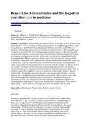
levels of DHEA immediately after birth <strong>and</strong> up to 6<br />
months postnatally as well as at adrenarche may be<br />
responsible for acne infantum <strong>and</strong> prepubertal acne<br />
(Fig 1). These types of acne often present not only with<br />
comedonal acne, but also later with inflammatory lesions,<br />
because DHEA exhibits a proinflammatory activity<br />
opposing the anti-inflammatory action of glucocorticoids.<br />
70 <strong>Acne</strong> neonatorum, which is present at birth or<br />
appears 2–4 weeks after birth, is not uncommon, with a<br />
prevalence of approximately 20% in newborns. 71 It usually<br />
presents with mostly mild facial lesions, is selflimited,<br />
<strong>and</strong> has a male predominance (4.5–5:1). 12 Histological<br />
findings from newborns with acne<br />
neonatorum demonstrate hyperplasic sebaceous gl<strong>and</strong>s<br />
with keratin-plugged orifices. 71<br />
<strong>Acne</strong> in childhood has been suggested to be strongly<br />
associated with the development of severe acne during<br />
adolescence. 26,71 On the other h<strong>and</strong>, high sebum production<br />
rates are a predisposing factor associated with<br />
the formation of primary acne lesions in adolescence. 47<br />
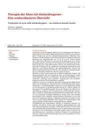
DHEA also may be responsible for acne tarda in females,<br />
which presents with inflammatory lesions of the<br />
lower part of the face (Fig 2). 43<br />
Inflammation <strong>and</strong> <strong>Acne</strong> Vulgaris<br />
It is widely accepted that inflammation in acne vulgaris<br />
may be induced mainly by an immunologic reaction to<br />
extracellular products of P. acnes. 72 However, it is by no<br />
means clear that either bacteria or bacterial products<br />
initiate follicular inflammation. Ingham et al 73 investigated<br />
the presence of proinflammatory cytokines in 108
Clinics in Dermatology Y 2004;22:360–366 ACNE AND SEBACEOUS GLAND FUNCTION 363<br />
Figure 1. <strong>Acne</strong> infantum.<br />
open acne comedones from 18 untreated acne patients.<br />
Bioactive IL-1�–like material was demonstrated in 76%<br />
of open comedones; in 58%, levels exceeded 100 pg/<br />
mg. Most of the open comedones (97%) also contained<br />
microorganisms, but there was no significant correlation<br />
between levels of any cytokine (in particular, IL-1�)<br />
<strong>and</strong> the numbers of microorganisms.<br />
Figure 2. <strong>Acne</strong> tarda.<br />
Additional results have shown that the sebaceous<br />
gl<strong>and</strong> expresses a number of different cytokines at<br />
steady state, without the influence of any external factors.<br />
Zouboulis et al 47 stressed sebocytes in vitro by<br />
maintaining them in serum-free medium <strong>and</strong> detected<br />
IL-1� expression at the mRNA <strong>and</strong> protein levels. Antilla<br />
et al 74 showed that IL-1 is present in normal sebaceous<br />
gl<strong>and</strong>s, <strong>and</strong> Boehm et al 75 used in situ hybridization<br />
techniques to demonstrate that messenger RNA<br />
(mRNA) for IL-1�, IL-1�, <strong>and</strong> tumor necrosis factor-�<br />
are present at multiple sites in normal skin, including<br />
the sebaceous gl<strong>and</strong>s. Thus, whereas the presence of<br />
bacteria, most notably P. acnes, may stimulate up-regulation<br />
of cytokine expression in sebaceous gl<strong>and</strong>s <strong>and</strong> in<br />
monocytes, 32,76 inflammatory cells are also present<br />
around the sebaceous duct, <strong>and</strong> substance P is expressed<br />
in the nerve endings at the vicinity of healthylooking<br />
gl<strong>and</strong>s of acne patients in the absence of bacteria<br />
or their agents. 55,77<br />
Proinflammatory Cytokines <strong>and</strong><br />
Comedone Development<br />
Guy et al 78 assessed the action of IL-1 in the human<br />
pilosebaceous infundibulum isolated by microdissection<br />
<strong>and</strong> maintained in keratinocyte serum-free culture<br />
for 7 days. The addition of 1 ng/mL IL-1� resulted in<br />
hypercornification of the infundibulum similar to that<br />
seen in comedones. The dependence of this effect on a<br />
specific action of IL-1� was confirmed in an additional<br />
experiment demonstrating that the IL-1� effect could be<br />
nullified by the administration 1000 ng/mL of IL-1<br />
receptor antagonist. They further demonstrated that<br />
spontaneous hypercornification of the infundibulum<br />
(which occurred in about 20% of isolated pilosebaceous<br />
units) could also be blocked by the IL-1� receptor antagonist.<br />
These results are compatible with the data of<br />
Zouboulis et al, 47 Boehm et al, 75 Jeremy et al, 77 <strong>and</strong><br />
Ingham et al 73 <strong>and</strong> provide evidence for the involvement<br />
of endogenous inflammatory processes in the initiation<br />
of acne. In addition, they offer logical support to<br />
the current concept of anti-inflammatory treatment of<br />
acne 18,79,80 (Fig 3).<br />
Therefore, acne vulgaris is a genuine inflammatory<br />
disease, <strong>and</strong> evidence exists indicating that appropriate<br />
anti-inflammatory therapy has the potential to effectively<br />
treat this condition. Future compounds targeting<br />
acne vulgaris should be able to reduce proinflammatory<br />
lipids in sebum, down-regulate proinflammatory signals<br />
in the pilosebaceous unit, <strong>and</strong> inhibit LTB 4-induced<br />
accumulation of inflammatory cells <strong>and</strong> PPAR regulation,<br />
as well as significantly reduce sebum production.<br />
Retinoids, the <strong>Sebaceous</strong> Gl<strong>and</strong> <strong>and</strong> <strong>Acne</strong><br />
Isotretinoin is the most effective compound in reducing<br />
sebaceous gl<strong>and</strong> size by decreasing proliferation of
364 ZOUBOULIS Clinics in Dermatology Y 2004;22:360–366<br />
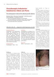
Figure 3. Efficacy of Zileuton, an oral 5-lipoxygenase inhibitor<br />
in inflammatory acne. Significant reduction is seen in (A) the<br />
number of inflammatory lesions, (B) the acne severity index, <strong>and</strong><br />
(C) the total sebum lipid levels after 12 weeks of treatment. The<br />
mean values � st<strong>and</strong>ard error are presented. Values are compared<br />
with baseline. (Modified with permission. 80 )<br />
basal sebocytes, suppressing sebum production (up to<br />
90%) <strong>and</strong> inhibiting sebocyte differentiation in vivo <strong>and</strong><br />
in vitro (reviewed in 81,82 ). A marked decrease in wax<br />
esters, a slight decrease in squalene <strong>and</strong> a relative increase<br />
in cholesterol level have been detected in skin<br />
surface lipids. 83 Oral isotretinoin was also shown to<br />
decrease the triglyceride fraction, whereas, free sterols<br />
<strong>and</strong> total ceramides were found increased in comedonal<br />
lipids. In contrast, other non-aromatic retinoids, such as<br />
tretinoin <strong>and</strong> alitretinoin have been found to be inferior<br />
to isotretinoin in reducing sebocyte proliferation <strong>and</strong><br />
suppressing sebum production, whereas most aromatic<br />
retinoids do not reduce sebum synthesis. 83 Since retinoids<br />
exert most of their effects by modulating gene<br />
expression <strong>and</strong>/or activating nuclear retinoid receptors,<br />
the high antisebotropic activity of isotretinoin is<br />
particularly surprising because it exhibits low binding<br />
affinities for both cellular retinoic acid-binding proteins<br />
I <strong>and</strong> II as well as for nuclear retinoid receptors, retinoic<br />
acid receptors (RAR) <strong>and</strong> retinoid X receptors. Tsukada<br />
et al 84 have partially elucidated this apparent contradiction<br />
by showing that isotretinoin undergoes significant<br />
<strong>and</strong> selective isomerization to tretinoin in cultured<br />
sebocytes. Intracellular tretinoin acts then via RAR to<br />
exert its antiproliferative effect on these cells. Therefore,<br />
isotretinoin acts probably as a prodrug that becomes<br />
active in the sebaceous gl<strong>and</strong>s after isomerization to<br />
tretinoin.<br />
Oral isotretinoin has revolutionized the treatment of<br />
severe acne. 82,83 It is the only drug available that affects<br />
all four pathogenic factors of the disease. A 6- to 12month<br />
course of isotretinoin 0.5–1 mg/kg/day in most<br />
cases with severe acne, to reach a �150 mg/kg total<br />
cumulative dose is recommended. 85 Individual risk factors<br />
must be taken into account for establishing the<br />
exact dosage. As a rule, after 2 to 4 weeks of treatment,<br />
a 50% reduction of the pustules can be expected. A<br />
6-month treatment course is sufficient for the majority<br />
of the patients but a continuation of low-dose treatment<br />
(0.2–0.5 mg/kg/day) may optimize the therapeutic<br />
outcome. Improvement continues during the post-treatment<br />
period. Relapses may occur after a single 6-month<br />
course. Relapses necessitating re-treatment occur significantly<br />
more frequently under low-doses among patients<br />
with severe acne. A 22 to 30% relapse rate was<br />
noted in patients followed for 10 years after having<br />
received isotretinoin 1 mg/kg/day (or cumulative dose<br />
�120 mg/kg), as compared to 39 to 82% with lower<br />
dose schedules. 86 In female patients, contraception is<br />
required <strong>and</strong> has to be enforced by the physician, because<br />
of the strong teratogenicity of isotretinoin.<br />
Isotretinoin can be well combined with a contraceptive<br />
pill which includes a hormonal anti-<strong>and</strong>rogen.<br />
References<br />
1. Benfenati A, Brillanti F. Sulla distribuzione delle ghi<strong>and</strong>ole<br />
sebacee nella cute del corpo umino. Arch Ital Dermatol<br />
1939;15:33–42.<br />
2. Montagna W. An introduction to sebaceous gl<strong>and</strong>s. J Invest<br />
Dermatol 1974;62:120–3.<br />
3. Thody AJ, Shuster S. Control <strong>and</strong> function of sebaceous<br />
gl<strong>and</strong>s. Physiol Rev 1989;69:383–416.<br />
4. Deplewski D, Rosenfield RL. Role of hormones in pilosebaceous<br />
unit development. Endocr Rev 2000;21:363–92.<br />
5. Fujita H, Asagami C, Murota S, et al. Ultrastructural study<br />
of embryonic sebaceous cells, especially of their sebum<br />
droplet formation. Acta Derm Venereol 1972;52:99–115.<br />
6. Sato S, Hiraga K, Nishijima A, et al. Neonatal sebaceous<br />
gl<strong>and</strong>s: fine structure of sebaceous <strong>and</strong> dendritic cells.<br />
Acta Derm Venereol 1977;57:279–87.<br />
7. Tosti A. A comparison of the histodynamics of sebaceous<br />
gl<strong>and</strong>s <strong>and</strong> epidermis in man: a microanatomic <strong>and</strong> morphometric<br />
study. J Invest Dermatol 1974;62:147–52.<br />
8. Fenske NA, Lober CW. Structural <strong>and</strong> functional changes<br />
of normal aging skin. J Am Acad Dermatol 1986;15:571–<br />
85.<br />
9. Zouboulis ChC, Boschnakow A. Chrono- <strong>and</strong> photoaging<br />
of the human sebaceous gl<strong>and</strong>. Clin Exp Dermatol 2001;<br />
26:600–7.<br />
10. Serri F, Huber WM. The development of sebaceous gl<strong>and</strong>s<br />
in man. In: Montagna W, Ellis RA, Silver AF, editors.<br />
Advances in biology of skin, vol IV: sebaceous gl<strong>and</strong>s.<br />
Oxford, UK: Pergamon, 1963:1–18.<br />
11. Plewig G, Kligman AM. Proliferative activity of the sebaceous<br />
gl<strong>and</strong>s of the aged. J Invest Dermatol 1978;70:314–7.<br />
12. Zouboulis ChC, Fimmel S, Ortmann J, et al. <strong>Sebaceous</strong><br />
gl<strong>and</strong>s. In: Hoath SB, Maibach HI, editors. Neonatal skin:<br />
structure <strong>and</strong> function (ed 2). New York: Marcel Dekker,<br />
2003:59–88.<br />
13. Zouboulis ChC. Human skin: an independent peripheral<br />
endocrine organ. Horm Res 2000;54:230–42.<br />
14. Fritsch M, Orfanos CE, Zouboulis ChC. Sebocytes are the<br />
key regulators of <strong>and</strong>rogen homeostasis in human skin.<br />
J Invest Dermatol 2001;116:793–800.
Clinics in Dermatology Y 2004;22:360–366 ACNE AND SEBACEOUS GLAND FUNCTION 365<br />
15. Thiboutot D, Jabara S, McAllister JM, et al. Human skin is<br />
a steroidogenic tissue: steroidogenic enzymes <strong>and</strong> cofactors<br />
are expressed in epidermis, normal sebocytes, <strong>and</strong> an<br />
immortalized sebocyte cell line (SEB-1). J Invest Dermatol<br />
2003;120:905–14.<br />
16. Zouboulis ChC, Seltmann H, Hiroi N, et al. Corticotropinreleasing<br />
hormone: an autocrine hormone that promotes<br />
lipogenesis in human sebocytes. Proc Natl Acad Sci U S A<br />
2002;99:7148–53.<br />
17. Wille JJ, Kydonieus A. Palmitoleic acid isomer (C16:1�6) is<br />
the active antimicrobial fatty acid in human skin sebum.<br />
Skin Pharmacol Appl Skin Physiol 2003;16:176–87.<br />
18. Zouboulis ChC. Is acne vulgaris a genuine inflammatory<br />
disease? Dermatology 2001;203:277–9.<br />
19. Böhm M, Schiller M, Ständer S, et al. Evidence for expression<br />
of melanocortin-1 receptor in human sebocytes in<br />
vitro <strong>and</strong> in situ. J Invest Dermatol 2002;118:533–9.<br />
20. Nikkari T. Comparative chemistry of sebum. J Invest Dermatol<br />
1974;62:257–67.<br />
21. Pochi P. The sebaceous gl<strong>and</strong>. In: Maibach HI, Boisits EK,<br />
editors. Neonatal skin: structure <strong>and</strong> function. New York:<br />
Marcel Dekker, 1982:67–80.<br />
22. Wheatley VR. Sebum: its chemistry <strong>and</strong> biochemistry. Am<br />
Perfumer 1956;68:37–47.<br />
23. Cunliffe WJ. <strong>Acne</strong>. London: Martin Dunitz, 1989.<br />
24. Niemann C, Unden AB, Lyle S, et al. Indian hedgehog <strong>and</strong><br />
�-catenin signaling: role in the sebaceous lineage of normal<br />
<strong>and</strong> neoplastic mammalian epidermis. Proc Natl<br />
Acad Sci USA2003;100(Suppl 1):11873–80.<br />
25. Agache P, Blanc D, Barr<strong>and</strong> C, et al. Sebum levels during<br />
the first year of life. Br J Dermatol 1980;103:643–9.<br />
26. Henderson CA, Taylor J, Cunliffe WJ. Sebum excretion<br />
rates in mothers <strong>and</strong> neonates. Br J Dermatol 2000;142:<br />
110–1.<br />
27. Ramasastry P, Downing DT, Pochi PE, et al. Chemical<br />
composition of human skin surface lipids from birth to<br />
puberty. J Invest Dermatol 1970;54:139–44.<br />
28. Pochi PE, Strauss JS, Downing DT. Sebum, acne <strong>and</strong><br />
<strong>and</strong>rogens in children [abstract]. Clin Res 1977;25:531A.<br />
29. Sharp F, Calman KC, Milne JA, et al. The demonstration<br />
of hydroxysteroid dehydrogenase activity in human foetal<br />
skin in the first 32 weeks of gestation. Br J Dermatol<br />
1970;83:177–81.<br />
30. Sharp F, Hay JB, Hodgins MB. Metabolism of <strong>and</strong>rogens<br />
in vitro by human foetal skin. J Endocrinol 1976;70:491–9.<br />
31. Billich A, Rot A, Lam C, et al. Immunohistochemical<br />
localization of steroid sulfatase in acne lesions: implications<br />
for the contribution of dehydroepi<strong>and</strong>rosterone sulfate<br />
to the pathogenesis of acne [abstract]. Horm Res<br />
2000;53:99.<br />
32. Kim MH, Herrmann WL. In vitro metabolism of dehydroepi<strong>and</strong>rosterone<br />
sulphate in foreskin, abdominal skin <strong>and</strong><br />
vaginal mucosa. J Clin Endocrinol Metab 1969;28:187–91.<br />
33. Labrie F, Luu-The V, Labrie C, et al. Intracrinology <strong>and</strong><br />
the skin. Horm Res 2000;54:218–29.<br />
34. Chen W, Zouboulis ChC, Orfanos CE. The 5�-reductase<br />
system <strong>and</strong> its inhibitors: recent development <strong>and</strong> its<br />
perspective in treating <strong>and</strong>rogen-dependent skin disorders.<br />
Dermatology 1996;193:177–84.<br />
35. Luu-The V, Sugimoto Y, Puy L, et al. Characterization,<br />
expression, <strong>and</strong> immunohistochemical localization of 5�-<br />
reductase in human skin. J Invest Dermatol 1994;102:<br />
221–6.<br />
36. Thiboutot D, Harris G, Iles V, et al. Activity of the type I<br />
5�-reductase exhibits regional differences in isolated sebaceous<br />
gl<strong>and</strong>s <strong>and</strong> whole skin. J Invest Dermatol 1995;<br />
105:209–14.<br />
37. Chen W, Zouboulis ChC, Fritsch M, et al. Evidence of<br />
heterogeneity <strong>and</strong> quantitative differences of the type 1<br />
5�-reductase expression in cultured human skin cells.<br />
Evidence of its presence in melanocytes. J Invest Dermatol<br />
1998;110:84–9.<br />
38. Cunliffe WJ, Forster RA. Androgen control of the pilosebaceous<br />
duct (abstract). Br J Dermatol 1987;116:449.<br />
39. Thiboutot D, Knaggs H, Gillil<strong>and</strong> K, et al. Activity of type<br />
15�-reductase is greater in the follicular infrainfundibulum<br />
compared with the epidermis. Br J Dermatol 1997;<br />
136:166–71.<br />
40. Bläuer M, Vaalasti A, Pauli SL, et al. Location of <strong>and</strong>rogen<br />
receptor in human skin. J Invest Dermatol 1991;97:264–8.<br />
41. Choudhry R, Hodgins MB, Van der Kwast TH, et al.<br />
Localization of <strong>and</strong>rogen receptors in human skin by<br />
immunohistochemistry: implications for the hormonal<br />
regulation of hair growth, sebaceous gl<strong>and</strong>s <strong>and</strong> sweat<br />
gl<strong>and</strong>s. J Endocrinol 1992;133:467–75.<br />
42. Liang T, Hoyer S, Yu R, et al. Immunocytochemical localization<br />
of <strong>and</strong>rogen receptors in human skin using monoclonal<br />
antibodies against the <strong>and</strong>rogen receptor. J Invest<br />
Dermatol 1993;100:663–6.<br />
43. Orfanos CE, Adler YD, Zouboulis ChC. The SAHA syndrome.<br />
Horm Res 2000;54:251–8.<br />
44. Rosenfield RL, Deplewski D, Kentsis A, et al. Mechanisms<br />
of <strong>and</strong>rogen induction of sebocyte differentiation. Dermatology<br />
1998;196:43–6.<br />
45. Chen W, Yang C-C, Sheu H-M, et al. Expression of peroxisome<br />
proliferator-activated receptor <strong>and</strong> CCAAT/enhancer<br />
binding protein transcription factors in cultured<br />
human sebocytes. J Invest Dermatol 2003;121:441–7.<br />
46. Brun R, Tontonoz P, Forman B, et al. Differential activation<br />
of adipogenesis by multiple PPAR isoforms. Genes<br />
Dev 1996;10:974–84.<br />
47. Zouboulis ChC, Xia L, Akamatsu H, et al. The human<br />
sebocyte culture model provides new insights into development<br />
<strong>and</strong> management of seborrhoea <strong>and</strong> acne. Dermatology<br />
1998;196:21–31.<br />
48. Strauss JS, Kligman AM, Pochi PE. Effect of <strong>and</strong>rogens<br />
<strong>and</strong> estrogens on human sebaceous gl<strong>and</strong>s. J Invest Dermatol<br />
1962;39:139–55.<br />
49. Guy R, Ridden C, Kealey T. The improved organ maintenance<br />
of the human sebaceous gl<strong>and</strong>: modeling in vitro<br />
the effects of epidermal growth factor, <strong>and</strong>rogens, estrogens,<br />
13-cis retinoic acid, <strong>and</strong> phenol red. J Invest Dermatol<br />
1996;106:454–60.<br />
50. Pochi PE, Strauss JS, Mescon H. The role of adrenocortical<br />
steroids in the control of human sebaceous gl<strong>and</strong> activity.<br />
J Invest Dermatol 1963;41:391–9.<br />
51. Goolamali SK, Plummer N, Burton JL, et al. Sebum excretion<br />
<strong>and</strong> melanocyte stimulating hormone in hypoadrenalism.<br />
J Invest Dermatol 1974;63:253–5.<br />
52. Glickman SP, Rosenfield RL, Bergenstal RM, et al. Multiple<br />
<strong>and</strong>rogenic abnormalities, including elevated free tes-
366 ZOUBOULIS Clinics in Dermatology Y 2004;22:360–366<br />
tosterone, in hyperprolactinemic women. J Clin Endocrinol<br />
Metab 1982;55:251–7.<br />
53. Slominski A, Wortman J, Luger T, et al. Corticotropinreleasing<br />
hormone <strong>and</strong> propiomelanocortin involvement<br />
in the cutaneous response to stress. Physiol Rev 2000;80:<br />
979-1020.<br />
54. Stewart ME, McDonnell MW, Downing DT. Possible genetic<br />
control of the proportions of branched-chain fatty<br />
acids in human sebaceous wax esters. J Invest Dermatol<br />
1986;86:706–8.<br />
55. Toyoda M, Nakamura M, Makino T, et al. <strong>Sebaceous</strong><br />
gl<strong>and</strong>s in acne patients express high levels of neutral<br />
endopeptidase. Exp Dermatol 2002;11:241–7.<br />
56. Toyoda M, Morohashi M. Pathogenesis of acne. Med Elect<br />
Microsc 2001;34:29–40.<br />
57. Nicolaides N, Wells GC. On the biogenesis of the free fatty<br />
acids in human skin surface fat. J Invest Dermatol 1957;<br />
29:423–33.<br />
58. Shalita AR. Genesis of free fatty acids. J Invest Dermatol<br />
1974;62:332–5.<br />
59. Pochi PE, Strauss JS, Downing DT. Skin surface lipid<br />
composition, acne, pubertal development, <strong>and</strong> excretion<br />
<strong>and</strong> urinary excretion of testosterone <strong>and</strong> 17-ketosteroids.<br />
J Invest Dermatol 1977;69:485–98.<br />
60. Yamamoto A, Ito M. <strong>Sebaceous</strong> gl<strong>and</strong> activity <strong>and</strong> urinary<br />
<strong>and</strong>rogen levels in children. J Dermatol Sci 1992;4:98–104.<br />
61. Stewart ME, Downing DT. Measurement of sebum secretion<br />
rates in young children. J Invest Dermatol 1985;84:<br />
59–61.<br />
62. Jacobsen E, Billings JK, Frantz RA, et al. Age-related<br />
changes in sebaceous wax ester secretion rates in men <strong>and</strong><br />
women. J Invest Dermatol 1985;85:483–5.<br />
63. Akamatsu H, Zouboulis ChC, Orfanos CE. Control of<br />
human sebocyte proliferation in vitro by testosterone <strong>and</strong><br />
5-�-dihydrotestosterone is dependent on the localization<br />
of the sebaceous gl<strong>and</strong>s. J Invest Dermatol 1992;99:509–11.<br />
64. Zouboulis ChC. <strong>Sebaceous</strong> gl<strong>and</strong> in human skin: the fantastic<br />
future of a skin appendage. J Invest Dermatol 2003;<br />
120:xiv–xv.<br />
65. Pilgram GS, van der Meulen J, Gooris GS, et al. The<br />
influence of two azones <strong>and</strong> sebaceous lipids on the lateral<br />
organization of lipids isolated from human stratum<br />
corneum. Biochim Biophys Acta 2001;1511:244–54.<br />
66. Fluhr JW, Mao-Qiang M, Brown BE, et al. Glycerol regulates<br />
stratum corneum hydration in sebaceous gl<strong>and</strong> deficient<br />
(a sebia) mice. J Invest Dermatol 2003;120:728–37.<br />
67. Ge L, Gordon JS, Hsuan C. Identification of the �-6 desaturase<br />
of human sebaceous gl<strong>and</strong>s: expression <strong>and</strong> enzyme<br />
activity. J Invest Dermatol 2003;120:707–14.<br />
68. Thiele JJ, Weber SU, Packer L. <strong>Sebaceous</strong> gl<strong>and</strong> secretion<br />
is a major physiologic route of vitamin E delivery to skin.<br />
J Invest Dermatol 1999;113:1006–10.<br />
69. Nicolaides N, Fu HC, Ansari MNA, et al. The fatty acids<br />
of esters <strong>and</strong> sterol esters from vernix caseosa <strong>and</strong> from<br />
human surface lipid. Lipids 1972;7:506–17.<br />
70. Bradlow HL, Murphy J, Byrne JJ. Immunological properties<br />
of dehydroepi<strong>and</strong>rosterone, its conjugates, <strong>and</strong> metabolites.<br />
Ann N Y Acad Sci 1999;876:91–101.<br />
71. Katsambas AD, Katoulis AC, Stavropoulos P. <strong>Acne</strong> neonatorum:<br />
a study of 22 cases. Int J Dermatol 1999;38:128–<br />
30.<br />
72. Burkhart CG, Cantrill J, Butcher CL, et al. Propionibacterium<br />
acnes: interaction with complement <strong>and</strong> development<br />
of an enzyme-linked immunoassay for the detection<br />
of antibody. Int J Dermatol 1999;38:200–3.<br />
73. Ingham E, Eady EA, Goodwin CE, et al. Pro-inflammatory<br />
levels of interleukin-1alpha–like bioactivity are present in<br />
the majority of open comedones in acne vulgaris. J Invest<br />
Dermatol 1992;98:895–901.<br />
74. Antilla HS, Reitamo S, Saurat JH. Interleukin 1 immunoreactivity<br />
in sebaceous gl<strong>and</strong>s. Br J Dermatol 1992;127:<br />
585–8.<br />
75. Boehm KD, Yun JK, Strohl KP, et al. Messenger RNAs for<br />
the multifunctional cytokines interleukin-1�, interleukin-1�<br />
<strong>and</strong> tumor necrosis factor-� are present in adnexal<br />
tissues <strong>and</strong> in dermis of normal human skin. Exp Dermatol<br />
1995;4:335–41.<br />
76. Vowels BR, Yang S, Leyden JJ. Induction of proinflammatory<br />
cytokines by a soluble factor of Propionibacterium<br />
acnes: implications for chronic inflammation acne. Infect<br />
Immun 1995;63:3158–65.<br />
77. Jeremy AHT, Holl<strong>and</strong> DB, Roberts SG, et al. Inflammatory<br />
events are involved in acne lesion initiation. J Invest Dermatol<br />
2003;121:20–7.<br />
78. Guy R, Green MR, Kealey T. Modeling acne in vitro.<br />
J Invest Dermatol 1996;106:176–82.<br />
79. Zouboulis ChC. Exploration of retinoid activity <strong>and</strong> the<br />
role of inflammation in acne: issues affecting future directions<br />
for acne therapy. J Eur Acad Dermatol Venereol<br />
2001;15(Suppl 3):63–7.<br />
80. Zouboulis ChC, Nestoris S, Adler YD, et al. A new concept<br />
for acne therapy: a pilot study with zileuton, an oral<br />
5-lipoxygenase inhibitor. Arch Dermatol 2003;139:668–70.<br />
81. Orfanos CE, Zouboulis ChC, Almond-Roesler B, et al.<br />
Current use <strong>and</strong> future potential role of retinoids in dermatology.<br />
Drugs 1997;53:358–88.<br />
82. Zouboulis ChC, Orfanos CE. Retinoids. In: Millikan LE,<br />
editor. Drug Therapy in Dermatology. New York Basel:<br />
Marcel Dekker, 2000:171–233.<br />
83. Orfanos CE, Zouboulis ChC. Oral retinoids in the treatment<br />
of seborrhoea <strong>and</strong> acne. Dermatology 1998;196:140–7.<br />
84. Tsukada M, Schröder M, Roos TC, et al. 13-cis Retinoic<br />
acid exerts its specific activity on human sebocytes<br />
through selective intracellular isomerization to all-trans<br />
retinoic acid <strong>and</strong> binding to retinoid acid receptors. J Invest<br />
Dermatol 2000;115:321–7.<br />
85. Zouboulis ChC, Piquero-Martin J. Update <strong>and</strong> future of<br />
systemic acne treatment. Dermatology 2003;206:37–53.<br />
86. Goulden V, Layton AM, Cunliffe WJ. Longterm safety of<br />
isotretinoin as a treatment for acne vulgaris. Br J Dermatol<br />
1994;131:360–3.