Management of Venous Thoracic Outlet Syndrome - Methodist ...
Management of Venous Thoracic Outlet Syndrome - Methodist ...
Management of Venous Thoracic Outlet Syndrome - Methodist ...

You also want an ePaper? Increase the reach of your titles
YUMPU automatically turns print PDFs into web optimized ePapers that Google loves.
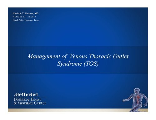
Heitham T. Hassoun, MD<br />
AUGUST 20 – 22, 2010<br />
Hotel ZaZa, Houston, Texas<br />
<strong>Management</strong> <strong>of</strong> <strong>Venous</strong> <strong>Thoracic</strong> <strong>Outlet</strong><br />
<strong>Syndrome</strong> (TOS)
<strong>Venous</strong> TOS – “Effort Thrombosis”<br />
Pathophysiology<br />
• Mechanical abnormality at the costoclavicular portion <strong>of</strong><br />
the axillosubclavian vein<br />
• Most <strong>of</strong>ten acute thrombosis is in an area <strong>of</strong> chronic<br />
compression and stricture - between the scalene or<br />
subclavius tendon and the first rib
Multiple treatment algorithms for successful<br />
outcomes in venous thoracic outlet syndrome<br />
Ricardo A. de Leon, MD, David C. Chang, PhD, MPH, MBA, Heitham T. Hassoun, MD, James H. Black, MD,<br />
Glen S. Roseborough, MD, Bruce A. Perler, MD, Lisa Rotini-Coltvet, MA, MMS, PAC, Diana Call, BS, RVT,<br />
Christopher Busse, BS, RVT, RDMS, and Julie A. Freischlag, MD<br />
Background. We sought to determine the outcomes in patients presenting with venous thuracic outlet syndrome. Methods.<br />
Prospectively collected data from 67 patients between October 2003 and December 2007. The average age was<br />
31 years (range, 16-54); the 371lUlleS and 30females presented on average 9.2 months (range, 1 month to 6 years) after acute<br />
thrombosis. Four treatment algorithms were utilized. Results. In group 1, 3 patients presented with acute<br />
occlusion and received tissue plasminogen activatur (tPA) and immediate first rib resection with scalenectomy (FRRS). One<br />
vein rethrombosed and was treated fly intravenous tPA postoperatively. In group 2, 39 patients presented with<br />
stenotic subclavian veins an average <strong>of</strong> 22 weeks after their initial thrombosis, all <strong>of</strong> whom underwent FRRS followed by a<br />
venogram 2 weeks postoperatively: 25 had a tight stenosis and underwent venoplasty with anticoagulation; 13 had patent,<br />
nonstenotic subclavian veins, and 1 patient required tPA and venoplasty owing to rethrombosis. Two patients had their subclavian<br />
vein thrombose after venoplasty and were treated with anticoagulation, tPA, and venoplasty. In group 3, 11 patients<br />
presented with intermittent venous obstruction without thrombosis and underwent FRRS; 3 underwent<br />
venograms because <strong>of</strong> concerns <strong>of</strong> residual stenosis, 2 <strong>of</strong> whom required venoplasty postoperatively. Finally, in group 4, 14<br />
patients presented with occluded subclavian veins and underwent FRRS with long-term anticoagulation. Eleven<br />
have recanalized at an average <strong>of</strong> 6 months (range, 2-12). Conclusion. Overall, 64 <strong>of</strong> 67 patients have patent<br />
subclavian veins after a median follow-up <strong>of</strong> 10 months, and all patients are asymptomatic for a<br />
success rate <strong>of</strong> 96 %. Tailored treatment algorithms including FRRS, postoperative venograms with or without intervention, and the<br />
use <strong>of</strong> long-term anticoagulation seems to be required in this complicated group <strong>of</strong> young patients to achieve optimal results.<br />
(Surgery 2009)<br />
From the Department <strong>of</strong> Surgery, Division <strong>of</strong> Vascular Surgery, Johns Hopkins Medical Institutions, Baltimore, MD
Treatment # 1: Acute Vein Occlusion<br />
• Three patients (4.5%) presented with acute occlusion <strong>of</strong><br />
the subclavian vein (2,3 and 7 days)<br />
• All were lysed, underwent first rib resection and<br />
scalenectomy followed by 2 week postoperative<br />
venogram +/- intervention<br />
• One re-thrombosed and was lysed<br />
• Postoperative anticoagulation was given to all patients<br />
de Leon et al, Surgery 2009
Paget-Schroetter <strong>Syndrome</strong><br />
“Effort Thrombosis”
Primary Trombolysis
Post-Thrombolysis
Post-Thrombolysis PTA
Transaxillary 1 st Rib Resection
Treatment # 2: Subclavian Vein Stenosis<br />
• Comprised <strong>of</strong> 39 (58%) patients who presented<br />
with symptomatic subclavian vein stenosis<br />
• Presented on average 27 weeks following acute<br />
event<br />
• All underwent first rib resection and scalenectomy<br />
followed by 2 week postoperative venogram<br />
de Leon et al, Surgery 2009
Treatment # 2: Subclavian Vein Stenosis<br />
• Postoperative venograms<br />
– 25/39 (64%) had significant stenosis and<br />
underwent PTA<br />
– 13 (33%) were non-stenotic and required no<br />
further intervention<br />
– 1 presented with an occluded subclavian vein<br />
and was treated with lysis then PTA<br />
• Overall, 26 (67%) patients underwent PTA and<br />
were maintained on anticoagulation for 3 months<br />
– 2 (5%) pts required re-intervention at 1 and 5 weeks<br />
de Leon et al, Surgery 2009
Ultrasound-Guided Access
Subclavian Vein Stenosis<br />
“Focal”<br />
• Usually cross with .035 hydrophilic wire and glidecath<br />
• Sometimes need lower pr<strong>of</strong>ile catheter (ie. QuickCross)
Post-operative Subclavian Vein PTA
Post-operative Subclavian Vein PTA
Post-operative Subclavian Vein PTA
Treatment # 3: Intermittent Vein Obstruction<br />
“McCleery <strong>Syndrome</strong>”<br />
• 11/67 (16%) patients presented on average 57 weeks<br />
after onset <strong>of</strong> symptoms<br />
• All presented with swelling but no thrombosis<br />
• All demonstrated vein occlusion with abduction<br />
• All underwent first rib resection and scalenectomy<br />
• Post-operative venograms in 3 patients for persistent<br />
symptoms<br />
- 2 required PTA<br />
• All are currently asymptomatic<br />
de Leon et al, Surgery 2009
Imaging <strong>of</strong> Intermittent Vein Obstruction
Treatment # 4: Chronically Occluded Veins<br />
• 14/67 (21%) patients presented on average 54 weeks<br />
since their initial thrombosis<br />
• All underwent first rib resection and scalenectomy<br />
followed by venograms in 12 patients<br />
• 6-month course <strong>of</strong> coumadin<br />
• Duplex scan was utilized in the follow-up period to<br />
assess subclavian vein patency<br />
de Leon et al, Surgery 2009
Complications/Outcomes<br />
• No vascular injury<br />
• No brachial plexus or long thoracic nerve<br />
injury<br />
• 10/67 (15%) pneumothorax<br />
• 64/67 (96%) patients have patent subclavian<br />
veins and are asymptomatic
Failure <strong>of</strong> stents as primary therapy<br />
Initial experience with venous stents in exertional<br />
axillary-subclavian vein thrombosis<br />
Presented at the Tenth Annual Meeting <strong>of</strong> the Eastern Vascular Society, Washington, D.C., May 3-5, 1996.<br />
George H. Meier MD, Jeffrey S. Pollak MD, Melvin Rosenblatt MD, Kevin W. Dickey MD and Richard J. Gusberg MD<br />
New Haven, Conn.<br />
JVS 1996
Failure <strong>of</strong> stents as primary therapy<br />
Complication <strong>of</strong> a venous wallstent<br />
Richard Dowling,1 Peter Mitchell,1 Ge<strong>of</strong>frey S Cox2 and Ken R Thomson1<br />
1Department <strong>of</strong> Radiology and 2Vascular Surgical Unit, Royal Melbourne Hospital, Parkville, Victoria,<br />
Australia<br />
Australasian Radiology (1999) 43, 246–248
Top 3 Future Directions<br />
• Anticoagulation protocol<br />
• IVUS?<br />
• Secondary use <strong>of</strong> stents?