DeBAKEy CARDIOvASCuLAR JOuRNAL - Methodist Hospital
DeBAKEy CARDIOvASCuLAR JOuRNAL - Methodist Hospital
DeBAKEy CARDIOvASCuLAR JOuRNAL - Methodist Hospital

Create successful ePaper yourself
Turn your PDF publications into a flip-book with our unique Google optimized e-Paper software.
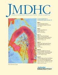
Volume 7, Number 1 • Jan. – Mar. 2011 Official publication of <strong>Methodist</strong> DeBakey Heart & Vascular Center<br />
METHODIST<br />
<strong>DeBAKEy</strong> <strong>CARDIOvASCuLAR</strong> <strong>JOuRNAL</strong><br />
Looking down on the 36 in x 42 in multi-user<br />
virtual surgical table in Plato’s CAvE using Siemens<br />
flash low-dose CT scan patient data with TeraRecon,<br />
Inc. volume visualization.<br />
PAGE 2 Four Decades of Clinical Experience<br />
Jimmy F. Howell, M.D.<br />
PAGE 19 Private Practice of Cardiac Surgery at The <strong>Methodist</strong> <strong>Hospital</strong><br />
Walter S. Henly, M.D.; John B. Fitzgerald, M.D.<br />
PAGE 21 Three-Dimensional Blood Flow Dynamics: Spiral/Helical<br />
Laminar Flow<br />
Peter A. Stonebridge, Ch.M.<br />
PAGE 27 Plato’s CAvE – Knowledge-Based Medicine or Black Swan<br />
Technology?<br />
Paul E. Sovelius, Jr.<br />
PAGE 35 What Are the Current Risks of Cardiac Catheterization?<br />
Michel E. Bertrand, M.D.<br />
PAGE 40 Initial Clinical Experience of Total Cardiac Replacement with<br />
Dual Heartmate-II ® Axial Flow Pumps for Severe<br />
Biventricular Heart Failure<br />
Matthias Loebe, M.D., Ph.D.; Brian Bruckner, M.D.; Michael J. Reardon, M.D.;<br />
Erika van Doorn, M.D.; Jerry Estep, M.D.; Igor Gregoric, M.D.; Faisel<br />
Masud, M.D.; William Cohn, M.D.; Tadashi Motomura, M.D., Ph.D.;<br />
Guillermo Torre-Amione, M.D.; O.H. Frazier, M.D.<br />
PAGE 45 Pumps and Pipes<br />
Alan B. Lumsden, M.D.; Stephen R. Igo, B.Sc.<br />
PAGE 49 TAvI: Transcatheter Aortic valve Implantation<br />
Neal Kleiman, M.D.; Michael J. Reardon, M.D.<br />
PAGE 53 In Tribute: Ernest Stanley Crawford, M.D.<br />
Hazim J. Safi, M.D.<br />
PAGE 55 Museum of TMH Multimodality Imaging Center<br />
PAGE 57 Abstracts<br />
PAGE 63 My Years with Michael E. DeBakey<br />
Louis H. Green, M.D.<br />
PAGE 64 My Years with Michael E. DeBakey<br />
Larry L. Mathis, M.H.A.<br />
PAGE 65 Poet’s Pen: Why I Am Not a Buddhist<br />
Molly Peacock<br />
PAGE 66 In Memoriam: Michael Thomas McDonough, M.D.<br />
William L. Winters Jr., M.D.<br />
PAGE 67 Letters to the Editor
We Welcome Your<br />
Questions and Comments<br />
W.L. Winters Jr., M.D.<br />
Inquiries, letters to the editor and original<br />
contributions can be directed to MDCvJ@tmhs.org.<br />
Correction Notice<br />
Please note the following corrections for Volume 6, Number 4:<br />
On page 59, the title and institutional affiliation of William<br />
L. Winters Jr., M.D. was incorrect. It states “Professor of<br />
Medicine and Executive Dean, Cleveland Clinic Lerner College<br />
of Medicine of Case, Western Reserve University, George<br />
and Linda Kaufman Chair Chairman, Endocrinology and<br />
Metabolism Institute, Cleveland Clinic, Cleveland, Ohio,” which<br />
are the credential for James B. Young, M.D. It should have<br />
simply read: <strong>Methodist</strong> DeBakey Heart & Vascular Center,<br />
Houston, Texas.<br />
The statements and opinions expressed in the articles and<br />
editorials included in the <strong>Methodist</strong> DeBakey Cardiovascular<br />
Journal are those of their authors and are not necessarily<br />
those of the <strong>Methodist</strong> DeBakey Heart & Vascular Center, The<br />
<strong>Methodist</strong> <strong>Hospital</strong> System or affiliated institutions, unless this<br />
is clearly specified.<br />
<strong>Methodist</strong> DeBakey<br />
Cardiovascular Journal<br />
Volume 7, Number 1, 2011<br />
ISSN 1947-6094<br />
debakeyheartcenter.com/journal<br />
Editor-in-Chief<br />
William L. Winters Jr., M.D.<br />
Managing Editor<br />
Sheshe Giddens, B.A.<br />
Contributing Editor: Poet’s Pen<br />
Michael W. Lieberman, M.D., Ph.D.<br />
Editorial Board<br />
Michel Bertrand, M.D.<br />
Lois DeBakey, Ph.D.<br />
Selma DeBakey, B.A.<br />
Kim A. Eagle, M.D.<br />
Robert A. Guyton, M.D.<br />
Gerald Lawrie, M.D.<br />
Alan B. Lumsden, M.D.<br />
Joseph Naples, M.D.<br />
Craig Pratt, M.D.<br />
Miguel Quiñones, M.D.<br />
Albert Raizner, M.D.<br />
Michael J. Reardon, M.D.<br />
Frank J. Veith, M.D.<br />
James B. Young, M.D.<br />
<strong>Methodist</strong> DeBakey Cardiovascular Journal (MDCVJ)<br />
provides an update from <strong>Methodist</strong> DeBakey Heart &<br />
Vascular Center specialists about leading-edge research,<br />
diagnosis and treatments, as well as occasional articles<br />
or commentary by others.<br />
U.S.News & World Report ranks the <strong>Methodist</strong> DeBakey<br />
Heart & Vascular Center’s cardiology, cardiothoracic and<br />
vascular surgery programs among the best in the nation.<br />
MDCVJ is written for physicians and should be relied<br />
upon for medical education purposes only. It does not<br />
provide a complete overview of the topics covered<br />
and should not replace the independent judgment of<br />
a physician about the appropriateness or risks of a<br />
procedure or treatment for a given patient.<br />
© 2011 The <strong>Methodist</strong> <strong>Hospital</strong><br />
Houston, Texas<br />
<strong>Methodist</strong> DeBakey Heart & Vascular Center<br />
6565 Fannin Street<br />
Houston, Texas 77030<br />
Telephone: 713-DEBAKEY (713-332-2539)<br />
debakeyheartcenter.com
W.L. Winters Jr., M.D.<br />
EDITOR’S NOTE<br />
editorial<br />
William L. Winters Jr., M.D.<br />
<strong>Methodist</strong> DeBakey Heart & Vascular Center, Houston, Texas<br />
In this issue, we are privileged to publish the entire<br />
cardiovascular surgical experience of Dr. Jimmy F.<br />
Howell between the years of 1964 and 2004. He was<br />
but one of a remarkable cadre of cardiovascular surgeons<br />
trained by Dr. Michael E. DeBakey during that<br />
period and who worked at The <strong>Methodist</strong> <strong>Hospital</strong> in<br />
the academic cardiovascular surgical program at Baylor<br />
College of Medicine (BCM).<br />
The next article by Dr. Henly describes the parallel<br />
development — the first of its kind at The <strong>Methodist</strong><br />
<strong>Hospital</strong> and in Houston — of a private practice cardiovascular<br />
surgical team of surgeons all trained by<br />
the same Michael E. DeBakey, M.D. Simultaneously,<br />
the private practice cardiology community was enlarging<br />
faster than the academic cardiology community<br />
because of the existence of few and small local cardiology<br />
training programs until well into the 1970s. Thus<br />
was laid the groundwork for a strong private practice<br />
medical/surgical cardiovascular network working<br />
and competing in the same close clinical environment<br />
with the academic cardiovascular programs of BCM at<br />
<strong>Methodist</strong>. As one of our early colleagues commented,<br />
“That environment led to a natural grist between the<br />
private practice groups and the academic Baylor group.”<br />
On the one hand, there was the perceived view by some<br />
of a more personal approach in patient care provided in<br />
the private practice scheme compared to the academic<br />
group. yet in the setting of high tech demands and complex<br />
team requirements, Michael E. DeBakey placed no<br />
barrier to the development of a competing cardiovascular<br />
surgical team. They were, after all, his own trainees<br />
providing the competition. And let no one doubt that<br />
if they had not met the standards set by Dr. DeBakey,<br />
they would not have continued to practice and operate<br />
at The <strong>Methodist</strong> <strong>Hospital</strong>. So, as the bar stood high for<br />
cardiovascular surgeons, so it did for cardiologists as<br />
cardiology training programs at BCM, <strong>Methodist</strong>, and<br />
the Texas Heart Institute began to expand after the<br />
mid-1970s.<br />
As academic surgeons and academic cardiologists<br />
absorbed some of each other’s attributes, private practice<br />
cardiologists and surgeons increasingly became contributors<br />
to the academic programs by participating in<br />
the teaching and research opportunities. The side-byside<br />
growth at The <strong>Methodist</strong> <strong>Hospital</strong> of both types of<br />
practices, tolerated by Dr. DeBakey, encouraged by The<br />
<strong>Methodist</strong> <strong>Hospital</strong>, and to a lesser degree, by Baylor<br />
College of Medicine, was unique in those early years of<br />
cardiac surgery.<br />
MDCvJ | vII (1) 2011 1
J.F. Howell, M.D.<br />
Foreward<br />
FOuR DECADES OF CLINICAL<br />
EXPERIENCE<br />
JIMMY F. HOWELL, M.D.<br />
Adapted by William L. Winters Jr., M.D., from a monograph published by<br />
Baylor College of Medicine in 2009<br />
Baylor College of Medicine, Houston, Texas<br />
This compilation presents a unique look at the first 40 years of the career of a remarkable cardiovascular<br />
surgeon. One who has refined surgical skills to an unparalleled degree in addressing whatever different<br />
surgical challenges he might encounter in the course of his work. He has maintained a meticulous<br />
and comprehensive record of his surgical experience, which stands as a tribute to him, to the surgical<br />
standards he set and to which he adhered. As one of many of his cardiology colleagues, I feel privileged to<br />
have known and worked with him — as fine a person and physician as I have ever met.<br />
Jimmy Frank Howell, known to The <strong>Methodist</strong> <strong>Hospital</strong> community simply as “Jimmy,” was trained in<br />
cardiovascular surgery under the watchful eye of Dr. Michael E. DeBakey, whose strict discipline and<br />
adherence to a high standard of excellence shaped the entire career of Dr. Howell. He accepted an<br />
invitation to join Dr. DeBakey on completion of his training and very quickly developed his own surgical<br />
following.<br />
One of my criteria for assessing the skill of a surgeon is to query nurses in the operating room, intensive<br />
care unit, and the surgical floors. “Still the best hands and judgment around,” is the inevitable assessment<br />
I hear. We all will look back on a career of the remarkable, the Howells and others, with awe, appreciation,<br />
and respect. I know you will enjoy and appreciate reading about the career of a remarkable surgeon —<br />
Dr. Jimmy Frank Howell.<br />
William L. Winters Jr., M.D.<br />
Professor of Medicine<br />
Weill Cornell Medical College<br />
Chief Education Officer<br />
The <strong>Methodist</strong> <strong>Hospital</strong> Education Institute<br />
Editor-in-Chief, <strong>Methodist</strong> DeBakey Cardiovascular Journal<br />
2 vII (1) 2011 | MDCvJ
Every generation has its heroes. Dr. Jimmy Howell vigorously participated in the surge of interest<br />
and the spectacular growth of cardiac and vascular surgery during the DeBakey era and went on to<br />
establish himself as one of the most accomplished and prolific clinical surgeons of his generation. This<br />
text is an important addition to the medical and surgical literature in that it chronicles one of the truly<br />
remarkable individuals in the history of cardiac surgery. In addition to active participation in the explosion<br />
of cardiovascular surgery — with its technological advances including coronary bypass, management<br />
of aneurysmal and peripheral vascular disease, carotid endarterectomy, valve, pacemaker, heart/lung<br />
technology and many more — Dr. Howell contributed to the education, over decades, of generations<br />
of future cardiothoracic and vascular surgeons. His commitment to excellence across four decades of<br />
surgical practice is beyond reproach as evidenced by the outstanding clinical results chronicled on the<br />
following pages. Despite his voluminous surgical schedule and clinical activities, he always has and still<br />
manages to devote the individual attention and care that each patient deserves. In his personal life, he is<br />
a dedicated family man in his roles as husband, father, and grandfather. To this day, Dr. Howell continues<br />
to draw on his wealth of experiences to provide his patients with the highest quality surgical care. We all<br />
remain enormously grateful to these pioneers who provided the foundation for our specialty and for their<br />
careers in cardiac surgery that will never be duplicated.<br />
Joseph S. Coselli, M.D.<br />
Cullen Foundation Endowed Chair<br />
Chief, Division of Cardiothoracic Surgery<br />
Michael E. DeBakey Department of Surgery<br />
Baylor College of Medicine<br />
This issue of the <strong>Methodist</strong> DeBakey Cardiovascular Journal (MDCVJ) contains a tribute to Dr. Jimmy<br />
Howell in the form a description of his immense cardiovascular experience. Cardiovascular surgery at<br />
The <strong>Methodist</strong> <strong>Hospital</strong> was led for many years by Dr. Michael E. DeBakey who is justly remembered by<br />
many as the father of cardiovascular surgery. While many are familiar with Dr. DeBakey’s technical skills<br />
and achievements as a surgeon, fewer people are aware of his keen incite into recognizing and fostering<br />
surgical talent. Dr. Jimmy Howell was one of those surgeons recognized and chosen by Dr. DeBakey to<br />
participate as one of his early partners in developing cardiovascular surgery at <strong>Methodist</strong>. The work of<br />
Dr. DeBakey, Dr. Howell and others placed <strong>Methodist</strong> on the world map as a center for cardiovascular<br />
surgery. That legacy has been passed down to shape the <strong>Methodist</strong> DeBakey Heart & Vascular Center<br />
where Dr. Howell has spent his entire career. I had the singular privilege to have Dr. Howell as one of my<br />
teachers and personally stand in awe of his stellar career. Indeed, at the MDCVJ, we are both extremely<br />
proud and honored to recognize Dr. Howell’s many contributions to the growth of our center and<br />
recognize his importance in shaping its future.<br />
Michael J. Reardon, M.D.<br />
Professor of Cardiothoracic Surgery<br />
Weill Cornell Medical College<br />
Vice Chair, Department of Cardiovascular Surgery<br />
The <strong>Methodist</strong> <strong>Hospital</strong><br />
MDCvJ | vII (1) 2011 3
Introduction<br />
For decades, Baylor College of Medicine’s Department of Surgery, chaired by Dr. Michael E. DeBakey,<br />
has led the way in developing treatments for cardiac and vascular diseases (Figures 1, 2). This document,<br />
presents the personal surgical results of Jimmy F. Howell, M.D., who over the course of 40 years<br />
has performed more than 27,500 cardiac or vascular procedures excluding general and thoracic surgical<br />
opportunities. To this day, now six years beyond the scope of this report, he maintains an active surgical<br />
schedule albeit at a lesser pace (Figures 3, 4). This impressive volume of work was conducted at The<br />
<strong>Methodist</strong> <strong>Hospital</strong>, known for its excellence in the treatment of cardiac and vascular diseases — where<br />
the origins of modern vascular surgery came alive with the pioneering abdominal aorta and carotid artery<br />
operations performed by Dr. DeBakey in the early 1950s. The majority of Dr. Howell’s surgical procedures<br />
were performed during the late 1970s to early 1990s, the formative years of cardiac and vascular surgery<br />
at Baylor College of Medicine and The <strong>Methodist</strong> <strong>Hospital</strong> (Figures 5, 6).<br />
Acquired heart disease accounted for 43% of patients in this cardiovascular series. Coronary artery<br />
bypass (CABG) operations were the most common procedures. This was due in part to the pioneering<br />
work conducted by the cardiovascular surgeons at Baylor College of Medicine and The <strong>Methodist</strong><br />
<strong>Hospital</strong> in the early 1960s, including the first successful bypass operation performed by Harvey E.<br />
Garrett, M.D. with the assistance of Jimmy F. Howell, M.D. in 1964 with encouragement and support of<br />
Dr. DeBakey (Figure 7). Their work, and the work of others at the Cleveland Clinic Foundation, established<br />
the basis for what is now the most frequently performed heart operation in the world.<br />
Figure 1. Michael E.<br />
DeBakey, M.D.<br />
Figure 4. Jimmy E. Howell, M.D.<br />
Figure 2. Jimmy E. Howell, M.D. Figure 3. Jimmy E. Howell, M.D.<br />
4 vII (1) 2011 | MDCvJ
OTHER CHEST<br />
8.96%<br />
Figure 5. Surgical procedures<br />
Figure 7. 7-year follow-up of the still-functioning bypass graft from<br />
the first successful coronary artery bypass by Drs. Garrett and<br />
Howell at The <strong>Methodist</strong> <strong>Hospital</strong> in 1964.<br />
800<br />
700<br />
600<br />
500<br />
400<br />
300<br />
200<br />
100<br />
0<br />
VALVES<br />
8.28%<br />
PVI<br />
10.33%<br />
CVI<br />
10.67%<br />
137<br />
16<br />
10.4%<br />
1965-1975<br />
Cases 153<br />
UPPER EXTREMITY<br />
3.56%<br />
689<br />
GENERAL<br />
SURGERY<br />
17.13%<br />
85<br />
10.9%<br />
1976-1985<br />
Cases 774<br />
CABG<br />
35.07%<br />
CABG with associated<br />
procedures = 2,093<br />
OP survival = 1,851<br />
OP death = 242 11.5%<br />
698<br />
100<br />
12.5%<br />
1986-1995<br />
Cases 798<br />
Figure 9. CABG with associated procedures<br />
327<br />
41<br />
10%<br />
1996-2005<br />
Cases 368<br />
Figure 6. Total cases by year<br />
Figure 8. CABG only; STS benchmark 2002 = 5%, 2006 = 4%<br />
Primary Coronary Artery Bypass<br />
Dr. Howell performed 9,634 primary coronary artery<br />
bypass procedures. There were 9,425 survivors and 209<br />
30-day operative deaths for a 30-day mortality of 2.1%<br />
(Figure 8). During the second and third decades of Dr.<br />
Howell’s practice, the mortality rate for his patients<br />
ranged between 1.7 and 1.9%, well below the national<br />
STS benchmark of 2.6%. The mortality for isolated CAB<br />
in the fourth decade was 2.4%, a slight increase from the<br />
previous three decades attributed to increased complexity<br />
of the cases.<br />
Coronary Artery Bypass with Associated<br />
Procedures<br />
Coronary artery bypass combined with other major<br />
surgical operations comprised 2,093 cases in this series<br />
(Figure 9). The most common additional procedures<br />
were valve replacement or repair, carotid artery recon-<br />
MDCvJ | vII (1) 2011 5<br />
4,000<br />
3,500<br />
3,000<br />
2,500<br />
2,000<br />
1,500<br />
1,000<br />
500<br />
0<br />
1,600<br />
51<br />
3%<br />
1965-1975<br />
Cases 1,651<br />
3,933<br />
80<br />
1.9%<br />
1976-1985<br />
Cases 4,013<br />
CABG only = 9,634<br />
OP survival = 9,425<br />
OP death = 209 2.1%<br />
3.0%<br />
2,726<br />
49<br />
1.7%<br />
1986-1995<br />
Cases 2,775<br />
1,166<br />
29<br />
2.4%<br />
1996-2005<br />
Cases 1,195
Figure 10. Multi-vessel reconstruction employing bilateral internal<br />
thoracic artery and venous grafts.<br />
200<br />
150<br />
100<br />
50<br />
0<br />
0<br />
1<br />
100%<br />
1965-1975<br />
Cases 1<br />
40<br />
10<br />
20%<br />
1976-1985<br />
Cases 50<br />
Redo CABG with<br />
associated procedures = 361<br />
OP survival = 287<br />
OP death = 74 20.4%<br />
Figure 12. Redo CABG with associated procedures<br />
struction, and left ventricular aneurysm repair. (Other<br />
associated procedures included arch aneurysm resection,<br />
lung resection, cholecystectomy, utilization of the<br />
intraaortic balloon pump IABP and 233 other miscellaneous<br />
operations.) The operative mortality remained<br />
little changed over four decades, averaging 11.5% for<br />
these higher risk, complex procedures. Long-term graft<br />
patency studies demonstrated arterial grafts to be superior<br />
to venous grafts at 5- and 10-year follow up. In the<br />
last two decades of the series, almost 100% of patients<br />
received a combination of arterial and venous grafts<br />
particularly in redo coronary artery operations (Figure<br />
10). Internal thoracic and radial arteries were most<br />
frequently utilized.<br />
176<br />
45<br />
20.3%<br />
1986-1995<br />
Cases 221<br />
71<br />
18<br />
18.3%<br />
1996-2005<br />
Cases 87<br />
Figure 11. Redo CABG only; STS benchmark 2002 = 5%;<br />
2006 = 4%<br />
Bjork Shiley<br />
Carpentier<br />
0 250 500 750 1,000<br />
Total operative procedures = 1,516<br />
OP deaths = 96<br />
Figure 13. Aortic valves<br />
Combined carotid and coronary artery revascularization<br />
were performed in 293 patients presenting with<br />
symptoms of both cerebral vascular insufficiency and<br />
coronary artery ischemia or with severe structural<br />
abnormalities of the carotid artery; mortality and morbidity<br />
were 6% in this high-risk group. The rationale of<br />
the combined procedure was to reduce the risk of stroke<br />
near and long term.<br />
Redo Coronary Artery Bypass Alone<br />
Reoperative coronary artery bypass procedures<br />
accounted for 1,077 cases (Figure 11), the majority of<br />
which occurred in the third decade of Dr. Howell’s<br />
practice. There were 1,047 survivors and 30 hospital<br />
6 vII (1) 2011 | MDCvJ<br />
600<br />
500<br />
400<br />
300<br />
200<br />
100<br />
0<br />
20<br />
0<br />
1965-1975<br />
Cases 20<br />
Cutter<br />
DeBakey<br />
Duramedics<br />
Hancock<br />
McGovern<br />
Pericardial<br />
Bioprosthesis<br />
Starr Edwards<br />
St. Jude<br />
Redo CABG only = 1,077<br />
OP survival = 1,047<br />
OP death = 30 2.7%<br />
Unnamed<br />
Values<br />
8<br />
1<br />
8<br />
6<br />
6<br />
25<br />
10<br />
8<br />
271<br />
130<br />
11<br />
3.9%<br />
1976-1985<br />
Cases 282<br />
460<br />
563<br />
14<br />
2.4%<br />
1986-1995<br />
Cases 77<br />
854<br />
193<br />
5<br />
2.5%<br />
1996-2005<br />
Cases 198
Baxter Edwards<br />
Beall<br />
Bjork Shiley<br />
Carpentier<br />
Cooley Cutter<br />
Cutter<br />
Duramedics<br />
Gott<br />
Hancock<br />
Kay Shiley<br />
Pericardial<br />
Bioprothesis<br />
Starr Edwards<br />
St. Jude<br />
Unnamed<br />
Values<br />
1<br />
24<br />
9<br />
6<br />
1<br />
1<br />
6<br />
3<br />
2<br />
44<br />
58<br />
64<br />
111<br />
0<br />
Operative procedures = 789<br />
OP deaths = 78<br />
Figure 14. Mitral valves<br />
250 500<br />
Image provided courtesy of St. Jude Medical Inc.<br />
459<br />
Figure 15. St. Jude mechanical valve and St. Jude biological<br />
tissue valve.<br />
deaths for a 30-day mortality of 2.7%. This mortality<br />
remained well below the STS benchmark of 5% over all<br />
four decades.<br />
Redo Coronary Artery Bypass<br />
with Associated Procedures<br />
Redo coronary artery bypass surgery with associated<br />
procedures was performed on 361 patients (Figure 12).<br />
There were 287 survivors with 74 operative deaths for a<br />
30-day mortality of 20.4%. The added degree of surgical<br />
complexity accounted for the higher risk compared to<br />
redo coronary artery bypass alone. valve replacement,<br />
reconstruction of left ventricular chamber, and carotid<br />
artery revascularization accounted for the majority of<br />
the associated procedures. Intra-aortic balloon pump<br />
was employed in 50% of these procedures.<br />
Valve Operations<br />
Operations on heart valves were performed in 2,745<br />
AVR only = 695<br />
OP survival = 667<br />
OP death = 28 4.0%<br />
Figure 16. AVR only; STS benchmark 2001 = 4.0%<br />
MVR only = 455<br />
OP survival = 424<br />
OP death = 31 6.8%<br />
Figure 17. MVR only; STS benchmark 2001 = 6.5%<br />
cases and consisted of replacement, repair, or a combined<br />
procedure utilizing mechanical or bioprosthetic<br />
valves. A variety of valves were utilized over the four<br />
decades (Figures 13 and 14), with St. Jude mechanical<br />
being the most frequently used (Figure 15).<br />
Isolated aortic valve replacement procedures were<br />
performed on 695 patients. There were 667 30-day survivors<br />
and 28 deaths for a 30-day operative mortality of<br />
4.0% for the four decades. This is comparable to the STS<br />
benchmark of 4% in 2001. However, over the last three<br />
decades, the average mortality averaged 2.7% and distinctly<br />
less than the STS benchmark (Figure 16).<br />
There were 455 operations for isolated mitral valve<br />
replacement; 424 survived and 31 expired for a total<br />
30-day operative mortality of 6.8% compared to the STS<br />
benchmark in 2001 of 6.5%. The 30-day mortality over<br />
the last three decades steadily improved (Figure 17).<br />
Approximately one-sixth of the valve procedures<br />
MDCvJ | vII (1) 2011 7<br />
200<br />
150<br />
100<br />
50<br />
0<br />
200<br />
150<br />
100<br />
50<br />
0<br />
142<br />
13<br />
8.3%<br />
1965-1975<br />
Cases 155<br />
103<br />
12<br />
10.4%<br />
1965-1975<br />
Cases 115<br />
196<br />
5<br />
2.4%<br />
1976-1985<br />
Cases 201<br />
186<br />
13<br />
6.5%<br />
1976-1985<br />
Cases 199<br />
187<br />
6<br />
3.1%<br />
1986-1995<br />
Cases 193<br />
110<br />
5<br />
4.3%<br />
1986-1995<br />
Cases 115<br />
142<br />
4<br />
2.7%<br />
1996-2005<br />
Cases 146<br />
25<br />
1<br />
3.8%<br />
1996-2005<br />
Cases 26
Image provided courtesy of W.L. Gore & Associates, Inc.<br />
Figure 18. Left: Mitral valve repair involving quadrangular resection<br />
of posterior leaflet. Right: Mitral valve repair involving chordal<br />
replacement with Gortex.<br />
150<br />
120<br />
90<br />
60<br />
30<br />
0<br />
123<br />
6<br />
4.6%<br />
1965-1975<br />
Cases 129<br />
111<br />
4<br />
3.4%<br />
1976-1985<br />
Cases 115<br />
Aortic, mitral and tricuspid<br />
Valve repair = 456<br />
OP survival = 435<br />
OP death = 21 4.6%<br />
Figure 19. Aortic, mitral and tricuspid valve repair; STS benchmark<br />
for mitral valve repair 1991 = 4.2%<br />
involved valve repair or reconstruction of the aortic,<br />
mitral (Figure 18), or tricuspid valve. In this group, the<br />
30-day mortality averaged 4.6%, favorably compared<br />
to the STS benchmark for mitral valve repair in 1991 of<br />
4.2% (Figure 19).<br />
Aortic Valve With Associated Procedures<br />
Aortic valve replacements with one or more associated<br />
procedures numbered 806 accounting for more<br />
than 50% of the total Av valve procedures performed.<br />
The most common associated procedures included<br />
CAB, reconstruction of an ascending aortic aneurysm,<br />
or some other valve procedures. There were 741 survivors<br />
and 65 operative deaths for a 30-day operative<br />
mortality of 8.0%, more than doubling the operative<br />
mortality for isolated aortic valve replacement (Figure<br />
20). Similarly with mitral valve replacement, there was<br />
an increase in complexity and comorbidity. There were<br />
334 mitral valve replacements with associated procedures<br />
and 47 deaths for 30-day operative mortality of<br />
14% (Figure 21).<br />
In this series, there were 23 tricuspid valve replacements,<br />
always associated with concomitant aortic or<br />
115<br />
4<br />
3.1%<br />
1986-1995<br />
Cases 119<br />
86<br />
7<br />
7.5%<br />
1996-2005<br />
Cases 93<br />
Figure 20. AVR with associated procedures<br />
Figure 21. MVR with associated procedures<br />
LVA = 376<br />
OP survival = 348<br />
OP death = 28 7.4%<br />
Figure 22. LVA<br />
8 vII (1) 2011 | MDCvJ<br />
250<br />
200<br />
150<br />
100<br />
50<br />
0<br />
150<br />
120<br />
90<br />
60<br />
30<br />
0<br />
200<br />
150<br />
100<br />
50<br />
0<br />
91<br />
15<br />
14.0%<br />
1965-1975<br />
Cases 106<br />
41<br />
239<br />
22<br />
8.4%<br />
1976-1985<br />
Cases 261<br />
232<br />
10<br />
4.1%<br />
1986-1995<br />
Cases 242<br />
AVR with associated procedures = 806<br />
OP survival = 741<br />
OP death = 65 8.0%<br />
13<br />
24%<br />
1965-1975<br />
Cases 54<br />
80<br />
124<br />
20<br />
13.8%<br />
1976-1985<br />
Cases 144<br />
89<br />
9<br />
9.2%<br />
1986-1995<br />
Cases 98<br />
MVR with associated procedures = 334<br />
OP survival = 287<br />
OP death = 47 14%<br />
7<br />
8%<br />
1965-1975<br />
Cases 87<br />
159<br />
12<br />
7%<br />
1976-1985<br />
Cases 171<br />
83<br />
5<br />
5.6%<br />
1986-1995<br />
Cases 88<br />
179<br />
18<br />
9.1%<br />
1996-2005<br />
Cases 197<br />
33<br />
5<br />
13%<br />
1996-2005<br />
Cases 38<br />
26<br />
4<br />
13%<br />
1996-2005<br />
Cases 30
mitral valve operations. In most cases, the tricuspid<br />
valve was replaced with a biologic tissue valve. Two of<br />
the 23 patients receiving tricuspid valve replacement<br />
expired.<br />
Left Ventricular Aneurysm<br />
Reconstruction for left ventricular aneurysm (LvA)<br />
formation following left ventricular (Lv) infarction was<br />
performed in 376 patients with an overall mortality of<br />
7.4% (Figure 22). This operation was performed most<br />
often in conjunction with CABG and repair of postinfarction<br />
ventricular septal defects (vSD). However,<br />
included in this series were 50 patients who had surgery<br />
to repair only the Lv aneurysm, and no mortality<br />
occurred in this group (Figure 23).<br />
In 301 patients, LvA resection was combined with<br />
myocardial revascularization, resulting in improved<br />
30<br />
25<br />
20<br />
15<br />
10<br />
5<br />
0<br />
LVA only = 52<br />
OP survival = 52<br />
OP death = 0<br />
Figure 23. LVA only<br />
150<br />
120<br />
90<br />
60<br />
30<br />
0<br />
29<br />
1965-1975<br />
Cases 29<br />
46<br />
20<br />
0 0 2 0 1 0<br />
4<br />
8%<br />
1965-1975<br />
Cases 50<br />
1976-1985<br />
Cases 20<br />
134<br />
Figure 24. LVA with CABG<br />
8<br />
5.6%<br />
1976-1985<br />
Cases 142<br />
LVA with CABG = 301<br />
OP survival = 281<br />
OP death = 20 6.6%<br />
1986-1995<br />
Cases 2<br />
80<br />
4<br />
4.7%<br />
1986-1995<br />
Cases 84<br />
1996-2005<br />
Cases 1<br />
21<br />
4<br />
16%<br />
1996-2005<br />
Cases 25<br />
long-term results and reducing mortality from 20%<br />
(STS Benchmark) without revascularization to 6.6% with<br />
revascularization (Figure 24).<br />
In this series, 325 patients underwent LvA reconstruction<br />
associated with other procedures such as repair of<br />
a ruptured ventricular wall (Figure 25). There were 298<br />
operative survivors with an 8.3% operative mortality<br />
(Figure 26).<br />
Figure 25. Top: Acute posterior wall infarction with contained<br />
ruptured posterior wall aneurysm. Bottom: Repair of ruptured<br />
posterior wall aneurysm and associated myocardial revascularization.<br />
Figure 26. LVA with associated procedures<br />
MDCvJ | vII (1) 2011 9<br />
150<br />
120<br />
90<br />
60<br />
30<br />
0<br />
51<br />
7<br />
12%<br />
1965-1975<br />
Cases 58<br />
141<br />
11<br />
7.2%<br />
1976-1985<br />
Cases 152<br />
81<br />
5<br />
5.8%<br />
1986-1995<br />
Cases 86<br />
LVA with associated procedures = 325<br />
OP survival = 298<br />
OP death = 27 8.3%<br />
25<br />
4<br />
13.7%<br />
1996-2005<br />
Cases 29
3<br />
Table 1. Post infarction ventricular septal defect<br />
Figure 27. Top: Post infarct VSD with patch graft repair.<br />
Bottom: Surgical exposure of post infarction.<br />
13<br />
11<br />
9<br />
7<br />
5<br />
3<br />
1<br />
0<br />
Operative<br />
Procedure Cases Deaths Mortality<br />
VSD + LVA 11 6 55%<br />
VSD + VA + CAB 8 2 25%<br />
VSD + VA + VALVE 3 0 0%<br />
VSD + CAB 2 0 0%<br />
ALL CASES 24 8 33%<br />
1<br />
1<br />
50%<br />
1965-1975<br />
Cases 3<br />
Figure 28. LVA with VSD<br />
4<br />
2<br />
3.3%<br />
4<br />
1976-1985<br />
Cases 6<br />
LVA with VSD = 29<br />
OP survival = 22<br />
OP death = 7 24%<br />
4<br />
50%<br />
1986-1995<br />
Cases 5<br />
13<br />
0<br />
0%<br />
1996-2005<br />
Cases 4<br />
Post Infarction Ventricular Septal Defect<br />
Mortality after post-infarction ventricular septal<br />
rupture exceeds 90% at one year if untreated. Twentyfour<br />
such defects were surgically repaired during four<br />
decades (Table 1). Acute intervention was the choice in<br />
most of the patients in this series, with outcomes better<br />
than anticipated. More than 70% of these patients were<br />
alive at 24 months. Meticulous surgical techniques minimize<br />
the likelihood of disruption of the suture line or<br />
incomplete repair, which can lead to recurrence of the<br />
defect with resulting high mortality (Figure 27 and 28).<br />
Associated procedures such as coronary revascularization<br />
(10 patients) (Figure 29) and valvular<br />
reconstruction or replacement (3 patients) greatly<br />
improved operative results (Table 1) with overall<br />
33% mortality. Myocardial preservation techniques<br />
improved survival, especially during the last decade<br />
with no operative deaths (13 pts.).<br />
Tumors of the Heart<br />
Since the advent of echocardiography, cardiac neoplasms<br />
have become readily recognizable. And with<br />
cardiac MRI now available, tissue characterization differentiating<br />
benign and malignant cardiac tumors and<br />
thrombus is increasingly possible. This series includes<br />
31 patients with cardiac tumors that were treated surgically.<br />
Cardiac myxoma accounted for 23 of these cases,<br />
with no surgical mortality (Figure 30). (Editor’s Note: I<br />
recall one day when there were three left atrium myxomas<br />
on Dr. Howell’s surgical schedule.) There was one<br />
benign fibroma obstructing the left ventricular outflow<br />
track that was surgically removed and seven patients<br />
operated on for primary sarcoma of the heart. Out of<br />
the 31 patients, the only mortality occurred in two cases<br />
of inoperable sarcoma.<br />
Figure 29. Left: Myocardial infarction with anterior wall aneurysm<br />
and distal ventricular septal rupture. Right: Aneurysm resection and<br />
repair with associated patch graft repair of VSD and myocardial<br />
revascularization.<br />
10 vII (1) 2011 | MDCvJ
Figure 30. Myxoma filling the left atrial cavity with obstruction of<br />
the mitral valve.<br />
Figure 32. Left: Giant right coronary artery aneurysm and associated fistula to the right atrium. Right: Post operative reconstruction with<br />
resection of aneurysm, coronary artery, and closure of fistula to the right atrium.<br />
Miscellaneous Cardiac Operations<br />
The subset of this overall series categorized as miscellaneous<br />
cardiac operations was composed of 852<br />
procedures (Figure 31) that included one or more of<br />
the following: repair of a congenital heart defect (atrial<br />
septal defect, ventricular septal defect, Tetralogy of<br />
Fallot), septal myomectomy for idiopathic hypertrophic<br />
subaortic stenosis (IHSS), subaortic ring, coronary<br />
artery malformation, and pacemaker insertion<br />
(Figure 32).<br />
Pacemaker insertion had the largest number of<br />
patients in this series (568), and the 12 total deaths were<br />
all associated with a myocardial infarction or stroke<br />
occurring after the pacemaker insertion. There were<br />
two deaths in the IHSS group for a mortality of 5.5%.<br />
There was no mortality associated with any of the other<br />
procedures.<br />
Isolated Coronary<br />
Endarterectomy<br />
43<br />
Tetrology<br />
40<br />
Figure 31. Miscellaneous cardiac operations<br />
Carotid Endarterectomy<br />
Coronary Artery<br />
Abnormalities<br />
Supravalvular<br />
Aortic Stenosis<br />
3<br />
Surgery for cerebral vascular insufficiency was first<br />
performed at this institution by Michael E. DeBakey,<br />
M.D., and has continued to evolve over time. Dr. Howell<br />
has performed operations on 3,523 patients for this<br />
condition during the past four decades. Out of those,<br />
3,313 patients were operated on for carotid artery insufficiency<br />
and the remaining cases for vertebral artery<br />
insufficiency. During these four decades, electroencephalography<br />
(EEG) monitoring, first adopted by Dr.<br />
Howell at The <strong>Methodist</strong> <strong>Hospital</strong>, has continued as the<br />
gold standard for monitoring cerebral blood flow, thus<br />
avoiding routine carotid artery shunting.<br />
Right or left carotid endarterectomy was performed<br />
in 2,874 patients. There were 26 deaths and 2,848 survivors<br />
with a mortality rate of 0.9% over the four decades.<br />
The mortality after the first decade never exceeded 0.8%,<br />
and the last decade mortality rate was 0.3% (Figure 33).<br />
MDCvJ | vII (1) 2011 11<br />
ASD<br />
119<br />
IHSS<br />
32<br />
VSD<br />
29<br />
18<br />
Pacemaker<br />
568
1000<br />
800<br />
600<br />
400<br />
200<br />
0<br />
450<br />
11<br />
2.3%<br />
1965-1975<br />
Cases 461<br />
Figure 33. RCE & LCE<br />
791<br />
7<br />
0.8%<br />
1976-1985<br />
Cases 798<br />
RCE & LCE only = 2,874<br />
OP survival = 2,848<br />
OP death = 26 0.9%<br />
Figure 34. EEG monitoring during carotid endarterectomy.<br />
Figure 35. Left: Acute occlusion of the left caratid artery with extensive<br />
thrombus formation. Right: Post operative reconstruction with<br />
endarterectomy, thrombectomy, and patch graft angioplasty.<br />
825<br />
5<br />
0.6%<br />
1986-1995<br />
Cases 830<br />
782<br />
3<br />
0.3%<br />
1996-2005<br />
Cases 785<br />
Figure 36. RCE & LCE with associated procedures<br />
This improvement in mortality is attributed to using<br />
intraoperative EEG (Figure 34) while performing patch<br />
graft reconstruction utilizing either vein or bioprosthetic<br />
material (Figure 35).<br />
Right or left carotid endarterectomy with associated<br />
procedures was performed in 439 patients. The most<br />
common associated operations encompassed a CAB<br />
or valve surgery. Operative mortality and morbidity<br />
increased with the combined operation, but even with<br />
this change, the mortality has steadily decreased over<br />
the four decades (Figure 36).<br />
Aneurysmal Disease<br />
Between 1965 and 2005, great vessel aortic resections<br />
were completed in 2,023 patients for aneurysmal disease.<br />
Abdominal aortic resections accounted for 1,532<br />
patients in this series.<br />
Abdominal Aortic Aneurysm<br />
In this series, 964 patients had resection for isolated<br />
aneurysms of the abdominal aorta (AAA) and 568 had<br />
abdominal aortic aneurysm section in combination with<br />
associated procedures (Figures 37 and 38). In the AAA<br />
only group, after the initial decade, the 30-day operative<br />
mortality was less than 2.0% and declined each decade<br />
thereafter (Figure 39). For the AAA group with associated<br />
procedures, the mortality rate declined steadily<br />
after the first decade (Figure 40).<br />
Great Vessels<br />
Great vessel operations on the ascending aorta, aortic<br />
arch, descending aorta, and thoracoabdominal repairs<br />
were performed on 953 patients. Another 31 patients<br />
were operated on for coarctation of the thoracic aortic<br />
for a total of 984 patients.<br />
12 vII (1) 2011 | MDCvJ<br />
200<br />
150<br />
100<br />
50<br />
0<br />
26<br />
2<br />
7.1%<br />
1965-1975<br />
Cases 28<br />
169<br />
10<br />
5.5%<br />
1976-1985<br />
Cases 179<br />
140<br />
6<br />
4.1%<br />
1986-1995<br />
Cases 146<br />
RCE & LCE with associated procedures = 439<br />
OP survival = 418<br />
OP death = 21 4.7%<br />
83<br />
3<br />
3.4%<br />
1996-2005<br />
Cases 86
Figure 37. Left: Preoperative aortogram of abdominal aneurysm with<br />
involvement of renal and visceral vessels with erosion of the spine.<br />
Right: Post operative aortogram following resection of aneurysm with<br />
reconstruction of renal and visceral artery with Dacron graft<br />
and stablization of the spine.<br />
350<br />
300<br />
250<br />
200<br />
150<br />
100<br />
50<br />
0<br />
Figure 39. AAA only<br />
200<br />
150<br />
100<br />
50<br />
0<br />
177<br />
10<br />
5.3%<br />
1965-1975<br />
Cases 187<br />
93<br />
303<br />
6<br />
1.9%<br />
1976-1985<br />
Cases 309<br />
AAA only = 964<br />
OP survival = 940<br />
OP death = 24 2.4%<br />
12<br />
11.4%<br />
1965-1975<br />
Cases 105<br />
152<br />
10<br />
6.1%<br />
1976-1985<br />
Cases 162<br />
295<br />
5<br />
1.6%<br />
1986-1995<br />
Cases 300<br />
198<br />
6<br />
2.4%<br />
1986-1995<br />
Cases 204<br />
AAA with associated procedures = 569<br />
OP survival = 536<br />
OP death = 33 5.7%<br />
Figure 40. AAA with associated procedures<br />
165<br />
3<br />
1.7%<br />
1996-2005<br />
Cases 168<br />
93<br />
5<br />
5.1%<br />
1996-2005<br />
Cases 98<br />
Figure 38. Top: Aneurysm of abdominal aorta with rupture<br />
into inferior vena cava. Bottom: Post operative study following<br />
resection of aneurysm and repair of inferior vena cava.<br />
Uncomplicated Aneurysm of Ascending Aorta<br />
and Aortic Arch<br />
Simple aneurysms of the ascending aorta or the aortic<br />
arch accounted for 153 patients during this four-decade<br />
period. During the formative phase of operations for<br />
complex aortic disease, the operative mortality peaked<br />
at 15% in the third decade before declining drastically<br />
in the fourth decade after the operation became more<br />
refined and standard (Figure 41).<br />
Ascending aorta and aortic arch aneurysm operations<br />
with associated procedures were more the norm, with<br />
a total of 475 patients receiving surgery with this combination.<br />
Coronary bypass and valve replacement were<br />
the most common associated procedures. In this group<br />
as well as that above, the operative mortality declined<br />
during the fourth decade (Figure 42).<br />
Acute dissection of the ascending aorta and arch<br />
were encountered in 61 patients, and all were operated<br />
on emergently (Figure 43). The operative mortality was<br />
31.1% (Figure 44), with 19 of the 61 patients dying from<br />
rupture or complications of rupture. An example of an<br />
acute ascending aortic dissection and rupture into the<br />
right ventricle is displayed in Figures 45 and 46.<br />
MDCvJ | vII (1) 2011 13
60<br />
50<br />
40<br />
30<br />
20<br />
10<br />
0<br />
Figure 41. Aortic aneurysms Figure 42. Ascending aortic aneurysms with associated procedures<br />
Figure 43. Left: Acute type II dissecting aneurysm with subsequent rupture at base of heart. Middle: Method of reconstruction.<br />
Right: Postoperative aortogram and depiction following reconstruction.<br />
15<br />
12<br />
9<br />
6<br />
3<br />
0<br />
19<br />
7<br />
2<br />
9.5%<br />
1965-1975<br />
Cases 21<br />
1<br />
12.5%<br />
1965-1975<br />
Cases 8<br />
12<br />
21<br />
2<br />
8.6%<br />
1976-1985<br />
Cases 23<br />
Ascending aneurysms = 153<br />
OP survival = 138<br />
OP death = 15 9.8%<br />
5<br />
29%<br />
1976-1985<br />
Cases 17<br />
Figure 44. Dissecting ascending aortic aneurysms<br />
8<br />
45<br />
8<br />
50%<br />
1986-1995<br />
Cases 16<br />
Dissecting ascending aneurysms = 61<br />
OP survival = 42<br />
OP death = 19 31.1%<br />
8<br />
15%<br />
1986-1995<br />
Cases 53<br />
15<br />
53<br />
3<br />
5.3%<br />
1996-2005<br />
Cases 56<br />
5<br />
25%<br />
1996-2005<br />
Cases 20<br />
Figure 45. Series depicting the reconstruction of a type II<br />
dissection with rupture into he right ventricle.<br />
14 vII (1) 2011 | MDCvJ<br />
150<br />
120<br />
90<br />
60<br />
30<br />
0<br />
64<br />
15<br />
18.9%<br />
1965-1975<br />
Cases 79<br />
88<br />
15<br />
14.5%<br />
1976-1985<br />
Cases 103<br />
127<br />
28<br />
18%<br />
1986-1995<br />
Cases 155<br />
122<br />
16<br />
11.5%<br />
1996-2005<br />
Cases 138<br />
Ascending aortic aneurysms with associated procedures = 475<br />
OP survival = 401<br />
OP death = 74 15.5%
15<br />
12<br />
9<br />
6<br />
3<br />
0<br />
10<br />
1<br />
9%<br />
1965-1975<br />
Cases 11<br />
Figure 48. Thoracoabdominal aneurysms<br />
7<br />
2<br />
22%<br />
1976-1985<br />
Cases 9<br />
Figure 46.<br />
Patient status post<br />
CABG with acute<br />
type II dissection<br />
with rupture 35 into<br />
the right ventricle<br />
with fistula. 25<br />
5<br />
Resection of<br />
type II dissection<br />
and replacement<br />
of ascending aorta<br />
with Dacron graft,<br />
reimplantation of<br />
venous bypass<br />
grafts, and closure<br />
of the fistula to the<br />
right ventricle.<br />
Thoracic Aneurysms<br />
Descending thoracic aortic aneurysms accounted for<br />
a lesser number of aneurysms in this patient population,<br />
with 142 operated on in this series. Of this number,<br />
37 patients underwent surgery for acute dissection<br />
either because of rupture or acute expansion. In those<br />
patients operated on for a non-dissecting aortic aneurysm<br />
(105), the average mortality rate over the four<br />
decades was 14.2% — ranging from 2.6% in the second<br />
decade to 31.2% in the first decade (Figure 47).<br />
For those operated on for acute dissection of the<br />
descending thoracic aorta (37), the 30-day operative<br />
mortality ranged from 9–23.5% for the first three<br />
decades (Figure 48). There were none performed in<br />
the fourth decade, partly due to the development of<br />
endovascular interventional techniques with graft<br />
13<br />
4<br />
23.5%<br />
1986-1995<br />
Cases 17<br />
Dissecting descending aneurysms = 37<br />
OP survival = 30<br />
OP death = 7 18.9%<br />
15<br />
0<br />
0%<br />
1996-2005<br />
Cases 0<br />
Figure 47. Thoracic aneurysms<br />
Figure 49.<br />
Type III dissecting aneurysm<br />
of the descending thoracic<br />
aorta with post operative<br />
reconstruction utilizing a<br />
Dacron graft.<br />
placement. An example of a type-three dissecting<br />
aneurysm of the descending thoracic aorta is seen in<br />
Figure 49.<br />
Thoracoabdominal Aneurysms<br />
Thoracoabdominal aneurysms present the most<br />
challenging aspects of aortic surgery. Few surgeons<br />
were inclined to make them a routine practice until<br />
E. Stanley Crawford at Baylor College of Medicine<br />
refined the technical aspects to yield a marked<br />
reduction in mortality and spinal cord paralysis. He<br />
publicized his principles in his book, Diseases of the<br />
Aorta. 1 312<br />
There were 122 patients operated on for thoracoabdominal<br />
aneurysms in Dr. Howell’s 40-year<br />
series, with the majority performed during the last<br />
decade. The mortality and morbidity for this operation<br />
has continued to decline (Figure 50). An example<br />
of a thoracoabdominal aneurysm reconstruction is<br />
seen in Figure 51.<br />
MDCvJ | vII (1) 2011 15<br />
40<br />
30<br />
20<br />
10<br />
0<br />
22<br />
10<br />
31%<br />
1965-1975<br />
Cases 32<br />
37<br />
1<br />
2.6%<br />
1976-1985<br />
Cases 38<br />
Descending aneurysms = 105<br />
OP survival = 90<br />
OP death = 15 14.2%<br />
24<br />
2<br />
7.6%<br />
1986-1995<br />
Cases 26<br />
7<br />
2<br />
22%<br />
1996-2005<br />
Cases 9
60<br />
50<br />
40<br />
30<br />
20<br />
10<br />
0<br />
5<br />
2<br />
28.5%<br />
1965-1975<br />
Cases 7<br />
Figure 50. TAAA<br />
12<br />
5<br />
29%<br />
1976-1985<br />
Cases 17<br />
TAAA = 122<br />
OP survival = 104<br />
OP death = 18 14.7%<br />
Vascular Occlusive Disease<br />
Surgical reconstruction for both aortic and peripheral<br />
vascular occlusive disease was performed in 2,383<br />
patients in this series. Reconstruction of the aortic or<br />
iliac segment of the aorta for occlusive disease was<br />
accomplished in 1,016 patients (Figure 52).<br />
Leriche Syndrome<br />
In this situation, indications for surgery were either<br />
significant claudication or tissue loss in the lower<br />
extremities. Dacron bifurcated grafts were utilized in<br />
the majority of these cases, with endarterectomy and<br />
patch grafting to a lesser degree. Overall 30-day operative<br />
mortality of 1.1% was constant over four decades.<br />
The most common associated procedures were renal<br />
artery and visceral reconstruction with Dacron grafts<br />
(Figures 53 and 54).<br />
Figure 51. Left: Extent III TAAA involving the thoracic and abdominal aorta. Middle: Method of reconstruction with reimplantaton of intercostal,<br />
visceral and renal arteries. Right: Post operative aortogram and depiction of completed repair.<br />
Figure 52. Patient with aortoiliac<br />
occlusive disease with associated<br />
occlusion of celiac and SMA and chronic<br />
visceral insufficiency. Post operative<br />
aortogram demonstrating a functioning<br />
aorta, bilateral femoral, celiac and<br />
superior mesenteric arteries.<br />
36<br />
5<br />
12%<br />
1986-1995<br />
Cases 41<br />
51<br />
6<br />
10.5%<br />
1996-2005<br />
Cases 57<br />
Figure 53. Total occlusion of<br />
the abdominal aorta. Thromboendarterectomy<br />
and reconstruction<br />
of the aorta with a bifurcated Dacron<br />
graft to the femoral arteries.<br />
Figure 54. LERICHE<br />
16 vII (1) 2011 | MDCvJ<br />
400<br />
350<br />
300<br />
250<br />
200<br />
150<br />
100<br />
50<br />
0<br />
312<br />
5<br />
1.5%<br />
1965-1975<br />
Cases 317<br />
362<br />
3<br />
0.8%<br />
1976-1985<br />
Cases 365<br />
LERICHE = 1,016<br />
OP survival = 1,004<br />
OP death = 12 1.1%<br />
242<br />
3<br />
1.2%<br />
1986-1995<br />
Cases 245<br />
88<br />
1<br />
1.7%<br />
1996-2005<br />
Cases 1,195
Lower Extremity Revascularization<br />
Revascularization of the femoral, popliteal, and tibial<br />
segments was accomplished in 1,367 patients with a 1%<br />
30-day operative mortality over four decades (Figure<br />
55). Femoral popliteal and femoral tibial bypasses were<br />
performed with a multitude of graft materials including<br />
Dacron, Gore-Tex, homologous saphenous vein, and<br />
autologous saphenous vein. Other patch graft material<br />
included pericardium. Autologous saphenous vein<br />
proved to be the material of choice and has been used<br />
with excellent long-term results for the past 30 years<br />
(Figures 56 and 57).<br />
Tibial vessel reconstruction is usually reserved for<br />
gangrene or tissue loss of the lower extremities. The<br />
material of choice is autologous saphenous vein<br />
(Figure 58).<br />
Figure 56. Superficial femoral artery obstruction with saphenous vein bypass<br />
graft reconstruction.<br />
Figure 58. Concepts of reconstruction of<br />
anterior or posterior tibial vessels utilizing<br />
saphenous vein grafts.<br />
Figure 55. FEM-POP’s with associated procedures<br />
Figure 57. The first documented (1964)<br />
anterior tibial bypass and post operative<br />
arteriogram demonstrating functioning graft.<br />
MDCvJ | vII (1) 2011 17<br />
400<br />
350<br />
300<br />
250<br />
200<br />
150<br />
100<br />
50<br />
0<br />
383<br />
3<br />
0.7%<br />
1965-1975<br />
Cases 386<br />
338<br />
2<br />
0.5%<br />
1976-1985<br />
Cases 340<br />
365<br />
6<br />
1.6%<br />
1986-1995<br />
Cases 371<br />
FEM-POP’s with associated procedures = 1,367<br />
OP survival = 1,353<br />
OP death = 14 1%<br />
267<br />
3<br />
1.1%<br />
1996-2005<br />
Cases 270
Visceral Artery Reconstruction<br />
visceral artery reconstruction (bypass graft, patch<br />
graft) for acute or chronic visceral artery insufficiency<br />
was performed in 70 cases. Superior mesentary artery<br />
bypass was the most common operation, and the least<br />
performed was a combination of superior mesentary<br />
artery and celiac artery reconstruction (Figure 59). There<br />
was no operative mortality with this series.<br />
Isolated renal artery reconstruction for intractable<br />
hypertension was performed in 275 patients with no<br />
operative mortality.<br />
Miscellaneous Vascular Operations<br />
Other miscellaneous vascular operations were performed<br />
in 1,975 patients. These procedures included<br />
embolectomy, thromboembolectomy, sympathectomy,<br />
upper extremity reconstruction for occlusive vascular<br />
disease, and/or aneurysm resection and arterial venous<br />
malformations (Figure 60). There were 19 deaths following<br />
embolectomy or thromboembolectomy of the aorta<br />
or lower extremities. Another 17 died of postoperative<br />
myocardial infection and two of stroke in this series.<br />
One death occurred following pulmonary embolization<br />
secondary to repair of an Av fistula of the lower<br />
extremity. The total of 20 deaths in this series represents<br />
a 0.01% mortality rate.<br />
References<br />
1. Crawford, ES, Crawford, JL; Diseases of the aorta.<br />
Williams and Wickens. Baltimore, MD. 1984.<br />
Figure 59 Top: Localized obstruction of the aorta and involvement<br />
of the SMA with visceral artery insufficiency. Bottom: Follow up<br />
aortogram demonstrating post operative endarterectomy with<br />
functioning SMA bypass graft.<br />
Figure 60. Top: Subclavian artery obstruction with embolization<br />
secondary to cervical rib compression. Bottom: Resection of<br />
cervical rib and segment of 1st rib with resection of subclavian<br />
obstruction with graft interposition.<br />
18 vII (1) 2011 | MDCvJ
W.S. Henly, M.D.<br />
PRIvATE PRACTICE OF CARDIAC SuRGERy<br />
AT THE METHODIST HOSPITAL<br />
Walter S. Henly, M.D. a ; John B. Fitzgerald, M.D. b<br />
From a The <strong>Methodist</strong> <strong>Hospital</strong> and b St. Joseph <strong>Hospital</strong>, Houston, Texas<br />
In the mid-1960s, the practice of cardiac surgery outside<br />
of a medical school environment was a foreign<br />
concept. Graduates of approved university programs<br />
had to look to other academic programs in order to<br />
practice their newly acquired skills. In 1965, four surgeons,<br />
recently trained within the Baylor College of<br />
Medicine’s residency program, entered into an association<br />
that would venture into this new frontier.<br />
Surgical Associates was the third professional medical<br />
association formed in the city of Houston; the first<br />
was a radiological group practicing at St. Joseph’s<br />
<strong>Hospital</strong>, and the second was the obstetrical/gynecological<br />
physicians practicing in the Texas Medical Center.<br />
The purpose of Surgical Associates was to provide<br />
quality care to patients needing cardiac, thoracic, and<br />
vascular surgery within an environment of private practice.<br />
The founding partners of this association were Drs.<br />
Robert C. Overton Jr., John B. Fitzgerald, Don C. Quast,<br />
and Walter S. Henly. All were trained in their specialty<br />
under the supervision of Dr. Michael E. DeBakey, professor<br />
of surgery and chairman of the Department of<br />
Surgery at Baylor.<br />
Since Baylor owned the heart-lung machines within<br />
The <strong>Methodist</strong> <strong>Hospital</strong> and paid the perfusionists, the<br />
decision was made not to avail these services to this<br />
new group in private practice. A request for support<br />
was made to the administration of the hospital and<br />
found favor with President Ted Bowen and members of<br />
<strong>Methodist</strong>’s board of directors.<br />
To his credit, Dr. DeBakey did not try in any way to<br />
prevent this direct competition with the Baylor surgery<br />
department. While he stopped short of possibly endorsing<br />
our endeavor, he made no overt effort to hinder our<br />
progress. Perhaps he was secretly proud that a group<br />
of his trainees was taking this step, which obviously<br />
had to come some day. Thus, necessary equipment was<br />
ordered in preparation for the first patient. The day<br />
after the heart-lung machine was uncrated and tested,<br />
an atrial septal defect was successfully repaired with<br />
Dr. Fitzgerald acting as perfusionist.<br />
Dr. Fitzgerald has described this initial event in<br />
this manner: “The pump was delivered to the loading<br />
dock at The <strong>Methodist</strong> <strong>Hospital</strong> the afternoon<br />
before our first case was scheduled. We worked well<br />
into the night uncrating and assembling the equipment.<br />
I had learned to operate a pump-oxygenator<br />
while serving in the Air Force when assigned to the<br />
Experimental Surgery Department of the School of<br />
Aerospace Medicine. To this day, I have no clue as to<br />
why the School of Aerospace Medicine owned a heartlung<br />
machine, but they did, and I had plenty of time to<br />
tinker with it and learn how to set it up and operate it.<br />
Therefore, by default, I became the perfusionist for our<br />
team. The following morning, the case got underway<br />
without difficulty. The heart was cannulated in preparation<br />
for bypass. However, when it came time to initiate<br />
cardiopulmonary bypass, we were unable to lower the<br />
oxygenator enough to obtain good venous drainage. We<br />
were left with only one alternative: to raise the patient.<br />
By elevating the operating table to its maximum height<br />
and placing the entire surgical team on the highest platforms<br />
available, we were able to institute adequate flow<br />
for bypass. The operation proceeded smoothly, and the<br />
patient recovered without incident.” 1<br />
Ms. Shirley Bryson, a former Baylor perfusionist,<br />
came out of retirement to assist with subsequent cases<br />
and to train additional pump technicians. One trainee,<br />
who served well for many years, was Mr. E. J. Donnelly.<br />
He trained numerous perfusionists to work in other<br />
institutions throughout the state.<br />
In the early days, patients needing valve replacement,<br />
those with aortic aneurysms and dissection, and<br />
those with coronary artery disease were accepted for<br />
treatment. Aortic and mitral valves were replaced with<br />
Starr-Edwards ball valve prostheses until the bileaflet<br />
St. Jude Medical valves were proven superior. various<br />
MDCvJ | vII (1) 2011 19
types of aortic dissections and thoracic aneurysms were<br />
managed, some with the aid of the heart-lung machine<br />
and others with left atrial-femoral bypass techniques.<br />
The earliest attempts to improve the myocardial circulation<br />
in patients with coronary artery disease were<br />
by utilizing the vineberg procedure. 2 This operation<br />
involved implanting a bleeding internal mammary<br />
artery into a left ventricular myocardial tunnel, trusting<br />
that vascular connections between the graft and the<br />
myocardium would develop. In 1964, Drs. DeBakey and<br />
H.E. Garrett performed the first successful vein bypass<br />
to a coronary artery. 3 The value of this was not appreciated<br />
until Drs. Rene Favalaro 4 and Dudley Johnson 5<br />
independently published their series of bypass procedures<br />
in 1967–1969. Surgical Associates performed their<br />
first coronary artery bypass successfully in 1969.<br />
Having a private cardiac service and a university<br />
cardiac service in the same institution provided competitive<br />
advantageousness — for example, the use of<br />
the internal mammary artery as a bypass conduit for<br />
CABG. Some cardiologists and cardiac surgeons were<br />
outspoken against this. At a meeting of the Houston<br />
Society of Cardiologists, this subject came under discussion.<br />
One Baylor surgeon stated that taking the<br />
mammary took excessive time and increased morbidity.<br />
It was Dr. Denton Cooley who spoke of the value of an<br />
arterial graft. This seemed to change the minds of some<br />
cardiologists, for patient referrals became easier for the<br />
private practice service after that. When Dr. Garrett<br />
left Houston for Memphis, he stated to one colleague<br />
at Surgical Associates that we should “continue to use<br />
mammaries and we will be doing a superior operation.”<br />
Not long after, mammary use became the keystone of<br />
coronary bypass surgery.<br />
Surgical Associates chose to stay as close as possible<br />
to new developments in the field of cardiac surgery.<br />
Newer techniques such as cardioplegia, antegrade<br />
coronary artery perfusion, retrograde coronary sinus<br />
perfusion, and numerous other advances were used<br />
in practice as soon as proven safe. It was necessary to<br />
maintain an active service within a quality hospital<br />
such as <strong>Methodist</strong> in order to permit scrutiny of our<br />
work by peers since private services were being developed<br />
in other Houston hospitals. As the practice grew,<br />
additional surgeons were added, including Drs. Richard<br />
K. Ricks, Charles H. McCollum, Antoinette C. Ripepi,<br />
Richard C. Geis, and Michael J. Reardon.<br />
As Houston was growing, Surgical Associates<br />
opened active cardiac services at Hermann <strong>Hospital</strong>, St.<br />
Joseph <strong>Hospital</strong> and Memorial <strong>Hospital</strong>. These major<br />
hospitals began to update their intensive care units<br />
and cardiac catheterization laboratories. The city-wide<br />
practice increased to the point that two surgeons were<br />
always available for surgical operations and night and<br />
weekend call. Two factors were important to our success:<br />
1) our primary service remained at The <strong>Methodist</strong><br />
<strong>Hospital</strong>, where all of our academic peers ensured that<br />
our work was subject to professional scrutiny, and 2)<br />
we had two trained surgeons physically present at<br />
the operating table for every procedure, and one of us<br />
would remain in the hospital with the patient until<br />
everything was stable. This often entailed sitting with<br />
the patient through the first 24 to 48 hours after surgery.<br />
Looking back over the past 45 years, 6 our practice flourished<br />
and, of necessity, our association began to divide.<br />
Some left to lead in other institutions, some returned to<br />
academia, and some remained working at <strong>Methodist</strong>.<br />
under the management of Dr. DeBakey’s successors,<br />
the private cardiac surgery service was merged<br />
with the university service in <strong>Methodist</strong>’s Fondren-<br />
Brown operating areas. This move seemed motivated by<br />
cost-control factors, although some of the competitive<br />
advantages of having an independent private service<br />
were lost in the process. Of great satisfaction is the realization<br />
that Surgical Associates opened doors for many<br />
cardiac surgeons to practice their skills. This proved to<br />
be of particular importance as techniques for successful<br />
coronary bypass operations were perfected, since this of<br />
itself produced an explosion in the number of patients<br />
needing cardiac surgery, a need that could not be met<br />
by academia alone.<br />
References<br />
1. Mattox KL. The History of Surgery in Houston. Austin,<br />
TX: Eakin Press; 1998.<br />
2. vineberg A. Coronary vascular anastomoses by internal<br />
mammary artery implantation. Can Med Assoc J. 1958 Jun<br />
1;78(11):871-9.<br />
3. Garrett HE, Dennis EW, DeBakey ME. Aortocoronary<br />
bypass with saphenous vein graft. Seven-year follow-up.<br />
JAMA. 1973 Feb 12;223(7):792-4.<br />
4. Favaloro RG. Saphenous vein autograft replacement of<br />
severe segmental coronary artery occlusion: operative<br />
technique. Ann Thorac Surg. 1968 Apr;5(4):334-9.<br />
5. Johnson WD, Flemma RJ, Lepley D Jr, Ellison EH.<br />
Extended treatment of severe coronary artery disease: a<br />
total surgical approach. Ann Surg. 1969 Sep;170(3):460-70.<br />
6. DeBakey ME, Henly WS. Surgical treatment of angina<br />
pectoris: a fifty year retrospective from Baylor/<strong>Methodist</strong>.<br />
<strong>Methodist</strong> Debakey Cardiovasc J. 2008;4(2):1-7.<br />
20 vII (1) 2011 | MDCvJ
P.A. Stonebridge, Ch.M.<br />
Introduction<br />
THREE-DIMENSIONAL BLOOD FLOW<br />
DyNAMICS: SPIRAL/HELICAL LAMINAR<br />
FLOW<br />
Peter A. Stonebridge, Ch.M.<br />
University of Dundee, Scotland<br />
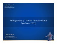
Recent work in cardiac and peripheral vascular blood flow has<br />
shown evidence for an elegant complexity to flow within the heart<br />
and in the large to medium arteries. Blood flow is normally described<br />
as laminar in that the blood travels smoothly or in regular paths. The<br />
velocity, pressure, and other flow properties at each point in the<br />
fluid remain constant, all parallel to each other. Our understanding<br />
has revolved around a two-dimensional representation of flow within<br />
three-dimensional (3-D) blood vessels. However, MRI and color<br />
Doppler flow imaging techniques have demonstrated that there is a<br />
spiral/helical/rotational property to laminar blood flow. (In this article,<br />
this blood flow profile will be termed spiral laminar flow though all are<br />
equally valid terms.) The column of blood turns on a central axis as it passes along the major<br />
arteries (Figure 1).<br />
Heart<br />
The heart is a remarkable structure that displays a<br />
twisting/wringing motion during emptying and early<br />
filling, in part due to counter-wound helical muscle<br />
fibers. Much of this underlying principle has been<br />
known for 500 years, the helical nature of myocardial<br />
fibers being described by Lower in the second half of<br />
the 17th century. More recently, Buckberg focused on<br />
linking cardiac helical anatomy with cardiac function<br />
based on work by Torrent-Guasp concerning the 3-D<br />
nature of left ventricular myocardial structure. 1, 2 This<br />
is encompassed in the concept of the “helical” pump<br />
or heart. The heart appears to operate as a spiral-compressive<br />
pump with the wall following a spiral descent<br />
rather than a direct linear move to the center (Figure 2). 3<br />
The spiral trabecular configuration of the internal surface<br />
of the left ventricle demonstrated by Gorodkov<br />
may also have a role. 4 This twisting motion during contraction<br />
is believed to result in more efficient cardiac<br />
Figure 1. Two-dimensional versus threedimensional<br />
representation of laminar flow in<br />
a tube.<br />
emptying, with blood ejected from the left ventricle<br />
having a rotational element. It can be argued that<br />
having a rotational element to flow has the additional<br />
advantage of imposing a laminar order rather than the<br />
natural “settling out” of turbulence. Eliminating turbulent<br />
flow may also be important with respect to cardiac<br />
work since turbulent flow is more resistant to pumping,<br />
requiring more energy to move the fluid and therefore<br />
more work for the heart. The concept of the ‘helical<br />
heart’ therefore could be anticipated to have advantages<br />
in terms of efficient emptying and efficient energy use.<br />
Interestingly, Leonardo da vinci appears to have<br />
come to a similar conclusion with respect to spiral<br />
cardiac emptying as he sketched what very much<br />
appears to be spiral flow being generated within the left<br />
ventricle of the heart in the 16th century.<br />
Aorta<br />
Modern imaging techniques have shown spiral<br />
MDCvJ | vII (1) 2011 21
LV<br />
Contraction / expansion<br />
Figure 2. MRI images of left ventricular myocardial motion and<br />
interpretation. From: Jung B, Markl M, Föll D, Hennig J. Investigating<br />
myocardial motion by MRI using tissue phase mapping. Eur J<br />
Cardiothorac Surg. 2006;29 Suppl 1:S150-7.<br />
laminar flow within the ascending, arch, descending<br />
thoracic and abdominal aorta. 5-11 A clockwise rotation<br />
has been demonstrated during systole, although a counter-clockwise<br />
rotation has been shown in diastole in the<br />
descending aorta. 12<br />
As there is spiral flow within the ascending aorta, the<br />
nonplanar arch of the aorta appears not to be the prime<br />
initiator of this flow profile. It is likely, however, that it<br />
reinforces and propagates this 3-D flow pattern through<br />
wall motion, compliance, and the tapering nature of the<br />
aorta. 13<br />
The spiral flow axis line is at the center line of the<br />
aorta. This is thought to play a significant part in maintaining<br />
the blood flow direction passing through the<br />
curved aortic arch, keeping the most effective ejection<br />
as well as in dispersing the shear stress in the aortic<br />
wall. 14<br />
Lee J. Frazin, 20 years ago, concluded that spiral<br />
flow may affect organ perfusion and that the rotational<br />
element of flow should be taken into account when<br />
studying flow within the aorta. 7 Recent numerical analyses<br />
of the impact of spiral flow within the aorta have<br />
concluded that the flow profile may indeed have physiological<br />
significance in the aorta. It is predicted to play<br />
a positive role in the transport of oxygen by enhancing<br />
oxygen flux to the arterial wall. It reduces the luminal<br />
surface LDL concentration in the aortic arch and probably<br />
plays a role in suppressing severe polarization of<br />
LDL at the origins of the three branches on the arch,<br />
therefore protecting them from atherogenesis. 15, 16 A<br />
study examining the effect of spiral flow on the uptake<br />
of LDL on a straight segment of rabbit aorta arrived at<br />
a very similar conclusion: that spiral flow in the arterial<br />
system plays a beneficial role in protecting the arterial<br />
wall from atherogenesis. 17<br />
Passing into the abdominal aorta, a further numerical<br />
study of spiral flow within the aorta showed a beneficial<br />
effect on the nature of flow within the iliac vessels.<br />
There were no regions of flow separation and decreased<br />
differences in the shear stress between the inner and<br />
outer walls in the iliac arteries. Spiral flow appears to<br />
impart a stabilizing effect on flow patterns in the downstream<br />
branches of the aorta. 18<br />
Peripheral Arterial<br />
In 1991, parallel work related to blood flow in arteries<br />
led to the suggestion that the normal physiological<br />
blood flow pattern may in fact be spiral laminar flow. 19<br />
The 3-D nature of blood flow was demonstrated in<br />
more peripheral and superficial arteries using colorflow<br />
Doppler. A true transverse plane, color Doppler<br />
interrogation of blood vessels at low velocity settings<br />
shows a characteristic appearance — the “red/blue<br />
split” (Figure 3). 20 This has been shown to represent<br />
spiral laminar flow with its axis at the centre of the<br />
artery, which has been termed “spiral laminar flow.” It<br />
is important to emphasize that the flow profile is not<br />
only rotating but also laminar for the characteristic profile<br />
to exist. using this methodology, this distinctive<br />
appearance and therefore flow profile has been reported<br />
in relatively superficial vessels such as the femoral and<br />
carotid arteries. 21 Magnetic resonance angiography<br />
(MRA) can be used in a similar way to interrogate the<br />
carotids, revealing the same 3-D flow profile. 22<br />
The peripheral arterial tree beyond the aortic arch<br />
also has significant elements of nonplanar construction<br />
that may play a role in the occurrence of spiral laminar<br />
flow. 23 It is also interesting to note that the arrangement<br />
of the muscle and elastin fibers of the arteries have been<br />
shown to be in helical arrangement. 24 Whether this is a<br />
coincidence or has a more direct relationship with the<br />
nature of flow in the vessel is as yet unknown.<br />
Figure 3. Arterial color Doppler of a transverse view of the common<br />
femoral artery and a graphic representation.<br />
22 vII (1) 2011 | MDCvJ
As a consequence of the above work, it is now possible<br />
to bring left ventricular anatomy and function<br />
together with blood flow within large-to-medium<br />
arteries as a coherent beneficial flow system. Cardiac<br />
architecture causes the left ventricle to “wring/squeeze”<br />
the heart empty, acting as a kind of spiral-compressive<br />
pump. This ejects the blood flow from the left ventricle<br />
through a nondiseased aortic valve with a spiral laminar<br />
flow profile. This flow pattern has been shown to be<br />
preserved as it passes around the arch of the aorta and<br />
into the descending/abdominal aorta, which may in<br />
part be due to the nonplanar nature of the aortic arch.<br />
Finally, spiral laminar flow has been identified within<br />
more distal direct line vessels (femoral arteries) and<br />
branch vessels (carotid arteries). Spiral flow may also<br />
have beneficial effects on cardiac work and tissue oxygenation<br />
and in the protection of the arterial wall from<br />
atherogenesis.<br />
Clinical Correlation<br />
When assessing the significance of spiral laminar<br />
flow, it is important to show a causal relationship with<br />
the development of arterial disease. There is very little<br />
work in this area and it is more circumstantial than<br />
definitive.<br />
The loss of spiral blood flow has been associated with<br />
the presence, severity and progression of atheromatous<br />
disease. 25, 26 However, it should be stressed that this is<br />
an association and not proof of a causal relationship. A<br />
long-term mapping of flow patterns against disease and<br />
its progression is required, and this has not been performed<br />
as yet.<br />
Flow Analysis and Vascular Stents and<br />
Grafts<br />
Hemodynamic forces are a key localizing factor in<br />
arterial endothelial dysfunction and, as a result, arterial<br />
pathology. 27 Low shear stress appears to be one of the<br />
key components of this flow/vessel wall interaction. 28 In<br />
1993, Zarins et al. demonstrated that intimal thickening<br />
and atherosclerosis develop in regions of relatively low<br />
shear stress and variation from axially aligned unidirectional<br />
flow. 29 Stonebridge and Brophy hypothesized that<br />
spiral flow could exert a beneficial effect on the mechanisms<br />
of endothelial damage and repair. 19 A study by<br />
Caro et al. supported this hypothesis and demonstrated<br />
that spiral flow may lead to relative uniformity of wall<br />
shear and inhibition of flow stagnation, separation and<br />
instability. 30<br />
Spiral flow has also been shown to preserve laminar<br />
flow through stenoses (“laminar stability”), markedly<br />
Figure 4. Laminar stability — nonspiral (upper) and spiral (lower)<br />
MRI image of flow through a stenosis showing loss of image with<br />
nonspiral flow and preservation of image with spiral flow. 31<br />
reducing turbulent kinetic energy (Figure 4). It also<br />
reduces laterally directed forces impacting on the vessel<br />
wall. Spiral laminar flow generates a thin, less-dense<br />
(probably acelluar) outer shell, which may act as a “fluid<br />
bearing” for the denser inner core, again assisting in<br />
31, 32<br />
reducing rotational decay.<br />
One study investigated the magnitude of oscillating<br />
shear stress in an aortocoronary bypass computer<br />
model in the presence of spiral flow. A linear inverse<br />
relationship was found between the oscillating shear<br />
index and the helical flow index for the models. The<br />
results indicated that spiral flow damped the wall shear<br />
stress temporal gradients within the proximal graft. The<br />
authors suggested that spiral flow might play a significant<br />
role in preventing plaque deposition by moderating<br />
the mechanotransduction pathways of cells. They further<br />
concluded that the strength of the spiral flow<br />
signal could be used to risk stratify for the activation of<br />
mechanical and biological pathways leading to fibrointimal<br />
hyperplasia. 33<br />
A graft or stent that recreates spiral flow at the distal<br />
anastomosis/outflow might be anticipated to have some<br />
benefits. A numerical analysis based on a spiral flow<br />
inducer for endovascular stents showed that the inducer<br />
could create sufficiently strong spiral flow, effectively<br />
MDCvJ | vII (1) 2011 23
educing turbulence created by the stent, so that arterial<br />
restenosis due to the stent implantation might be suppressed.<br />
34<br />
The manner in which these findings can be of importance<br />
to prosthetic graft design relate to the outflow<br />
from prosthetic grafts. Many prosthetic grafts fail due to<br />
neointimal hyperplasia at the distal anastomosis, which<br />
eventually occludes outflow. One hypothesis tested by<br />
stents and grafts, which engender spiral flow, is that the<br />
endothelial cells at the distal outflow are sensitive to the<br />
flow environment. Neointimal hyperplasia may in part<br />
or whole be a normal distal anastomotic endothelial cell<br />
mechanosensory response to an abnormal flow environment<br />
(nonlaminar flow, i.e., turbulence, stagnation,<br />
oscillatory shear stress). Reducing any flow-mediated<br />
drive to neointimal hyperplasia would be anticipated to<br />
prolong graft patency.<br />
Spiral flow has also been shown to increase the blood<br />
velocity near the vessel wall and the wall shear rate.<br />
This is thought to potentially reduce acute thrombus<br />
formation and intimal hyperplasia and thereby improve<br />
graft patency rates. 35 A second study, using a simplified<br />
model of a stent within a straight segment of an<br />
artery in which spiral flow was introduced, showed<br />
that this flow reduced the size of the disturbed flow<br />
zones, enhanced the average wall shear stress, and lowered<br />
oscillatory shear index in the stent. All of these<br />
are believed to be adverse factors in the development of<br />
arterial restenosis after stent deployment. 36 Interestingly,<br />
the minimum rotational velocity of the spiral flow<br />
required was approximately 6.5 cms/sec, which is very<br />
similar to that shown in femoral arteries of healthy<br />
volunteers. 21<br />
One of the major causes of early failure of smallcaliber<br />
artificial vascular grafts is acute thrombus<br />
formation, in which the interaction of platelets with<br />
the grafts’ thrombogenic surfaces is the initiating step.<br />
According to Sauvage, the action of shear forces can<br />
prevent thrombus from forming on the graft wall if the<br />
blood flow in the graft is higher than the thrombotic<br />
threshold velocity. 37 unfortunately, blood flow rates in<br />
small-diameter grafts are small, usually resulting in a<br />
time average velocity of blood flow in these grafts to<br />
be below this thrombotic threshold velocity. Enhancing<br />
blood velocity near the wall of a graft by inducing spiral<br />
flow may overcome this acute thrombus problem and<br />
may be a solution to increasing the patency of smalldiameter<br />
vascular grafts. A recent study showed that<br />
spiral flow generated less adhesion of platelets to the<br />
inner surface of a tube when compared with nonspiral<br />
flow. The authors concluded that intentionally introducing<br />
spiral flow in small-caliber arterial grafts has no<br />
adverse effect on platelet activation and may indeed be a<br />
solution to improving the patency of the grafts by<br />
suppressing acute thrombus formation. 38<br />
Whether the artificial engendering of spiral laminar<br />
flow has biological advantages has yet to be definitively<br />
answered. If endothelial cells are sensitive to juxtamural<br />
turbulence, areas of stagnation, stress gradients, and/<br />
or high oscillatory shear stress, then the stabilization of<br />
laminar flow is likely to be an advantage. The delivery<br />
of stabilized spiral laminar flow from prostheses has<br />
not been possible until recently and therefore could<br />
benefit from further research.<br />
Conclusion<br />
Spiral laminar flow is an elegant unified flow concept.<br />
There are a number of published features of spiral<br />
laminar flow, indicating that this flow profile may have<br />
advantages over nonspiral flow in both physiological<br />
(heart and the peripheral artery) and device-related flow<br />
(Table 1). It is possible to speculate about other potential<br />
beneficial properties but these have as yet not been<br />
tested. There is, therefore, a lot more work to be done in<br />
this field. Only more research and time will determine<br />
whether or not it is something of true significance.<br />
Laminar stability<br />
Reduced laterally directed forces<br />
Reduced near-wall turbulence<br />
Suppresses acute thrombus formation with no increase in<br />
platelet activation<br />
Enhances oxygen flux to the arterial wall<br />
Reduces luminal surface LDL concentration<br />
Dampens wall stress temporal gradients<br />
Lowers oscillatory shear stress index<br />
Table 1. Published features of spinal laminar flow<br />
24 vII (1) 2011 | MDCvJ
References<br />
1. Buckberg GD. Basic science review: the helix and the<br />
heart. J Thorac Cardiovasc Surg. 2002 Nov;124(5):863-83.<br />
2. Kocica MJ, Corno AF, Carreras-Costa F, Ballester-Rodes M,<br />
Moghbel MC, Cueva CN, Lackovic v, Kanjuh vI, Torrent-<br />
Guasp F. The helical ventricular myocardial band: global<br />
three-dimensional, functional architecture of the ventricular<br />
myocardium. Eur J Cardiothoracic Surg. 2006 Apr;29<br />
Suppl 1:S21-40.<br />
3. Jung B, Markl M, Föll D, Hennig J. Investigating myocardial<br />
motion by MRI using tissue phase mapping. Eur J<br />
Cardiothorac Surg. 2006 Apr;29 Suppl 1:S150-7.<br />
4. Bockeria LA, Gorodkov AJ, Dorofeev Av, Alshibaya<br />
MD; RESTORE Group. Left ventricular geometry reconstruction<br />
in ischemic cardiomyopathy patients with<br />
predominately hypokinetic left ventricle. Eur J Cardiothorac<br />
Surg. 2006 Apr;29 Suppl 1:S251-8.<br />
5. Segadal L, Matre K. Blood velocity distribution in the<br />
human ascending aorta. Circulation. 1987 Jul;76(1):90-100.<br />
6. Kilner PJ, yang GZ, Mohiaddin RH, Firmin DN, Longmore<br />
DB. Helical and retrograde secondary flow patterns<br />
in the aortic arch studied by three-directional magnetic<br />
resonance velocity mapping. Circulation. 1993 Nov;88<br />
(5 Pt 1):2235-47.<br />
7. Frazin LJ, Lanza G, vonesh M, Khasho F, Spitzzeri C,<br />
McGee S, Mehlman D, Chandran KB, Talano J, McPherson<br />
D. Functional chiral asymmetry in descending thoracic<br />
aorta. Circulation. 1990 Dec;82(6):1985-94.<br />
8. Weigang E, Kari FA, Beyersdorf F, Luehr M, Etz CD,<br />
Frydrychowicz A, Harloff A, Markl M. Flow-sensitive<br />
four-dimensional magnetic resonance imaging: flow<br />
patterns in ascending aortic aneurysms. Eur J Cardiothorac<br />
Surg. 2008 Jul;34(1):11-6.<br />
9. Frazin LJ, vonesh MJ, Chandran KB, Shipkowitz T,<br />
yaacoub AS, McPherson DD. Confirmation and initial<br />
documentation of thoracic and abdominal aortic<br />
helical flow. An ultrasound study. ASAIO J. 1996<br />
Nov-Dec;42(6):951-6.<br />
10. Markl M, Draney MT, Hope MD, Levin JM, Chan FP,<br />
Alley MT, Pelc NJ, Herfkens RJ. Time-resolved 3-dimensional<br />
velocity mapping in the thoracic aorta: visualization<br />
of 3-directional blood flow patterns in healthy volunteers<br />
and patients. J Comput Assist Tomogr. 2004<br />
Jul-Aug;28(4):459-68.<br />
11. Jin S, Oshinski J, Giddens DP. Effects of wall motion and<br />
compliance on flow patterns in the ascending aorta. J<br />
Biomech Eng. 2003 Jun;125(3):347-54.<br />
12. Frazin LJ, vonesh MJ, Chandran KB, Shipkowitz T,<br />
yaacoub AS, McPherson DD. Confirmation and initial<br />
documentation of thoracic and abdominal aortic<br />
helical flow. An ultrasound study. ASAIO J. 1996<br />
Nov-Dec;42(6):951-6.<br />
13. Shipkowitz T, Rodgers vG, Frazin LJ, Chandran KB.<br />
Numerical study on the effect of secondary flow in the<br />
human aorta on local shear stresses in abdominal aortic<br />
branches. J Biomech. 2000 Jun;33(6):717-28.<br />
14. Tanaka M, Sakamoto T, Sugawara S, Nakajima H,<br />
Kameyama T, Katahira y, Ohtsuki S, Kanai H. Spiral<br />
systolic blood flow in the ascending aorta and aortic<br />
arch analyzed by echo-dynamography. J Cardiol. 2010<br />
Jul;56(1):97-110. Epub 2010 May 13.<br />
15. Liu X, Fan y, Deng X. Effect of spiral flow on the transport<br />
of oxygen in the aorta: a numerical study. Ann Biomed<br />
Eng. 2010 Mar;38(3):917-26. Epub 2009 Dec 24.<br />
16. Liu X, Pu F, Fan y, Deng X, Li D, Li S. A numerical study<br />
on the flow of blood and the transport of LDL in the<br />
human aorta: the physiological significance of the helical<br />
flow in the aortic arch. Am J Physiol Heart Circ Physiol.<br />
2009 Jul;297(1):H163-70. Epub 2009 May 8.<br />
17. Ding Z, Fan y, Deng X, Zhan F, Kang H. Effect of swirling<br />
flow on the uptakes of native and oxidized LDLs in a<br />
straight segment of the rabbit thoracic aorta. Exp Biol Med<br />
(Maywood). 2010 Apr;235(4):506-13.<br />
18. Shipkowitz T, Rodgers vG, Frazin LJ, Chandran KB.<br />
Numerical study on the effect of secondary flow in the<br />
human aorta on local shear stresses in abdominal aortic<br />
branches. J Biomech. 2000 Jun;33(6):717-28.<br />
19. Stonebridge PA, Brophy CM. Spiral laminar flow in arteries?<br />
Lancet. 1991 Nov 30;338(8779):1360-1.<br />
20. Hoskins PR, Fleming AD, Stonebridge PA, Allan PL,<br />
Cameron DC. Scan-plane vector maps and secondary flow<br />
motions in arteries. Eur J ultrasound. 1994;1:159-69.<br />
21. Stonebridge PA, Hoskins PR, Allan PL, Belch JF. Spiral<br />
laminar flow in vivo. Clin Sci (Lond). 1996 Jul;91(1):17-21.<br />
22. Harloff A, Albrecht F, Spreer J, Stalder AF, Bock J, Frydrychowicz<br />
A, Schöllhorn J, Hetzel A, Schumacher M, Hennig<br />
J, Markl M. 3-D blood flow characteristics in the carotid<br />
artery bifurcation assessed by flow-sensitive 4D MRI at 3T.<br />
Magn Reson Med. 2009 Jan;61(1):65-74.<br />
23. Long Q, Xu Xy, Ariff B, Thom SA, Hughes AD, Stanton<br />
Av. Reconstruction of blood flow patterns in a human<br />
carotid bifurcation: a combined CFD and MRI study. J<br />
Magn Reson Imaging. 2000 Mar;11(3):299-311.<br />
24. Rhodin JAG. Architecture of the vessel wall. In: Bohr DF,<br />
Somylo AP, Sparks Hv Jr, editors. Handbook of Physiology,<br />
Sect 2. Bethesda, MD: American Physiology Society,<br />
1980.<br />
25. Houston JG, Gandy SJ, Sheppard DG, Dick JB, Belch<br />
JJ, Stonebridge PA. Two-dimensional flow quantitative<br />
MRI of aortic arch blood flow patterns: Effect of age, sex,<br />
and presence of carotid atheromatous disease on prevalence<br />
of spiral blood flow. J Magn Reson Imaging. 2003<br />
Aug;18(2):169-74.<br />
MDCvJ | vII (1) 2011 25
26. Houston JG, Gandy SJ, Milne W, Dick JB, Belch JJ, Stonebridge<br />
PA. Spiral laminar flow in the abdominal aorta:<br />
a predictor of renal impairment deterioration in patients<br />
with renal artery stenosis? Nephrol Dial Transplant. 2004<br />
Jul;19(7):1786-91. Epub 2004 May 25.<br />
27. Friedman MH, Krams R, Chandran KB. Flow interactions<br />
with cells and tissues: cardiovascular flows and<br />
fluid-structure interactions. Sixth International Bio-<br />
Fluid Mechanics Symposium and Workshop, March<br />
28-30, 2008, Pasadena, California. Ann Biomed Eng. 2010<br />
Mar;38(3):1178-87.<br />
28. Caro CG, Fitz-Gerald JM, Schroter RC. Arterial wall shear<br />
and distribution of early atheroma in man. Nature. 1969<br />
Sept 13;223(5211):1159-60.<br />
29. Zarins CK, Giddens DP, Bharadvaj BK, Sottiurai vS,<br />
Mabon RF, Glagov S. Carotid bifurcation atherosclerosis.<br />
Quantitative correlation of plaque localization with<br />
flow velocity profiles and wall shear stress. Circ Res. 1983<br />
Oct;53(4):502-14.<br />
30. Caro CG, Cheshire NJ, Watkins N. Preliminary comparative<br />
study of small amplitude helical and conventional<br />
ePTFE arteriovenous shunts in pigs. J R Soc Interface. 2005<br />
Jun 22;2(3):261-6.<br />
31. Stonebridge PA, Buckley C, Thompson A, Dick J, Hunter<br />
G, Chudek JA, Houston JG, Belch JJ. Non spiral and spiral<br />
(helical) flow patterns in stenoses. In vitro observations<br />
using spin and gradient echo magnetic resonance imaging<br />
(MRI) and computational fluid dynamic modeling. J Int<br />
Angiol. 2004 Sep;23(3):276-83.<br />
32. Paul MC, Larman A. Investigation of spiral blood flow<br />
in a model of arterial stenosis. Med Eng Phys. 2009<br />
Nov;31(9):1195-203. Epub 2009 Aug 11.<br />
33. Morbiducci u, Ponzini R, Grigioni M, Redaelli A. Helical<br />
flow as fluid dynamic signature for atherogenesis risk<br />
in aortocoronary bypass. A numeric study. J Biomech.<br />
2007;40(3):519-34. Epub 2006 Apr 19.<br />
34. Teng X, Deng X. [Optimization of a helical flow inducer of<br />
endovascular stent based on the principle of swirling flow<br />
in arterial system][Article in Chinese]. Sheng Wu yi Xue<br />
Gong Cheng Xue Za Zhi. 2010 Apr;27(2):429-34.<br />
35. Zhang Z, Fan y, Deng X, Wang G, Zhang H, Guidoin R.<br />
Simulation of blood flow in a small-diameter vascular<br />
graft model with a swirl (spiral) flow guider. Sci China C<br />
Life Sci. 2008 Oct;51(10):913-21. Epub 2008 Sept 26.<br />
36. Chen Z, Fan y, Deng X, Xu Z. Swirling flow can suppress<br />
flow disturbances in endovascular stents: a numerical<br />
study. ASAIO J. 2009 Nov-Dec;55(6):543-9.<br />
37. Sauvage LR, Walker MW, Berger K, Robel SB, Lischko<br />
MM, yates SG, Logan GA. Current arterial prostheses.<br />
Experimental evaluation by implantation in the carotid<br />
and circumflex coronary arteries of the dog. Arch Surg.<br />
1979 Jun;114(6):687-91.<br />
38. Zhan F, Fan y, Deng X. Swirling flow created in a glass<br />
tube suppressed platelet adhesion to the surface of the<br />
tube: its implication in the design of small-caliber arterial<br />
grafts. Thromb Res. 2010 May;125(5):413-8. Epub 2009<br />
Mar 21.<br />
26 vII (1) 2011 | MDCvJ
P.E. Sovelius Jr.<br />
Overview<br />
PLATO’S CAvE — KNOWLEDGE-BASED<br />
MEDICINE OR BLACK SWAN<br />
TECHNOLOGy?<br />
Paul E. Sovelius Jr.<br />
The <strong>Methodist</strong> <strong>Hospital</strong>, Houston, Texas<br />
Plato’s CAVE TM is a presurgical planning, multidimensional “life space situation room” designed, developed,<br />
and introduced to clinical practice by the Department of Radiation Oncology at The <strong>Methodist</strong><br />
<strong>Hospital</strong>, located in Houston’s Texas Medical Center. At approximately 500 square feet, Plato’s CAVE<br />
was specifically designed to permit a team of physicians to review all available diagnostic images of the<br />
patient (Figures 1a, 1b). The initial clinical focus was for interventions within the domain of surgical oncology,<br />
including radiation therapy, reconstructive surgery, and organ transplantation. This advanced clinical<br />
visualization process, supported by a novel and creative assemblage of FDA-approved, commercially<br />
available diagnostic imaging components, is available for all relevant patient care services within The<br />
<strong>Methodist</strong> <strong>Hospital</strong> System.<br />
Figure 1a. Inside Plato’s CAVE with large screen and virtual<br />
surgical table visualized with TeraRecon, Inc.<br />
Figure 1b. Inside Plato’s CAVE with large screen and laptop<br />
integrated visualization by BodyViz, Inc.<br />
The underlying concept is that of a military flight simulator. As opposed to a commercial flight simulator, a<br />
fighter pilot environment is a multi-body simulation that records all of the plane’s internal instruments and<br />
the pilot’s actions with the digital fly-by-wire avionics system; its external position in relation to its global<br />
position; and, with GPS, the absolute spatial (X, Y, Z) position of the squadron and the aggressor(s) within<br />
its three-dimensional sphere of influence. This well-vetted process of recording input/output variables and<br />
resultant differences along the flight path of interaction is critical for pilots to learn from each other’s experience<br />
what works well and what should be improved, all within a real-time frame of reference for action<br />
and reaction. The knowledge gained is now being used by unmanned aerial vehicles that are remotely<br />
piloted, not unlike the da Vinci surgical robot.<br />
MDCvJ | vII (1) 2011 27
Figure 2. “The Visible Human”<br />
from the1995 National Library of<br />
Medicine, rendered with Fovia, Inc.<br />
Figure 3. Archimedes<br />
Circulatory System from 287 B.C.<br />
Plato’s CAVE: Conceptual Beginnings<br />
I first designed the single-user surgeon computeraugmented<br />
virtual environment (CAvE) concept in 2006<br />
as the sole proprietor of a 20 ft x 15 ft glass-walled kiosk<br />
that was to be installed within one of The <strong>Methodist</strong><br />
<strong>Hospital</strong> System’s professional buildings. Initially<br />
called the Surgeons EDGE TM , it was designed for cardiovascular<br />
surgeons who did not have the time or<br />
tools necessary to manipulate and integrate a series of<br />
patient-specific CT angiography (CTA) studies into an<br />
interactive 3-D volume that could be used to measure<br />
aortic grafts for individual patients. With the encouragement<br />
of Drs. Richard Geis, Samuel Henly, and Alan<br />
Lumsden, I designed a kiosk near the OR where cardiovascular<br />
physicians could fit aortic grafts to their<br />
patients on a 24/7 basis. The kiosk was to be fitted with<br />
a state-of-the-art 3-D TeraRecon large-screen workstation,<br />
and my role evolved into an “aorta tailor” who<br />
works with the physicians and sets up the procedure for<br />
measurement and validation.<br />
In 2007, Drs. E. Brian Butler and Alan Lumsden were<br />
collaborating with IBM on a research project called the<br />
“virtual Patient.” They were attempting to use the<br />
National Library of Medicine’s MRI-based visible<br />
Human Project ® as a base map for a detailed, imageguided,<br />
auto-segmented atlas of patient-specific CTAs<br />
(Figures 2 and 3). The visible Human Project involved the<br />
creation of complete, detailed, 3-D representations of the<br />
male and female human bodies using CT and MRI slices.<br />
Lumsden and Butler built on this concept with the<br />
acquisition of transverse CT, MR, and cryosection<br />
images of representative male and female cadavers.<br />
The male was sectioned at one-millimeter intervals, the<br />
female at one-third of a millimeter intervals; this level<br />
of resolution still exceeds the standard of care today.<br />
The virtual Patient data set, with its detailed crosssectional<br />
pathology photographs, was coregistered<br />
with medical imaging data sets of the body. The 3-D<br />
reconstructed virtual cardiovascular images were then<br />
automatically labeled and subsequently tagged with<br />
SQuID (superconducting quantum interference device)<br />
detectable anatomical markers for the proposed intraoperative<br />
image-guided system.<br />
This process was designed to assist Drs. Lumsden<br />
and Butler in creating a vascular roadmap for the targeted<br />
delivery of a gene therapy “package” that could<br />
be activated with a time-release viral agent designed<br />
by Dr. Butler. Before it could be concluded, it was put<br />
on hold for two reasons: Dr. Butler was named chairman<br />
of the newly formed Department of Radiation<br />
Oncology at The <strong>Methodist</strong> <strong>Hospital</strong>, and Dr. Lumsden<br />
was named chair of <strong>Methodist</strong>’s Department of<br />
Cardiovascular Surgery. In order to keep the project<br />
moving, both doctors asked me to work full-time with<br />
Dr. Butler at The <strong>Methodist</strong> <strong>Hospital</strong> and to look outside<br />
the box for nonclinical engineering solutions to the<br />
image-guided delivery system based on my previous<br />
scientific visualization experience.<br />
I have worked for many years providing the visual<br />
system hardware and software for real-time fighter<br />
pilot training, 3-D seismic/geologic oil exploration, and<br />
pharmaceutical drug design using supercomputing<br />
tightly coupled to high-performance 3-D workstations.<br />
Previously, I had worked with Dr. Lumsden at Baylor<br />
College of Medicine on a peripheral arterial disease<br />
(PAD) multi-institutional clinical trial using 3T MRI<br />
with specialized bilateral coils for measuring plaque<br />
within the superficial femoral artery. Dr. Lumsden<br />
would repeat the phrase “it’s all about the imaging”<br />
like a mantra for the vascular residents and fellows. Dr.<br />
Butler’s vision for an interactive, real-time, 3-D radiation<br />
dose cloud for intensity modulated radiation therapy<br />
(IMRT) treatment planning was a logical and parallel<br />
extension of Dr. Lumsden’s vision.<br />
However, Dr. Butler needed to have a full integration<br />
of diagnostic imaging capabilities that would allow<br />
for clearer visualization and provide a more accurate,<br />
interactive tumor targeting and delineation system<br />
that could protect normal tissue from adverse radiation<br />
exposure. It was at this point that I decided to design<br />
an FDA-approved clinical diagnostic imaging integration<br />
that would be clinically relevant and make a<br />
difference in patient care.<br />
Why “Plato’s CAVE?”<br />
When I asked Dr. Butler to think about a name for<br />
this pilot clinical project, he responded with “Plato’s<br />
CAvE” from Book vII of Plato’s The Republic, “The<br />
Allegory of the Cave,” in which the prisoners see and<br />
28 vII (1) 2011 | MDCvJ
Figure 4. Plato, in The Republic, Book VII, 514a-520a. 1 Figure 5. “The Anatomy Lecture of Dr. Nicolaes Tulp” by Rembrandt,<br />
1632.<br />
hear shadows and echoes cast by objects that they<br />
cannot see clearly (Figure 4 and 5). 1<br />
It was the perfect code name for exploring the art and<br />
science of “seeing” the invisible.<br />
Dr. Butler’s research over the past decade has been<br />
focused on image-guided radiation and gene therapies<br />
for cancer. In the past year, he and I have broadened the<br />
focus to include visualization-guided clinical interventions<br />
that are within the domain of surgical oncology,<br />
including reconstructive surgery and transplantation.<br />
This expanded focus was made possible by the development<br />
of an N-dimensional (space, time, color, and<br />
multimodality images) image-guided intervention<br />
system, more correctly a visualization system, that has<br />
been operational in our laboratory since April 2009.<br />
Plato’s CAVE: The Advanced Clinical<br />
Visualization Process<br />
The current standard of care for medical imaging is<br />
to interpret slices of 2-D shades-of-gray images in an<br />
attempt to translate them into a physician’s “personal<br />
virtual 3-D anatomical and disease world,” then interpreting<br />
the problem and rendering a written diagnosis<br />
— granted, an expert professional diagnosis by a welltrained<br />
imaging specialist, but still an interpretation of<br />
a 2-D dataset nonetheless. Plato’s CAvE raises the bar<br />
with an advanced clinical visualization process that<br />
creates 3-D images derived from a patient’s DICOM<br />
image studies that have been pulled from <strong>Methodist</strong>’s<br />
Picture Archiving and Communication System (PACS),<br />
from other institutional PACS via electronic transfer, or<br />
from CDs that patients bring with them. Regardless of<br />
the image source, the 3-D visualizations are ready for<br />
projection and viewing on a variety of screen formats<br />
within minutes. The region of interest is measurable,<br />
can be manipulated in three dimensions, and can be<br />
presented stereoscopically like the movie Avatar. This<br />
approach makes full use of the strengths of each imaging<br />
modality by displaying and/or fusing all of the<br />
images available for the patient (CT, MRI, PET and,<br />
soon, HR ultrasound) in a sequence of the events that<br />
are relevant to the treatment team. The scene is adaptable<br />
to all surgical interventions, from reconstruction<br />
through transplantation, and capable of bringing quality<br />
improvements to the diagnosis and treatment<br />
planning for many medical conditions.<br />
Our technology is totally complementary to all<br />
standard-of-care imaging modalities currently in use<br />
and some that are in development, i.e., physiodynamic<br />
processes, beating heart, mitral valves opening and<br />
closing. There is a high level of clinical imaging and<br />
medical device industry interest in experimenting with<br />
or enhancing Plato’s CAvE with natural user interactions.<br />
Our surgeons have discovered, once they work<br />
with this system, that they cannot easily justify returning<br />
to the current standard of viewing images for their<br />
patients. We have designed and integrated the CAvE<br />
to function as a node/portal on a physician grid or<br />
network. It is not an island: the visualization can be<br />
distributed to the scientist’s or clinician’s desktop<br />
because it is server-based, thin-client enabled, and<br />
HIPAA compliant.<br />
The system offers enormous opportunity for educating<br />
primary care and referring physicians, patients and<br />
their families, medical students, residents and staff surgeons<br />
by narrowing the degree of uncertainty that each<br />
brings to the interaction (Figure 6 and 7). It also gives<br />
the medical team the ability to make a more informed<br />
decision regarding the best intervention or treatment<br />
of the disease. In some cases, the visualization has<br />
revealed previously unrecognized ancillary anatomical<br />
findings or additional disease complications.<br />
Development, testing, validation and translation of<br />
this technology into practice will shape the metrics<br />
MDCvJ | vII (1) 2011 29
Figure 6. Virtual surgical table inside Plato’s CAVE using TeraRecon,<br />
Inc. visualization<br />
necessary for adoption, and we anticipate that more<br />
refined tools will be developed to interact with the<br />
visualized patient. ultimately, the images that are<br />
fused to create the visualized patient will be registered<br />
and superimposed to the real patient, and the instruments<br />
will interact with the patient to provide clinical<br />
interventions of the highest quality and safety. At the<br />
fundamental level, this system has universal applicability<br />
across all medical specialties and subspecialties and<br />
will allow the patient and physician to understand disease<br />
processes in ways never before possible.<br />
We plan systematic evaluations of organ systems<br />
and their related disease processes. The hepatobiliary<br />
system provides a good example of the importance of<br />
doing this with high-fidelity visual data. Measuring the<br />
volume of this difficult-to-image organ is key to clinical<br />
decisions with respect to cancer diagnosis and therapy,<br />
among other medical conditions. yet even simple volumetric<br />
evaluation represents a paradigm shift in the<br />
way that cancers are staged. For example, conventional<br />
staging of liver cancer is based on two dimensions — X<br />
and y. Typically “size,” as measured on axial CT or MRI<br />
images, is a major consideration, e.g., a 5 cm lesion will<br />
be classified as stage T2. In contrast, volumetric evaluation<br />
introduces a third dimension, Z, and may change<br />
the staging in significant ways. We now evaluate all<br />
hepatobiliary tumors volumetrically and within the last<br />
few weeks have begun mapping the arterial and venous<br />
branches so that surgeons can clearly avoid them while<br />
performing a resection.<br />
Physician Response to Plato’s CAVE<br />
Plato’s CAvE has generated a great deal of interest in<br />
and respect for its current ability and potential capabilities<br />
among our surgical colleagues. Some have used it<br />
Figure 7. Virtual surgical table inside Plato’s CAVE using TeraRecon,<br />
Inc. visualization.<br />
multiple times to perform pre-surgery evaluation and<br />
planning, even to the extent of revising initial treatment<br />
plans or not doing an intervention based on the<br />
Figure 8a. Virtual surgical table inside Plato’s CAVE standard-of-care<br />
alongside interactive volume visualization.<br />
Figure 8b. Volume visualization with red finger tracings on virtual<br />
surgical table.<br />
30 vII (1) 2011 | MDCvJ
Figure 9. The Artery of Adamkiewicz identified by Dr. Sam Henly.<br />
Magnified virtual surgical table view inside Plato’s CAVE using<br />
Siemens flash CT scan study with TeraRecon, Inc. visualization.<br />
Figure 11. TeraRecon, Inc. visualization for planning the repair of<br />
esophageal varices that was more complicated than anticipated.<br />
additional information they gained from the 3-D visualization<br />
(Figures 8a and 8b).<br />
Some surgeons have used our visualization as an<br />
education tool for their patients (Figures 9 and 10). The<br />
surgeons are impressed with the quality of the visualization,<br />
the ease and speed with which positions of the<br />
images can be changed, the ability to strip away tissue<br />
that blocks the view of the subject tissue, the fact that<br />
no contrast agents are required to achieve the visualization,<br />
the saving of time, and the unexpected ancillary<br />
findings (Figures 11 and 12).<br />
On the other hand, some surgeons have said they<br />
do not see the need for such an environment because<br />
their experience overrides the real-time process and 3-D<br />
visualization that we provide, and that conventional<br />
imaging meets their needs. yet, these same surgeons<br />
admit that the CAvE would have great value for medi-<br />
Figure 10. The Artery of Adamkiewicz, identified by Dr. Sam Henly,<br />
on the virtual surgical table inside Plato’s CAVE using Siemens flash<br />
CTA scan with TeraRecon, Inc. visualization.<br />
Figure 12. Virtual surgical table view inside Plato’s CAVE using GE<br />
CTA scan study with TeraRecon, Inc. visualization for planning repair<br />
of esophageal varices.<br />
cal education because of its clarity and responsiveness<br />
for accurately viewing patient-specific images and associated<br />
disease.<br />
Clearly, N-dimensional visualization challenges<br />
current clinical practice paradigms. unlike many<br />
technological advances in science and medicine, the<br />
constraint on acceptance and adoption of Plato’s CAvE<br />
does not appear to be clinician resistance to change but<br />
rather the hospital’s need for 1) metrics that will satisfy<br />
institutional risk assessment requirements, and 2)<br />
an effective process for disseminating the innovation<br />
along with best practice standards and protocols that<br />
are accepted by the general clinical staff. We routinely<br />
ask surgeons, “When you review currently available<br />
CT or MR studies for pre-surgical planning, have the<br />
standard diagnostic imaging and viewing tools provided<br />
you with the confidence that your treatment plan<br />
MDCvJ | vII (1) 2011 31
Figure 13. Looking down on the 36 in x 42 in multi-user virtual surgical table in Plato’s CAVE using<br />
Siemens flash low-dose CT scan patient data with TeraRecon, Inc.<br />
is based on objective visual anatomical accuracy versus<br />
subjective image estimation?” Their response is invariably<br />
“No.”<br />
In my 30 years within the scientific visualization<br />
world, I believe the experience that Plato’s CAvE provides<br />
is approaching what some would call a black<br />
swan. The term “black swan” was a Latin term; its<br />
oldest reference is in the poet Juvenal’s expression that<br />
“a good person is as rare as a black swan” (“rara avis<br />
in terris nigroque simillima cygno”). 2 It was a common<br />
expression in 16th-century that derived from the oldworld<br />
presumption that all swans must be white<br />
because all historical records of swans reported that<br />
they had white feathers. After the 1697 discovery of<br />
black swans in Western Australia, 3 the term metamorphosed<br />
to connote that a supposed impossibility may<br />
later be found to exist. Writer Michael Mandel used<br />
the term succinctly in an online issue of Bloomberg<br />
Businessweek: “The world is consistently capable of generating<br />
‘black swans’ — outlier events, which have an<br />
extreme impact and retrospective (though not prospective)<br />
predictability.” 4<br />
For the last few months, the clinical focus of the<br />
CAvE has evolved from physician-to-physician collaboration<br />
to physician-to-patient consultation and<br />
consensus. Almost 200 patients have been reviewed in<br />
the CAvE, and a word-of-mouth, patient-driven referral<br />
process has evolved because the involved physicians say<br />
that Dr. Butler’s vision is providing an additional level<br />
of confidence, trust and understanding in what the physician<br />
and medical team can provide for each patient.<br />
The Future of Plato’s CAVE<br />
We have begun to develop, adapt and test dual-reality<br />
tools (physical devices synched with a virtual clone<br />
of the device) that enable operator interaction with the<br />
patient-specific volume. Modeled to reflect the way<br />
surgeons use the real device, these tools will allow measurement<br />
and quantification of disease processes within<br />
the visualized patient (e.g., volume of a liver or blood<br />
vessel, size and location of an obstruction within the<br />
bile duct, how a pancreatic carcinoma encases the superior<br />
mesenteric artery, etc.). The physician can interact<br />
with the visualization of the patient before performing<br />
a procedure and record a scenario that can act as a<br />
look-ahead avoidance system, something the current da<br />
vinci robots do not permit (Figure 13). This interaction<br />
will occur in numerous ways, one of which involves<br />
using a flat-panel, multi-user, multi-touch “surgical<br />
table” that allows multiple users to interact simultaneously<br />
with the visualized patient in a 1:1 mapped<br />
relationship. We are currently refining techniques for<br />
capturing a surgeon’ s eye-hand movements in the OR<br />
that will enable us to build feedback loops based on<br />
32 vII (1) 2011 | MDCvJ
Figure 14. Reconstructed left breast with implant failure and disease<br />
in right breast; CT from Siemens.<br />
haptic, hand-gesture, voice, and eye-tracking interaction<br />
between the operator and the patient visualization synchronously<br />
as it appears on the surgical table.<br />
The CAvE’s multi-dimensional interactive field of<br />
view includes the three spatial dimensions as well<br />
as motion, coordinated contrasting colors for different<br />
tissue types (such as muscle, organ, bone, nerve,<br />
blood vessels and ducts), time, and scale — in effect,<br />
it provides a virtual biopsy of any part of the visualized<br />
patient. Our first research and development project<br />
will soon enable Plato’s CAvE to integrate and visualize<br />
physiological and biochemical parameters at the cell<br />
and molecular levels for breast cancer pattern finding<br />
and refined surgical reconstruction (Figures 14 and 15).<br />
Plato’s CAvE will be tailored for an array of clinical<br />
practices, ranging from tumor board and surgical<br />
Figure 15. Toshiba breast MRI images without contrast, volume<br />
visualization on virtual surgical table. Please note clarity of IMAs.<br />
Figure 16. Virtual surgical table, large screen and iPad. Figure 17. iPad and iPhone with transplanted kidney running<br />
remotely from area code (650).<br />
review processes within a hospital or department to<br />
surgeons’ workstations linked to a high-performance<br />
computing cloud for new device development.<br />
If Plato’s CAvE is enabled to provide full support for<br />
physicians within The <strong>Methodist</strong> <strong>Hospital</strong> System, with<br />
the goal of using advanced clinical visualization techniques<br />
that integrate results of the patient’s EMR with<br />
global bioinformatic markers, then the secondary goal<br />
of using CAvE software techniques that reflect timesaving<br />
workflow templates for individual physicians<br />
will provide an enterprise system that offers increasing<br />
levels of interactivity, speed and accuracy. It will<br />
also provide many opportunities, with our worldwide<br />
partners, to explore international clinical practice, translational<br />
research, and medical education with greater<br />
specificity for local environments.<br />
MDCvJ | vII (1) 2011 33
We are currently using devices such as the iPhone<br />
and iPad with our surgeons and hope to include all<br />
physicians and medical students in a gradual, systematic,<br />
open-source process of sharing anatomic<br />
atlases and summaries of current specialized imaging<br />
protocols wherever they may be (Figure 16 and<br />
17). Dual-reality visualizing (DRv) of patients can<br />
become a part of this open-source knowledge base as<br />
Plato’s CAvE technology is developed, clinically vetted,<br />
released and able to record a visual record of surgical<br />
procedures.<br />
Lessons being learned in Plato’s CAvE are validating<br />
the clinical practicality of blending imaging, visualization<br />
and simulation with a CAvE measurement system<br />
that, with a dual-reality instrument or probe, offers six<br />
degrees of freedom within the virtual patient. What<br />
started with Dr. Lumsden’s and Dr. Butler’s mandate to<br />
“give me all of the imaging” is evolving into “calibrate,<br />
measure and record everything within a surgeon-specified<br />
360-degree field of view” and providing a more<br />
accurate, patient-specific, pre-surgical planning and<br />
visualization-guided surveillance system (Figures 18a<br />
and 18b).<br />
Conclusion<br />
Plato’s CAvE was built in 90 days and operational<br />
on April 1, 2009. Every day, it is refining an advanced<br />
clinical visualization acquisition process for viewing<br />
patient-specific, standard-of-care images within a multidimensional<br />
computer-augmented virtual environment.<br />
Its FDA-cleared technology is enhancing image acquisition<br />
protocols, diagnostic accuracy, treatment planning,<br />
post-treatment evaluation and follow-up while increasing<br />
patient safety and education, improving clinical<br />
productivity, and reducing costs.<br />
Dr. Butler’s vision of multiple physicians collaborating<br />
in a dedicated space and viewing all of the relevant<br />
diagnostic images on a large screen has been realized.<br />
Plato’s CAvE has made and will continue to make<br />
a quality difference in patient care at The <strong>Methodist</strong><br />
<strong>Hospital</strong> and soon will be deployed as a pre-surgical<br />
planning clinical imperative. As one young surgeon said,<br />
“It is the difference between knowing and guessing.”<br />
Figure 18a and b. Real-time 3-D virtual surgical table inside Plato’s<br />
CAVE; patient study courtesy of OSIRIX with TeraRecon, Inc.<br />
reconstruction and 3-D visualization.<br />
References<br />
1. Sir Luke Fildes: The Doctor 1887, oil on canvas. Br J Gen<br />
Pract. 2008 March 1;58(548):210–213.<br />
2. The Allegory of the Cave. In: Plato. The Republic, Book<br />
vII, 514a-517a. 360 B.C.E.<br />
3. Halsall P. Internet Ancient History Sourcebook [Internet].<br />
New york: Fordham university; c1998-2010. Juvenal: Satire<br />
vI. 1999 Jan [cited 2010 Nov 15]. Available from: http://<br />
www.fordham.edu/halsall/ancient/juvenal-satvi.html.<br />
4. The Constitutional Centre of Western Australia [Internet].<br />
Australia: Government of Western Australia; 2008 May 27<br />
[cited 2010 Nov 15]. Available from: http://www.ccentre.<br />
wa.gov.au/index.cfm?event=heritageIconsJanuary.<br />
5. Mandel M. The Reverse Black Swan, Part I. Bloomberg<br />
Businessweek [Internet]. 2010 Apr 1 [cited 2010 Nov 2].<br />
Available from: www.businessweek.com/the_thread/<br />
economicsunbound/archives/2009/04/the_reverse_bla.<br />
html.<br />
34 vII (1) 2011 | MDCvJ
M.E. Bertrand, M.D.<br />
Introduction<br />
WHAT ARE THE CuRRENT RISKS OF<br />
CARDIAC CATHETERIZATION?<br />
Michel E. Bertrand, M.D.<br />
Lille Heart Institute, Lille, France<br />
Since the first heart catheterization performed by Werner Forssmann on himself in 1929, this technique<br />
has undergone an extraordinary expansion and widespread application. Today, several million heart catheterizations<br />
are performed throughout the world each year. Heart catheterization is not only a fantastic<br />
investigative tool that provides precise information regarding anatomy and physiology, but it also offers<br />
a number of important and very effective interventions. Forssmann imagined heart catheterization as a<br />
delivery mechanism for drugs to enhance their efficacy, and heart catheterization has indeed become, in<br />
a large number of cases, a therapeutic tool.<br />
Cardiac catheterizations are performed in a large number of centers worldwide, and the complication rate<br />
is low. Nevertheless, zero risk does not exist in medicine. This analysis focuses on the procedural risks<br />
of cardiac catheterization and coronary angiography but does not discuss the risks related to specific<br />
interventions (e.g., the risks of stent implantation, atherectomy, or systemic anaphylactoid reactions to<br />
iodinated contrast media). Actually, vital risks of heart catheterization are very rare, and most complications<br />
are related to the access site.<br />
Recent Data Concerning the Risks of<br />
Procedure-Related Complications<br />
Many publications and books about complications<br />
during and after cardiac catheterization refer to the<br />
publication of a 1991 report prepared by Noto for the<br />
Society for Cardiac Angiography and Interventions. 1<br />
Ten years later, a single-center report, summarized in<br />
Table 1, was published by the Montreal Heart Institute. 2<br />
The report is a prospective analysis collected over a<br />
2-year period (April 1996 to March 1998) of 11,821<br />
procedures and includes in-hospital and one-month<br />
follow-up of 7,953 diagnostic and 3,868 therapeutic<br />
procedures.<br />
More recent data can be obtained from the ALKK<br />
registry (Arbeitsgemeinshaft Leitende Kardiologische<br />
Krakenhausärzte) initiated in 1992 in Germany. The<br />
last report, published in 2005, 3 covers the year 2003 and<br />
89,064 procedures performed in 75 German centers,<br />
including 58,935 invasive procedures, 23,867 invasive<br />
Complication Diagnostic Therapeutic Total<br />
n=7,953 n=3,868 n=11,821<br />
Death 34 (0.4%) 42 (1.1%) 76 (0.6%)<br />
Procedure-<br />
related death<br />
8 (0.1%) 21 (0.5%) 29 (0.2%)<br />
Q-wave MI 3 (0.04%) 24 (0.6%) 27 (0.2%)<br />
Non-Q-wave MI 5 (0.06%) 133 (3.4%) 138 (1.2%)<br />
Emergency<br />
CABG<br />
Pulmonary<br />
edema<br />
4 (0.05%) 13 (0.3%) 17 (0.1%)<br />
11 (0.1%) 21 (0.5%) 32 (0.3%)<br />
Table 1. Incidence of cardiac complications from a single center<br />
reporting 11,821 procedures (April 1996-March 1998). 2<br />
MDCvJ | vII (1) 2011 35
Complications Total Stable<br />
angina<br />
In-hospital<br />
death<br />
In cath-lab<br />
death<br />
Myocardial<br />
infarction<br />
procedures followed by PCI, and 6,802 lone PCIs. A<br />
summary of this registry is presented in Table 2.<br />
Two years ago, Mehta et al. published the rate of<br />
complications occurring in 11,703 pediatric cardiac catheterization:<br />
the rate was 7.3% of cases with a mortality<br />
risk of 0.23%. 4<br />
Major Vital Complications<br />
Unstable<br />
angina<br />
NSTEMI STEMI<br />
0.45% 0.21% 0.48% 1.74% 2.84%<br />
0.02% 0.01% 0% 0.03% 0.30%<br />
0.10% 0.08% 0.26% 0.25% 0.34%<br />
TIA/stroke 0.11% 0.01% 0.17% 0.25% 0.05%<br />
CPR 0.16% 0.10% 0.30% 0.43% 0.87%<br />
Pulmonary<br />
embolism<br />
0.01% 0% 0.10% 0% 0%<br />
Table 2. Mortality and procedure-related complications in a cohort of 57,581<br />
patients with coronary artery disease and lone diagnostic procedures (ALKK<br />
registry). 3<br />
The overall risk of death from heart catheterization<br />
is less than 0.5%, and only 0.02% occur in the catheterization<br />
laboratory. Death may be caused by perforation<br />
of the heart (extremely rare) or great vessels, cardiac<br />
arrhythmias, myocardial infarction, or systemic anaphylactic<br />
reactions to iodinated contrast media (1 death<br />
per 55,000). The risk of in-hospital death is significantly<br />
higher when catheterization is performed in the presence<br />
of acute coronary syndrome (2.84% in patients<br />
with ST elevation myocardial infarction) than when it is<br />
performed in stable patients (0.21%).<br />
The risk of myocardial infarction (MI) is 1 per 1,000<br />
patients (0.1%). However, these figures are certainly<br />
higher after coronary interventions, especially with<br />
the new definitions based upon elevation of troponin<br />
levels. Several studies have investigated the relationship<br />
between a rise in periprocedural levels, indicating myonecrosis,<br />
and the clinical outcome; an increase of more<br />
than three times the upper limit of normal for creatine<br />
kinase-MB has a significant clinical impact, and this<br />
cut-off is routinely used in many clinical trials concerning<br />
percutaneous coronary interventions.<br />
Large MI are caused by catheter-induced coronary<br />
injury or embolization from the catheter or left<br />
ventricular (Lv) thrombus. The most common site of<br />
catheter-induced injury is the ostium of the coronary<br />
vessel with occlusive dissection. Coronary<br />
embolization usually occurs when catheters are<br />
insufficiently rinsed, allowing a thrombus to be<br />
pushed into the vessel and leading to infarction<br />
and, even more likely, to cardiac arrest when the<br />
thrombus is expelled into the left main. Small<br />
air emboli are usually benign but may create<br />
ischemia for 5-10 minutes.<br />
The rate of cerebrovascular accidents ranges<br />
from 0.11 to 0.4%. 5-7 Major cerebrovascular accidents<br />
with permanent disabling disorders have<br />
been noted but with a very low incidence. In<br />
most cases, these cerebral ischemic events are<br />
transient and without sequel. Several reports 8,9<br />
have mentioned the high incidence of microemboli<br />
during left heart catheterization, the<br />
majority of which are probably of gaseous origin<br />
since they occurred predominantly during contrast<br />
media injection in this study. With the advent of diffusion-weighted<br />
MRI (DW-MRI), which is particularly<br />
sensitive in detecting acute ischemic lesion, 10 it has been<br />
shown that the rate of asymptomatic cerebral emboli<br />
might be much more frequent. Omran 11 observed that<br />
silent cerebral embolism after retrograde catheterization<br />
of the aortic valve in valvular aortic stenosis was present<br />
in 22% of cases, and Lund 12 and Busing 13 found an<br />
incidence of 13.5% and 15%, respectively. More recently,<br />
Hamon et al., 10 using DW-MRI, found an incidence of<br />
only 2.2% in patients with severe aortic valve stenosis.<br />
Serious, life-threatening arrhythmias are observed<br />
mainly during coronary angiography (0.5%). They were<br />
noted in the past when the large coronary catheters<br />
(>8F) were used, particularly during intubation of the<br />
right coronary artery. They generally occurred when<br />
the anatomical configuration of the first segment of the<br />
right coronary artery allowed untimely deep intubation<br />
and suppression of the flow by the wedged catheter.<br />
The resulting arrhythmia was most often a ventricular<br />
fibrillation, easily reducible by electrical defibrillation.<br />
In some other cases, it was an A-v block, which sometimes<br />
required a temporary pacing. These complications<br />
have become less frequent with the use of smaller<br />
catheters (5–6F).<br />
ventricular tachycardia is most likely to occur when<br />
introducing a catheter (most often a pigtail catheter)<br />
in the left ventricle and mainly in patients with Lv<br />
dysfunction or Lv aneurysm. Atrial fibrillation and<br />
supraventricular tachycardia might be observed when<br />
the catheter is introduced in the left atrium through an<br />
atrial septal defect or patent foramen ovale.<br />
36 vII (1) 2011 | MDCvJ
Vascular Access Complications<br />
A number of local vascular complications have been<br />
described and are observed in 0.5 to 1.5% of patients,<br />
although these complications are probably underreported.<br />
They occur after left heart catheterization and<br />
arterial access.<br />
Perforation of peripheral arteries by a guide wire or<br />
a catheter is uncommon but may occur in cases of very<br />
tortuous vessels and when stiff wires and catheters are<br />
used. A retroperitoneal hemorrhage can occur with or<br />
without hemodynamic compromise. The use of a J-type<br />
tip prevents this rare complication.<br />
Artery dissection is more frequent with stiff guide<br />
wires, but fortunately the flap is created against the<br />
flow of blood (from distal to proximal), and in most the<br />
cases the flap is sealed. However, dissection may promote<br />
local thrombosis and artery occlusion.<br />
Arterial occlusion is rare. It can be observed when<br />
the femoral artery puncture is done exactly at the site of<br />
an atherosclerotic plaque. This “iatrogenic” plaque rupture<br />
may lead to thrombosis in situ and occurrence of<br />
acute leg ischemia. In most cases, this peripheral acute<br />
leg ischemia is related to a distal embolism of a plaque<br />
or a fragment of plaque dislodged during the passage of<br />
the catheter in an atherosclerotic iliac artery. This complication<br />
requires rapid embolectomy with the Fogarty’s<br />
catheter.<br />
Pseudoaneurysm is an intraparietal artery hematoma<br />
14 characterized by swelling at the puncture site<br />
and a murmur at auscultation. Initially, surgical repair<br />
was recommended, but ultrasound-guided compression<br />
therapy (uGCT) is currently the nonsurgical recommended<br />
treatment. 15 Routine color duplex control of<br />
the puncture site the day following the removal of the<br />
sheath after percutaneous catheterization and uGCT<br />
increases the success rate of uGCT and minimizes the<br />
need for surgical repair. 14 Infection may occur at the site<br />
of pseudoaneurysm. 16<br />
Arteriovenous (Av) fistula occurred at a rate ranging<br />
from 0.017% in 1989 to 0.86% in 2002. 17, 18 In most cases,<br />
the fistulas are located below the bifurcation of the<br />
common femoral artery. Actually, the common femoral<br />
artery and the common femoral vein are located side by<br />
side, but after the bifurcation, the proximal femoral vein<br />
crosses the proximal superficial femoral artery laterally<br />
and lies posterior to the artery. Thus, a puncture at that<br />
site might interest both vessels, and this facilitates the<br />
connection. Between 30-38% of iatrogenic Av fistulas<br />
close spontaneously within one year. Cardiac volume<br />
overload leading to heart failure and limb damage is<br />
highly unlikely, so, in general, conservative management<br />
for at least one year is justified.<br />
Numerous closure devices have been developed<br />
in recent years to obtain an efficient arteriotomy closure<br />
immediately at the end of the procedure. For the<br />
moment, evidence-based data are disappointing, and<br />
all randomized studies included in a recently published<br />
meta-analysis 8 confirmed that, compared to manual<br />
management of the puncture site, these devices are<br />
unable to reduce complications; moreover, they sometimes<br />
induce additional complications such as infection<br />
or ischemia.<br />
Retroperitoneal hemorrhage requires a specific<br />
mention because it can affect the vital prognosis of the<br />
patient, and early recognition is especially critical. The<br />
clinical signs derived from the retrospective study of<br />
Farouque et al. are listed in Table 3. 19<br />
Symptoms<br />
Abdominal pain 42%<br />
Groin pain 46%<br />
Back pain 23%<br />
Diaphoresis 58%<br />
Physical signs<br />
Abdominal tenderness 69%<br />
External groin hematoma 31%<br />
Bradycardia 31%<br />
Hypotension 92%<br />
Table 3. Summary of clinical signs for retroperitoneal hemorrhage<br />
diagnosis (n=26 patients; incidence of 0.74%, adapted from<br />
Farouque et al. 19 ).<br />
Different mechanisms can lead to major bleeds,<br />
which represent an important complication after left<br />
heart catheterization. Within the last 10 years, diagnostic<br />
or therapeutic cardiac catheterizations have been<br />
performed in patients who received a combination of<br />
very powerful antithrombotic drugs. Large groin hematomas<br />
and retroperitoneal hemorrhage, which occur<br />
with the femoral approach, are associated with important<br />
blood loss that requires blood transfusion. These<br />
complications and related transfusions have been identified<br />
as a predictor of poor outcome. The transradial<br />
approach is associated with fewer bleeding events and<br />
transfusions than is the femoral approach. A meta-analysis<br />
of 15 randomized clinical trials (including 3,662<br />
patients) 20 showed that in terms of major adverse cardiovascular<br />
events (death, MI, stroke, emergency PCI or<br />
CABG), both the radial and femoral approach yielded<br />
MDCvJ | vII (1) 2011 37
similar results, with 2.3% and 2.6% of events respectively.<br />
The radial approach was significantly superior<br />
in the risk of entry site complications (0.3% versus 2.8%;<br />
odds ratio: 2.2, 95% confidence interval, 0.11–0.44) but<br />
at the price of a higher rate of procedural failure (odds<br />
ratio 3.45, 95% confidence interval, 1.63–6.71; p 75 years, female gender or obesity, and renal<br />
insufficiency or a prior history of bleeding represent the<br />
major predictive factors of complications.<br />
References<br />
1. Noto TJ Jr, Johnson LW, Krone R, Weaver WF, Clark DA,<br />
Kramer JR Jr, vetrovec GW. Cardiac catheterization 1990: a<br />
report of the Registry of the Society for Cardiac Angiography<br />
and Interventions (SCA&I). Cathet Cardiovasc Diagn.<br />
1991 Oct;24(2):75-83.<br />
2. Chandrasekar B, Doucet S, Bilodeau L, Crepeau J, deGuise<br />
P, Gregoire J, Gallo R, Cote G, Bonan R, Joyal M, Gosselin<br />
G, Tanguay JF, Dyrda I, Bois M, Pasternac A. Complications<br />
of cardiac catheterization in the current era: a<br />
single-center experience. Catheter Cardiovasc Interv. 2001<br />
Mar;52(3):289-95.<br />
3. Zeymer u, Zahn R, Hochadel M, Bonzel T, Weber M,<br />
Gottwik M, Tebbe u, Senges J. Incications and complications<br />
of invasive diagnostic procedures and percutaneous<br />
coronary interventions in the year 2003. Results of the<br />
quality control registry of the Arbeitsgemeinschaft<br />
Leitende Kardiologische Krankenhausarzte (ALKK).<br />
Z Kardiol. 2005 Jun;94(6):392-8.<br />
4. Mehta R, Lee KJ, Chaturvedi R, Benson L. Complications<br />
of pediatric cardiac catheterization: a review in the current<br />
era. Catheter Cardiovasc Interv. 2008 Aug 1;72(2):278-85.<br />
5. Brown DL, Topol EJ. Stroke complicating percutaneous<br />
coronary revascularization. Am J Cardiol. 1993 Nov<br />
15;72(15):1207-9.<br />
38 vII (1) 2011 | MDCvJ
6. Segal AZ, Abernethy WB, Palacios IF, BeLue R, Rordorf<br />
G. Stroke as a complication of cardiac catheterization:<br />
risk factors and clinical features. Neurology. 2001 Apr<br />
10;56(7):975-7.<br />
7. Wong SC, Minutello R, Hong MK. Neurological complications<br />
following percutaneous coronary interventions (a<br />
report from the 2000-2001 New york State Angioplasty<br />
Registry). Am J Cardiol. 2005 Nov 1;96(9):1248-50.<br />
8. Bladin CF, Bingham L, Grigg L, yapanis AG, Gerraty R,<br />
Davis SM. Transcranial Doppler detection of microemboli<br />
during percutaneous transluminal coronary angioplasty.<br />
Stroke. 1998 Nov;29(11):2367-70.<br />
9. Leclercq F, Kassnasrallah S, Cesari JB, Blard JM, Macia JC,<br />
Messner-Pellenc P, Mariottini CJ, Grolleau-Raoux R. Transcranial<br />
Doppler detection of cerebral microemboli during<br />
left heart catheterization. Cerebrovasc Dis. 2001;12(1):59-65.<br />
10. Hamon M, Gomes S, Oppenheim C, Morello R, Sabatier<br />
R, Lognoné T, Grollier G, Courtheoux P, Hamon M.<br />
Cerebral microembolism during cardiac catheterization<br />
and risk of acute brain injury: a prospective diffusionweighted<br />
magnetic resonance imaging study. Stroke. 2006<br />
Aug;37(8):2035-8. Epub 2006 Jun 22.<br />
11. Omran H, Schmidt H, Hackenbroch M, Illien S, Bernhardt<br />
P, von der Recke G, Fimmers R, Flacke S, Layer G, Pohl C,<br />
Lüderitz B, Schild H, Sommer T. Silent and apparent cerebral<br />
embolism after retrograde catheterisation of the aortic<br />
valve in valvular stenosis: a prospective, randomised<br />
study. Lancet. 2003 Apr 12;361(9365):1241-6.<br />
12. Lund C, Nes RB, ugelstad TP, Due-Tønnessen P, Andersen<br />
R, Hol PK, Brucher R, Russell D. Cerebral emboli during<br />
left heart catheterization may cause acute brain injury. Eur<br />
Heart J. 2005 Jul;26(13):1269-75. Epub 2005 Feb 16.<br />
13. Büsing KA, Schulte-Sasse C, Flüchter S, Süselbeck T,<br />
Haase KK, Neff W, Hirsch JG, Borggrefe M, Düber C.<br />
Cerebral infarction: incidence and risk factors after<br />
diagnostic and interventional cardiac catheterization —<br />
prospective evaluation at diffusion-weighted MR imaging.<br />
Radiology. 2005 Apr;235(1):177-83. Epub 2005 Feb 24.<br />
14. Wery D, Delcour C, Jacquemin C, Richoz B, Struyven J.<br />
[Iatrogenic femoral pseudo-aneurysm. Analysis of the<br />
causes, diagnosis and treatment. Study of 12,248 arterial<br />
catheterizations]. J Radiol. 1989 Nov;70(11):609-11.<br />
15. Loftus WK, Isaacs JL, Spyropoulos P. Femoral artery<br />
pseudo-aneurysm: ultrasound guided compression repair.<br />
Australas Radiol. 1993 Aug;37(3):256-8.<br />
16. McCready RA, Siderys H, Pittman JN, Herod GT,<br />
Halbrook HG, Fehrenbacher JW, Beckman DJ, Hormuth<br />
DA. Septic complications after cardiac catheterization<br />
and percutaneous transluminal coronary angioplasty. J<br />
vasc Surg. 1991 Aug;14(2):170-4.<br />
17. Glaser RL, McKellar D, Scher KS. Arteriovenous fistulas<br />
after cardiac catheterization. Arch Surg. 1989<br />
Nov;124(11):1313-5.<br />
18. Kelm M, Perings SM, Jax T, Lauer T, Schoebel FC, Heintzen<br />
MP, Perings C, Strauer BE. Incidence and clinical<br />
outcome of iatrogenic femoral arteriovenous fistulas:<br />
implications for risk stratification and treatment. J Am<br />
Coll Cardiol. 2002 Jul 17;40(2):291-7.<br />
19. Farouque HM, Tremmel JA, Raissi Shabari F, Aggarwal<br />
M, Fearon WF, Ng MK, Rezaee M, yeung AC, Lee<br />
DP. Risk factors for the development of retroperitoneal<br />
hematoma after percutaneous coronary intervention in<br />
the era of glycoprotein IIb/IIIa inhibitors and vascular<br />
closure devices. J Am Coll Cardiol. 2005 Feb 1;45(3):363-8.<br />
20. Hamon M, McFadden E, editors. Trans-radial approach<br />
for cardiovascular interventions. 2nd ed. France: Europa<br />
Stethoscope Media; 2010.<br />
21. Hamon M, McFadden E, editors. Trans-radial approach<br />
for cardiovascular interventions. 1st ed. Carpiquet,<br />
France: Europa Stethoscope Media; 2003.<br />
22. Ross J Jr., Braunwald E, Morrow AG. Transseptal<br />
left atrial puncture; new technique for the measurement<br />
of left atrial pressure in man. Am J Cardiol. 1959<br />
May;3(5):653-5.<br />
23. De Ponti R, Cappato R, Curnis A, Della Bella P, Padeletti<br />
L, Raviele A, Santini M, Salerno-uriarte JA. Trans-septal<br />
catheterization in the electrophysiology laboratory: data<br />
from a multicenter survey spanning 12 years. J Am Coll<br />
Cardiol. 2006 Mar 7;47(5):1037-42. Epub 2006 Feb 9.<br />
24. Swan HJ, Ganz W, Forrester J, Marcus H, Diamond G,<br />
Chonette D. Catheterization of the heart in man with use<br />
of a flow-directed balloon-tipped catheter. N Engl J Med.<br />
1970 Aug 27;283(9):447-51.<br />
25. Kelly TF Jr, Morris GC Jr, Crawford ES, Espada R, Howell<br />
JF. Perforation of the pulmonary artery with Swan-Ganz<br />
catheters: diagnosis and surgical management. Ann<br />
Surg. 1981 Jun;193(6):686-92.<br />
26. Zuffi A, Biondi-Zoccai G, Colombo F. Swan-Ganzinduced<br />
pulmonary artery rupture: Management with<br />
stent graft implantation. Catheter Cardiovasc Interv. 2010<br />
Mar 26.<br />
MDCvJ | vII (1) 2011 39
M. Loebe, M.D.<br />
Introduction<br />
INITIAL CLINICAL EXPERIENCE OF TOTAL<br />
CARDIAC REPLACEMENT WITH DuAL<br />
HEARTMATE-II ®<br />
AXIAL FLOW PuMPS FOR<br />
SEvERE BIvENTRICuLAR HEART FAILuRE<br />
Matthias Loebe, M.D., Ph.D. a ; Brian Bruckner, M.D. a ; Michael J. Reardon,<br />
M.D. a ; Erika van Doorn, M.D. a ; Jerry Estep, M.D. a ; Igor Gregoric, M.D. a ;<br />
Faisel Masud, M.D. a ; William Cohn, M.D. b ; Tadashi Motomura, M.D., Ph.D. a ,<br />
Guillermo Torre-Amione, M.D. a ; O.H. Frazier, M.D. b<br />
a <strong>Methodist</strong> DeBakey Heart & Vascular Center, and b Texas Heart Institute, Houston, Texas<br />
Continuous flow pumps are commonly used to support patients with acute cardiogenic shock and are<br />
increasingly used for chronic support in heart failure patients. In the past, pulsatile long-term left ventricular<br />
assist devices (LVADs), such as the Novacor and HeartMate-I, provided adequate cardiac support<br />
yet they were subject to complications including eventual device failure and infections. With improvements<br />
in technology and migration towards axial or continuous flow devices, device miniaturization has<br />
become possible, thereby leading to improved anatomical fitting, less vibration and driving noise, and<br />
more efficient power consumption. Furthermore, device durability, anti-thrombogenicity, and physiological<br />
adaptation to continuous flow pumps appear to be clinically feasible not only for bridge-to-transplant but<br />
1, 2<br />
also for extended longer-term support, namely “destination therapy.”<br />
In cases of severe biventricular heart failure, right heart support may be required in addition to left heart<br />
support. Using a biventricular assist device (BiVAD) to treat biventricular heart failure is one reasonable<br />
option; 3 however, its clinical feasibility remains controversial. 4 Another acceptable option for treating<br />
biventricular heart failure is total cardiac replacement with a total artificial heart (TAH). 5 According to<br />
clinical results reported by the INTERMACS Study group, the Syncardia pulsatile TAH (Syncardia<br />
Systems, Inc., Tucson, AZ, USA) whose implantation resulted in lower postoperative complications and<br />
better bridge-to-transplant rates compared to conventional BiVAD implants. 6<br />
As a similar technology transition from pulsatile to nonpulsatile LVAD, nonpulsatile mechanical circulatory<br />
support may be applicable for total cardiac replacement as well, i.e., continuous flow TAH. Unlike<br />
the conventional pulsatile TAH driven by pneumatic compression, a continuous flow total artificial heart<br />
(CFTAH) would generate absolutely no pulsatility at all. To bring CFTAH into the clinical arena, flow control<br />
including the right and left flow balance, inflow suction prevention, and overall physiological adaptation<br />
are still remaining issues to be solved. 7 Dr. O.H. Frazier (Texas Heart Institute, Houston, TX) reported<br />
the first animal study experiences using a CFTAH implant as a total heart replacement — with two<br />
HeartMate II ® axial flow LVADs — in 2009, which followed a study using dual Jarvik 2000 pumps<br />
in 2005. 8, 9 The animals implanted with the CFTAH in these studies maintained normal physiological<br />
parameters for up to seven weeks following device implant. These earlier results were encouraging and<br />
suggested the potential future role of a CFTAH for treating biventricular heart failure.<br />
This case report describes our initial clinical experience of total cardiac replacement (complete resection<br />
of the heart) with dual HeartMate II ® (HM II) axial flow pumps (Thoratec Corporation, Pleasanton, CA,<br />
USA) for treating severe biventricular heart failure.<br />
40 vII (1) 2011 | MDCvJ
Case Report<br />
Clinical course<br />
The patient was a 25-year-old male, originally diagnosed<br />
with nonischemic dilated cardiomyopathy,<br />
and was 4-year status post orthotopic heart transplant<br />
following chronic HM II LvAD support. 10 He<br />
was the first patient supported with the HM II, which<br />
is currently the most widely used LvAD. He had the<br />
pump implanted at the Texas Heart Institute at St.<br />
Luke’s Episcopal <strong>Hospital</strong> in Houston by Dr. Frazier<br />
in November 2003 and was supported for 749 days.<br />
Following his heart transplant, the patient developed<br />
end-stage renal disease and was maintained on chronic<br />
hemodialysis in addition to chronic LvAD support.<br />
He was electively admitted for dialysis access surgery,<br />
during which he suddenly developed cardiogenic shock<br />
with cardiac arrest during the operative procedure.<br />
He was urgently resuscitated and placed on femoral<br />
arteriovenous extracorporeal membrane oxygenation<br />
(ECMO) support. Transthoracic echocardiogram<br />
showed severely impaired biventricular function with<br />
ejection fraction less than 10%. Over the next 24 hours,<br />
he continued to be stabilized on full ECMO support and<br />
mechanical ventilation. The patient’s general condition,<br />
along with his hemodynamic parameters, gradually<br />
improved on ECMO support, however profound biventricular<br />
heart failure and multi-organ dysfunction<br />
remained as life-threatening conditions. His case was<br />
discussed at the transplant review board, and he was<br />
deemed to be too high-risk for urgent heart transplant;<br />
therefore, further mechanical support options were proposed.<br />
After discussions with the Institutional Review<br />
Board at The <strong>Methodist</strong> <strong>Hospital</strong>, the use of dual HM II<br />
devices was granted as a compassionate application for<br />
severe biventricular heart failure status post-orthotopic<br />
heart transplantation in this critically ill patient.<br />
Intra-operative findings and surgical procedures<br />
On May 3, 2010, the patient was taken to the operating<br />
room; upon entering the chest by redo sternotomy,<br />
there were noted to be numerous adhesions around the<br />
right ventricle. The ascending aorta was not recognizable<br />
and dense adhesions also surrounded it. During<br />
the dissection, a short period of hypothermic circulatory<br />
arrest (femoral cannulation) was performed to<br />
control surgical bleeding. The heart was observed to be<br />
diffusely enlarged and exhibited signs of severe biventricular<br />
failure. The cardiac tissue was extremely friable<br />
and the ventricular tissue would have not held any<br />
suture material. Therefore, both ventricles were completely<br />
excised and two HM II axial flow pumps were<br />
placed in order to achieve full hemodynamic support<br />
as a total cardiac replacement therapy. Both ventricles<br />
were excised and the atrial cuffs were then trimmed<br />
in order to prepare them for anastomosis (Figure 1).<br />
Prior to sewing ring and pump placement, two 20-mm<br />
Hemashield grafts (MAQuET, Inc., Wayne, NJ, uSA)<br />
were anastomosed to the pulmonary artery and the<br />
ascending aorta using a 5-0 Prolene running suture<br />
(Figure 2). Next, the left atrial cuff was then “buttressed”<br />
to the sewing ring by using interrupted 2-0<br />
Ti-Cron pledgeted sutures (Figure 3). Following the<br />
left sewing ring placement, another sewing ring was<br />
secured to a 38-mm Hemashield graft extension that<br />
had also been sewn to the right atrial cuff (Figure 4).<br />
To avoid collapse of the right inflow graft chamber<br />
by possible inflow suction, BioGlue ® surgical adhesive<br />
(CryoLife, Inc., Kennesaw, GA, uSA) was used by<br />
applying it to the outside walls of the Hemashield graft.<br />
The previously placed vascular graft “extensions” to<br />
the aorta and pulmonary artery were then sewn to the<br />
16-mm right and left pump outflow grafts included in<br />
the HM II LvAD package. Finally, the inflow cannulas<br />
for the HM II pumps were positioned into each sewing<br />
ring, and the previously placed outflow grafts were<br />
attached (Figure 5). Final orientation of both pumps is<br />
depicted in Figures 6 and 7. The patient tolerated weaning<br />
from cardiopulmonary bypass, and both pumps<br />
Figure 1. Schematic of the heart resection and atrial trimming for<br />
sewing ring placement. CPB was established by using the femoral<br />
ECMO venous cannula with another cannula directly inserted into the<br />
SVC through the right atrium. The previously placed femoral arterial<br />
cannula was used as the arterial return for the bypass circuit.<br />
MDCvJ | vII (1) 2011 41
Figure 2. Schematic of vascular graft “extensions” and preparation<br />
for the left atrial sewing ring attachment. 20-mm vascular grafts were<br />
sewn end-to-end and anastomosed to the ascending aorta and the<br />
pulmonary artery. Ti-Cron sutures with pledgets were placed around the<br />
circumference of the left atrial cuff.<br />
Figure 4. Surgical view of the reconstructed right atrium and<br />
HeartMate II ® placement for the right heart. A 38-mm vascular<br />
graft was sewn to the right atrial cuff and the other graft end was<br />
anastomosed to the sewing ring. The inflow conduit of the HeartMate<br />
II ® axial flow pump was inserted into the sewing ring and secured<br />
with tie bands..<br />
Figure 3. Schematic of the atrial sewing ring positions. The left inflow<br />
sewing ring was directly sewn to the left atrial cuff. A 38-mm vascular<br />
graft was interposed between the right inflow sewing ring and the right<br />
atrium to form a reservoir for venous return (neo-atrium).<br />
Figure 5. Schematic of the dual HeartMate II ® axial flow pump<br />
placement for total cardiac replacement. Two pumps were placed in the<br />
mediastinum and each outflow graft was sewn to the ascending aorta<br />
and pulmonary artery as previously described.<br />
42 vII (1) 2011 | MDCvJ
Figure 6. Surgical view of dual HeartMateII® axial flow pump<br />
placement. The outflow graft of the right pump and the inflow conduit of<br />
the left pump are hidden behind the chest retractor.<br />
were allowed to flow while the outflow grafts were<br />
needle de-aired. The LvAD was eventually run at 9,200<br />
rpm while the RvAD was run at 8,000 rpm. The initial<br />
flows were estimated by the device as 3.5 L/min on the<br />
right and 4.2 L/min on the left. During the rpm adjustment<br />
of the pumps in the operating room, it was noted<br />
that the RvAD had a tendency to “suction down” and<br />
partially collapse the right atrium/graft when the rpm<br />
was increased above 8,600 rpm. Therefore, the RvAD<br />
was only allowed to function at the lowest rpm settings<br />
(8,000-8,600) for the remainder of the patient’s support.<br />
Postoperative clinical course<br />
After several trips back to the operating room for<br />
bleeding, the patient’s hemodynamic parameters stabilized<br />
by post-op day 2. The LvAD was maintained at<br />
9,200–9,400 rpm and RvAD flow was limited to 8,000–<br />
8,600 rpm, which resulted in estimated flows of 4.8 L/<br />
min on the left and 3.8 L/min on the right. The mean<br />
arterial pressure was maintained around 70 mm Hg,<br />
and the patient’s end organ function slowly improved<br />
(i.e., improvement in liver function tests, arterial blood<br />
gas). Also of significance, the patient’s pulmonary func-<br />
Figure 7. Chest X-ray (A-P view) after total cardiac replacement with<br />
Dual HeartMate II ® devices.<br />
tion remained stable throughout the duration of his<br />
support with normal blood gases and oxygen requirement<br />
of 40–50% on the ventilator. Serial chest X-rays<br />
also demonstrated clear lung fields without evidence of<br />
congestion or infiltrate (Figure 7).<br />
Despite eventual hemodynamic stabilization, the<br />
patient developed severe brain injury and never<br />
regained consciousness, most likely secondary to his<br />
previous cardiac arrests. After one week of support<br />
with the dual HM II pumps, the patient was withdrawn<br />
from support because of irreversible neurologic injury.<br />
An autopsy was performed and confirmed the diffuse<br />
cerebral edema and brain injury. The pumps and outflow<br />
grafts were also analyzed and revealed no<br />
evidence of thrombus or any kinking of the grafts.<br />
Discussion<br />
The HM II axial flow LvAD is a FDA-approved continuous<br />
flow (nonpulsatile) mechanical circulatory<br />
assist device primarily designed for left ventricular<br />
assistance in classical bridge-to-heart transplant 10 and<br />
was also approved for destination therapy. In this case,<br />
our patient had severe biventricular heart failure and<br />
was dependent on ECMO support and high doses of<br />
vasopressors. Following placement of the devices, the<br />
vasopressor drips were weaned, liver tests improved,<br />
and pulmonary function remained stable. All aspects<br />
which indicate recovery of end organ function. This<br />
patient had total bilirubin levels over 40 mg/dL prior to<br />
MDCvJ | vII (1) 2011 43
cardiac replacement, but following biventricular replacement,<br />
the total bilirubin levels fell to 13.1 mg/dL within<br />
48 hrs of surgery.<br />
One of the major issues we had was determining<br />
the optimal rpm for each pump, especially on the right<br />
side. As mentioned before, the HM II displayed flows<br />
are only an estimated flow and can be unreliable, especially<br />
in a dual configuration. During the implantation<br />
surgery, as previously described, we were very careful<br />
with the right-sided flows or the remaining right atrial<br />
cuff and Hemashield graft (neo-atrium) would partially<br />
collapse secondary to “suction” from the pump.<br />
This tendency for the right side to collapse was another<br />
problem we encountered during the initial operation;<br />
however, strictly keeping the rpm on the RvAD at a<br />
lower level and reinforcing the graft wall with Bioglue<br />
appeared to correct this problem, because the dual HM<br />
IIs did provide adequate hemodynamic support for<br />
almost eight days. Additional caution was maintained<br />
with RvAD flows due to concern over the possibility of<br />
pulmonary congestion from high continuous flows and<br />
potential alveolar damage; however, as previously mentioned,<br />
the arterial blood gases remained within normal<br />
limits. It would appear that continuous flow on the<br />
right side was well tolerated by this patient and most<br />
likely directly affected the left-sided flows as well. From<br />
Fraizer’s experience with total cardiac replacement in<br />
calves, right-sided continuous flow support was well<br />
tolerated and in essence controlled the left-sided flows<br />
independent of the actual pump rpms. 8 These<br />
earlier studies and the current experience with our<br />
patient suggest that dual continuous flow pumps provide<br />
adequate and even “physiologic” support and<br />
automatically respond (by increasing or decreasing<br />
blood flow) to alterations in preload and afterload. In<br />
conclusion, the dual HM IIs did provide a continuous<br />
flow biventricular replacement strategy in this critically<br />
ill patient. Further refinements of this dual pump configuration,<br />
including optimization of the atrial cuffs,<br />
and determination of the optimal right-sided flows will<br />
be necessary before larger numbers of patients can be<br />
implanted in the setting of a clinical trial.<br />
References<br />
1. Kamdar F, Boyle A, Liao K, Colvin-adams M, Joyce L,<br />
John R. Effects of centrifugal, axial, and pulsatile left<br />
ventricular assist device support on end-organ function<br />
in heart failure patients. J Heart Lung Transplant. 2009<br />
Apr;28(4):352-9.<br />
2. Lahpor J, Khaghani A, Hetzer R, Pavie A, Friedrich I,<br />
Sander K, Strüber M. European results with a continuousflow<br />
ventricular assist device for advanced heart-failure<br />
patients. Eur J Cardiothorac Surg. 2010 Feb;37(2):357-61.<br />
Epub 1009 Jul 18.<br />
3. El-Banayosy A, Arusoglu L, Morshuis M, Kizner L, Tenderich<br />
G, Sarnowski P, Milting H, Koerfer R. CardioWest<br />
total artificial heart: Bad Oeynhausen experience. Ann<br />
Thorac Surg. 2005 Aug;80(2):548-52.<br />
4. Holman WL, Kormos RL, Naftel DC, Miller MA, Pagani<br />
FD, Blume E, Cleeton T, Koenig SC, Edwards L, Kirkin<br />
JK. Predictors of death and transplant in patients with a<br />
mechanical circulatory support device: a multi-institutional<br />
study. J Heart Lung Transplant. 2009 Jan;28(1):44-50.<br />
Epub 2008 Dec 12.<br />
5. Copeland JG, Smith RG, Arabia FA, Nolan PE, Sethi GK,<br />
Tsau PH, McClellan D, Slepian MJ, CardioWest Total Artificial<br />
Heart Investigators. Cardiac replacement with a total<br />
artificial heart as a bridge to transplantation. N Engl J<br />
Med. 2004 Aug 26;351(9):859-67.<br />
6. Stevenson LW, Pagani FD, young JB, Jessup M, Miller L,<br />
Kormos RL, Naftel DC, ulisney K, Desvigne-Nickens<br />
P, Kirklin JK. INTERMACS profiles of advanced heart<br />
failure: the current picture. J Heart Lung Transplant. 2009<br />
Jun;28(6):535-41.<br />
7. Myers TJ, Robertson K, Pool T, Shah N, Gregoric I, Frazier<br />
OH. Continuous flow pumps and total artificial hearts:<br />
management issues. Ann Thorac Surg. 2003 Jun;75(6<br />
Suppl):S79-85.<br />
8. Frazier OH, Tuzun E, Cohn WE, Conger JL, Kadipasaoglu<br />
KA. Total heart replacement using dual intracorporeal<br />
continuous-flow pumps in a chronic bovine model: a<br />
feasibility study. ASAIO J. 2006 Mar-Apr;52(2):145-9.<br />
9. Frazier OH, Cohn WE, Tuzun E, Winkler JA, Gregoric ID.<br />
Continuous-flow total artificial heart supports long-term<br />
survival of a calf. Tex Heart Inst J. 2009;36(6):568-74.<br />
10. Frazier OH, Delgado RM 3rd, Kar B, Patel v, Gregoric ID,<br />
Myers TJ. First clinical use of the redesigned HeartMate<br />
II left ventricular assist system in the united States: a case<br />
report. Tex Heart Inst J. 2004;31(2):157-9.<br />
44 vII (1) 2011 | MDCvJ
A.B. Lumsden, M.D.<br />
Introduction<br />
PuMPS AND PIPES<br />
Alan B. Lumsden, M.D.; Stephen R. Igo, B.Sc.<br />
<strong>Methodist</strong> DeBakey Heart & Vascular Center, Houston, Texas<br />
“The solution to our problems most likely lie in someone else’s toolbox.<br />
The challenge is in finding it.”<br />
A significant problem in medical technology development, and perhaps the energy business, is that<br />
developers are very in-bred — often exposed only to like-minded individuals, which in turn can prevent<br />
innovation and maturation of “out-of-the-box” ideas. We believe that great benefit may be gained by<br />
exposing cardiovascular and imaging researchers to technology currently available in the oil and gas world.<br />
Thus was created the “Pumps and Pipes” symposium, a collaboration between Houston’s two most<br />
prominent industries. Pumps and Pipes is a problem-focused forum analyzing issues relevant to both the<br />
energy and medical worlds. The goal is to stimulate discussion, spark ideas and brainstorm with industry<br />
counterparts to explore complementary technologies using common language and terminology, with such<br />
themes as “Docs and Rocks,” “The Other Guy’s Toolkit,” and “Better Together, and “No Boundaries.”<br />
Background<br />
Cardiovascular medicine and the oil and gas industry<br />
share remarkable similarities. Both deal in the business<br />
of “pumps and pipes.” Both use imaging to identify<br />
targets, navigate hollow tubes into those targets, create<br />
conduits for delivery of oil or blood, monitor and maintain<br />
those conduits, and intervene when they fail. Both<br />
consistently seek less expensive, less traumatic methods<br />
for achieving their goals. The research tools used to optimize<br />
our industries are similar as well: metallurgy, finite<br />
element analysis, computational fluid dynamics, stress<br />
testing, and search for new durable materials. Blood and<br />
oil are both non-Newtonian fluids that have remarkably<br />
similar flow characteristics. Computational fluid dynamics,<br />
a technique for analyzing how fluids flow, has been<br />
largely developed in the oil and gas business and used<br />
to optimize pipeline development. It is now emerging<br />
as a central tool in understanding the dynamics of how<br />
fluid flows in blood vessels in order to optimize device<br />
development. Indeed, one of the early Pumps and Pipes<br />
participants has evolved their CFD software for cardio-<br />
vascular diagnosis. Likewise, finite element analysis<br />
extensively employed for engineering of critical pump<br />
components is now used to predict how and when aneurysms<br />
rupture.<br />
Houston is uniquely positioned to benefit from collaboration<br />
between petroleum engineers and cardiovascular<br />
researchers, mainly because Houston’s two leading<br />
industries are oil and gas, and medicine. Both industries<br />
are national and international leaders. The Texas Medical<br />
Center, a consortium of medical schools, hospitals, and<br />
universities, is the single largest medical complex in the<br />
world. Houston also remains the unequivocal energy<br />
capital of the world, housing both Exxon Mobil and<br />
Shell’s largest research facilities. Although leadership<br />
crossover occurs at the board level in many medical and<br />
energy enterprises, this has not translated into a meaningful<br />
technology exchange. Houston unfortunately has<br />
virtually no medical device industry. Therefore, there is a<br />
unique opportunity for our community to integrate and<br />
leverage the synergy of these industries and technologies.<br />
There is precedent for integrating medical and petroleum<br />
know-how with resulting business success and a<br />
MDCvJ | vII (1) 2011 45
Figure 1. Lazar<br />
Greenfield, vascular<br />
surgeon<br />
positive impact on patient welfare. One such example<br />
is the Kimray-Greenfield inferior vena cava filter that<br />
is used clinically to prevent clots passing from the<br />
legs to the lungs. Dr. Lazar Greenfield (Figure 1) was<br />
prompted by a case of pulmonary embolus in a young<br />
trauma patient. After opening the chest and performing<br />
a pulmonary embolectomy, the patient died. Dr.<br />
Greenfield sought better techniques to prevent pulmonary<br />
embolus and asked Garman Kimmell (Figure 2),<br />
an entrepreneur-inventor from the oil and gas industry,<br />
for his help. Kimmell recognized the similar problem of<br />
sludge in oil pipelines and how a conical filter trapped<br />
the sludge at its center while still allowing flow around<br />
it on the sides. Together they designed a prototype and<br />
tested it in animals before implanting it in patients in the<br />
early 1970s. The modern descendant is the stainless steel<br />
Greenfield filter that has been implanted in more than<br />
200,000 patients to date (Figure 3).<br />
“Great invention is always metaphor,” says John Abele,<br />
a co-founder of Boston Scientific Corporation, which<br />
has manufactured the<br />
Greenfield filter since<br />
it acquired Kimmell’s<br />
medical-device company<br />
in 1980. “you look at the<br />
problem, and then you<br />
try to connect it to other<br />
areas which may have<br />
nothing to do with the<br />
problem you’re working<br />
on.”<br />
Figure 3. Greenfield vena cava<br />
filter.<br />
Figure 2. Garman Kimmel,<br />
oil-industry engineer, suggested<br />
an implantable filter for trapping<br />
blood clots before they can reach<br />
the lungs.<br />
Figure 4. Spiral Flow Vascular Access Graft (Tayside Flow<br />
Technologies).<br />
A. The novel graft design imparts a spiral laminar flow to the blood<br />
delivering less turbulent flow energy to the venous anastomosis site.<br />
B. Spiral Flow Inducer consists of an injection molded polyurethane<br />
component that forms one 360° helical turn (HT) running along the<br />
outside distal end of the graft.<br />
C. Pre-cut venous anastomosis cuff.<br />
D. Cross-section view of Spiral Flow Inducer showing injection molded<br />
component (*).<br />
E. Inside view of Spiral Flow Inducer showing ePTFE ridge (R) on the<br />
graft lumen that imparts a rotational force on the blood exiting the graft<br />
resulting in spiral laminar flow at the venous anastomosis site.<br />
A Synergy of Industries<br />
Tayside Flow Technologies, a small Scottish startup<br />
company and participant in the Pumps and Pipes 3<br />
Conference, has developed an artificial blood vessel that<br />
mimics naturally occurring spiral flow patterns (Figure<br />
4), thereby improving patency of bypass grafts. The same<br />
technology is being studied to move oil through pipelines<br />
with less frictional energy loss, thereby improving<br />
efficiency. Indeed, pipeline engineers have long understood<br />
the benefits of spiral flow patterns. There are other<br />
crossovers as well. Remote monitoring, remote visualization,<br />
3-D reconstruction of imaging data, automated<br />
data analyses, use of simulators, robotics, and quality<br />
improvement processes are all techniques common to<br />
both industries, where opportunities exist for accelerated<br />
learning.<br />
The public was recently amazed at the clarity of visualization<br />
and the dexterity of remote-controlled robots<br />
in severing and capping the leaking blowout preventer<br />
46 vII (1) 2011 | MDCvJ
one mile below the ocean surface following the Deepwater<br />
Horizon disaster. visualization, remote control, and<br />
tactile feedback are all key concepts in medical robotics.<br />
Partly as a result of Pumps and Pipes interactions, we<br />
have established the Cardiovascular Robotics Consortium<br />
at the <strong>Methodist</strong> DeBakey Heart & vascular Center<br />
where the principles of robot-assisted surgery can be<br />
studied and developed to treat cardiovascular diseases.<br />
The goals then of Pumps and Pipes 1, 2, 3 and 4 were<br />
to expose medical researchers and oil and gas engineers<br />
to similar technologies existing in the cardiovascular<br />
medicine and energy industries and to explore opportunities<br />
for developing “leap frog” technologies between<br />
these like industries. To do this, we provided an interdisciplinary<br />
platform to explore a series of topics of similar<br />
innovations and challenges. Each topic had a discussant<br />
from both the field of cardiovascular medicine and<br />
from numerous companies within the energy sector<br />
followed by an open discussion. Conference participants<br />
have included engineers from medical and imaging<br />
manufacturers, researchers and cardiovascular disease<br />
specialists from within the Texas Medical Center, and<br />
invited faculty with expertise in computational sciences<br />
and bioengineering from the university of Houston,<br />
Rice university, and Texas A&M university. The topics<br />
Section 1. Docs and Rocks<br />
Introduction and basic concepts. Let’s all talk the same language<br />
The Doc: anatomy and physiology of the cardiovascular system — Dr. Lumsden (MDHVC)<br />
The Rock: geology and physics of hydrocarbon production — Dr. Kline, Ph.D. (Exxon Mobil)<br />
Section 2. Hydraulics, Conduits, and Pumps<br />
Left ventricular assist devices — Dr. George Noon (MDHC)<br />
Subsurface pumps — Rodney Bane (Exxon Mobil)<br />
Staples, glues, and stitches — William E. Cohn (Texas Heart Institute)<br />
Joints, welds, and cements — Rustom Mody (Baker Hughes Inc)<br />
Atherosclerosis — chemical modification — Dr. Ballantyne (MDHVC)<br />
Corrosion and scale management — Gerald Brown PE (Brown Corrosion Services Inc.)<br />
Mechanical repair of blood vessels — Dr. Neil Kleiman (MDHVC)<br />
Through tubing workovers — Li GAO, Ph.D. (Halliburton Energy Services)<br />
Endovascular stents and stent grafts — Dr. Michael Silva<br />
Expandable casing and liners — Mark Holland (Enventure Global Technologies)<br />
Section 3. Accessing a Target<br />
Navigating and viewing — Dr. Lumsden (MDHVC)<br />
Geo-steering a drill bit — (Schlumberger)<br />
Atherectomy and plaque analysis — Eric Peden (MDHVC)<br />
Down hole coring and sampling — Fred Palumbo Jr. (Core Laboratories)<br />
Section 4. Imaging and Monitoring<br />
Medical imaging — Kin Li TMH, TMHRI<br />
Oil & Gas imaging — Bruce Verwerst (Veritas DGC Inc)<br />
Medical Image Computing — Ioannis A. Kakadriadis. Ph.D.(University of Houston)<br />
Patient monitoring online — Faisal Masud (MDHVC)<br />
Rapid flow stream analysis — Alan Schilowitz, Ph.D. (Exxon Mobil)<br />
Meeting Highlights<br />
Table 1. Topics in Pump and Pipes 1<br />
covered in the Pumps and Pipes Symposia are listed<br />
in Tables 1-3. The Pumps and Pipes conference, previously<br />
held at the university of Houston, was moved<br />
to the auditorium of the new The <strong>Methodist</strong> <strong>Hospital</strong><br />
Research Institute building for conference 4. The conference<br />
will become international in scope and will be held<br />
next in Qatar in April 2011.<br />
Summary<br />
The oil and gas industry is the world’s largest, with a<br />
demand for capital in excess of $150 billion each year. By<br />
collaborating with inventors, engineers, and other scientists<br />
in the oil and gas industry, medicine stands to gain<br />
from potential crossover ideas. One of the most tangible<br />
and useful outcomes of the Pumps and Pipes initiative<br />
was the widening of the researcher’s collaborative<br />
network for problem solving. Like the development of<br />
the Greenfield filter, some of our medical conundrums<br />
already have solutions discovered by others. However,<br />
many of the problems medical researchers face will<br />
not have ready-made transference from the oil and gas<br />
industry. The wealth of talent in different industries can<br />
trigger profitable associations and creative solutions.<br />
Pumps and Pipes provides the forum for this collaborative<br />
brainstorming.<br />
MDCvJ | vII (1) 2011 47
Welcome<br />
Renu Khator, Ph.D. —Chancellor & President (University of Houston)<br />
My Medical Toolkit — Alan B. Lumsden, M.D. (MDHVC)<br />
My Oilfield Toolkit — William E. Kline, Ph.D. (ExxonMobil)<br />
My Engineering Toolkit — Ioannis A. Kakadiaris, Ph.D. (University of Houston)<br />
Oilfield Robotics — Ruston Mody (Baker Hughes)<br />
Pipeline Robotic Connection — Kim Breaux (Quality Connector Systems)<br />
Medical Robotics — Nikolaos Tsekos, Ph.D. (University of Houston)<br />
Robot-Assisted High Throughout Experimentation — Rakesh Jain (Symyx Technologies)<br />
Use of Magnetics for Device Positioning — William Cohn, M.D. (Texas Heart Institute)<br />
Full Field Strain Mapping — John Tyson (Trillion Optical Systems)<br />
Practical Development of Medical Robotics — Fred Moll, M.D. (Hansen Medical)<br />
Inside Wall Imaging — Steven Hansen (Schlumberger Global Geology)<br />
Intravascular Imaging Catheters — Mark Davies, M.D., Ph.D. (The <strong>Methodist</strong> <strong>Hospital</strong>)<br />
Mapping Surfaces at the Molecular Level — Dalia Yablon, Ph.D. (ExxonMobil)<br />
(Structural) Health Monitoring Using (Vibration) Signature Changes — Kurt Steffen (ExxonMobil)<br />
Belief Networks in Geosciences — Osama Gabber, M.D. (The <strong>Methodist</strong> <strong>Hospital</strong>)<br />
Membranes and Filters — C. Vipulanandan (University of Houston)<br />
Nonmaterial’s: Present and Future — Alan Lumsden, M.D. (MDHVC); William E. Kline, Ph.D. (ExxonMobil); Ioannis A. Kakadiaris,<br />
Ph.D. (University of Houston)<br />
Vision: Pumps and Pipes III and Beyond<br />
Table 2. Topics in Pump and Pipes 2<br />
Session I. Pumps & Circuits—Tales From The Toolbox<br />
Indestructible Pumps—Here’s the Idea…<br />
Ideas Everywhere: My P&P Clippings File — William Kline, Ph.D. (ExxonMobi)<br />
A New Idea for Supporting the Heart — Basel Ramlawi, M.D. (MDHVC)<br />
Nanotechnology & Robotics — Fantastic Voyage<br />
Anything You Can Do, I Can Do Smaller — Li Sun, Ph.D. (University of Houstoni)<br />
I, Robot — Going New Places, Doing New Things — Jean Bismuth, M.D. (MDHVC)<br />
Distant Monitoring & Surveillance — Reach Out & Touch Someone<br />
You Think Your Long Distance Bill is High? — William Standifird (Halliburton)<br />
Sensors: Please Call Homes — Rebecca Seidel & Steven Glinski (Medtronic)<br />
Panel Roundtable: Pumps & Circuits — Billy Cohn, M.D. — Moderator (Texas Heart Institute)<br />
Interactive Break-out Workshops<br />
Lunch and Guest Speaker — Joe Cunningham, M.D. (Sante’ Ventures)<br />
Session II. Pumps And Pipes — Across the Backyard Fence<br />
Managing Imperfect Conduits — 40 Miles of Bad Roads<br />
Stent Technology: Keeping the Road Open — Michael Nilson (Gore Medical Products)<br />
Porous Media Flow: When Google Map is Not Enough — Karsten Thompson, Ph. D. (Louisiana State University)<br />
Intelligent Conduits — Where All Conduits are Above Average<br />
Field of the Future — Bill Blosser (British Petroleum)<br />
From Blood Vessels to Pipelines — Peter Stonebridge, M.D. (Dundee University and Flow Technology, Ltd.)<br />
Advanced Materials — The Devil Wears Prada<br />
Designer Materials I — Rustom Mody, Ph.D. (Baker Hughes)<br />
Designer Materials II — Ramanan Krishnamoorti, Ph.D. (University of Houston)<br />
Panel Roundtable: Pipes & Fluids — Heitham Hassoun, M.D., Moderator (MDHVC)<br />
Interactive Breakout Workshops<br />
Bringing People and Ideas Together: NSFI/UCRC on Pumps and Pipes — Ioannis Kakadiaris, Ph. D. (University of Houston)<br />
Closing Remarks — Renu Khator, Ph.D. (University of Houston Chancellor & President)<br />
Pumps & Pipes 3 Wrap–Up — William Kline, Ph.D.(ExxonMobil) & Alan Lumsden, M.D. (MDHVC)<br />
Table 3. Topics in Pump and Pipes 3<br />
48 vII (1) 2011 | MDCvJ
N. Kleiman, M.D. M.J. Reardon<br />
Introduction<br />
TAvI: TRANSCATHETER AORTIC<br />
vALvE IMPLANTATION<br />
Neal Kleiman, M.D.; Michael J. Reardon, M.D.<br />
<strong>Methodist</strong> DeBakey Heart & Vascular Center, Houston, Texas<br />
Aortic valvular stenosis is a disease with a<br />
long latent period followed by rapid progression<br />
to death after the onset of symptoms.<br />
The classic series by Ross and Braunwald<br />
reports an average survival of 2 to 5 years<br />
after symptom onset (Figure 1). 1 There is no<br />
medical therapy proven to extend survival.<br />
Fortunately, surgical aortic valve replacement<br />
(AVR) is now done with an operative<br />
mortality of 3% to 4% for isolated AVR and<br />
5.5% to 6.8% for AVR combined with coronary artery bypass (CAB) 2 and with a 10-year survival that<br />
averages a little over 60%. The success we have seen with surgical AVR is complicated by an increase in<br />
aortic stenosis with age combined with the aging of our population itself. It is estimated that by 85 years<br />
of age, 8% of the population will have aortic stenosis. 3 Surgical series have been reported with operative<br />
mortality of 2% for AVR in patients 80 years and older, 4 Figure 1. Survival in adults with aortic stenosis<br />
but increasing age is associated with increasing<br />
risk, and not all patients that meet guideline criteria for AVR are offered therapy.<br />
It was recently reported in a survey of European centers that 31.8% of patients with severe, isolated,<br />
symptomatic aortic stenosis were not offered surgical therapy due to risk level, comorbidities, or patient<br />
refusal. 5 A large academic medical center in the United States reported a review of echocardiographic<br />
results from their institution that showed only 453 out of 740 patients (61%) with severe aortic stenosis<br />
— defined as aortic valve area (AVA) of 0.8 cm 2 or less — received surgery. 6 In the United States, it is<br />
estimated that about 749,000 patients have aortic stenosis and, of these, 125,000 have severe stenosis.<br />
This can be compared to the estimated number of AVR operations done in the United States annually of<br />
70,000. It is clear that there is a substantial population with severe, life-threatening aortic stenosis that is<br />
underserved. This has led to the search for less morbid treatment options for aortic stenosis.<br />
MDCvJ | vII (1) 2011 49
Treatment of Aortic Stenosis<br />
Aortic stenosis is a mechanical obstruction to left<br />
ventricular outflow. As the impedance to outflow<br />
persists, left ventricular hypertrophy results from<br />
increased cardiac work and eventually leads to cardiac<br />
failure and decompensation. Effective treatment must<br />
incorporate relief of the mechanical obstruction. Surgical<br />
AvR provides complete excision of the stenotic native<br />
valve and replacement with a minimally obstructing<br />
prosthetic valve.<br />
Temporary relief of obstruction may be obtained by<br />
percutaneous balloon aortic valvuloplasty (BAv). The<br />
technique was first used in the 1980s, but its use was<br />
diminished when the high rate of recurrent valvular<br />
stenosis (>50%) became evident. Modest reduction of<br />
gradient and increase in AvA are seen following BAv,<br />
and the procedure is very effective in relieving the<br />
symptoms of congestive heart failure (CHF). However,<br />
the increments in AvA are small (valve rarely exceeds<br />
1.0 cm 2 ), recurrent symptoms usually occur within 6<br />
months, and the long-term survival is not different<br />
from untreated aortic stenosis. 7 In the last several years,<br />
BAv has seen increased use as a bridge to surgery for<br />
patients with severe heart failure and as a temporizing<br />
therapy in patients with severe symptomatic disease<br />
who are not candidates for operative valve replacement.<br />
The increase in its use is due at least in part to improvements<br />
in balloon technology, allowing lower profiles that<br />
are less likely to damage the iliofemoral vessels, and to<br />
the adaptation of rapid ventricular pacing to eliminate<br />
cardiac ejection during balloon inflation, thus allowing<br />
the balloon’s position to remain stable during inflation.<br />
As a result of the imperfect physiological and unsatisfactory<br />
clinical outcomes of BAv, the concept developed that<br />
a stent-mounted valve could be used to maintain its early<br />
success.<br />
In a proof-of-concept experiment, Andersen reported<br />
in 1992 the placement of a stent-mounted valve inside<br />
of the native valve of pigs. 8 In 2002, Cribier reported<br />
the first-in-man transcatheter aortic valve implantation<br />
(TAvI). 9 Since this first success, there has been an explosion<br />
of interest in and technology for TAvI. Currently<br />
there are 2 catheter-based aortic valve systems available<br />
in Canada and Europe, where more than 12,000<br />
implants have been performed. These are the SAPIEN<br />
valve (Edwards Lifesciences, Inc.) and the Corevalve<br />
(Medtronic, Inc.). The SAPIEN valve is balloon-expandable<br />
while the Corevalve is self-expanding. Both valve<br />
delivery systems are large (18–24 Fr), and insertion of<br />
either prosthesis requires a fair amount of operator skill.<br />
The Edwards SAPIEN valve has been studied in the<br />
Figure 2. SAPIEN Valve (Edwards, Inc.)<br />
Placement of Aortic Transcatheter valves (PARTNER)<br />
trial in the united States. The trial was a multicenter<br />
randomized clinical trial comparing TAvI with standard<br />
medical therapy (including BAv) in patients with severe<br />
aortic stenosis and high-risk surgical features. The first<br />
arm of the study compared TAvI to best medical therapy<br />
(including BAv) in patients thought not to be surgical<br />
candidates due to extreme risk. Mortality at the end<br />
of one year was 30.7% for TAvI and 50.7% for standard<br />
medical therapy, and heart failure symptoms of NyHA<br />
classes III or Iv were 25.3% versus 58%. 10 The results of<br />
the second arm of the study, in which TAvI is compared<br />
to surgical AvR, are pending. A second trial with the<br />
Medtronic Corevalve is underway. Trial design is very<br />
similar to the PARTNER trial with two trial arms. The<br />
first arm will compare TAvI with the Corevalve versus<br />
best medical therapy with patients randomized 2:1. The<br />
second arm will be TAvI versus open surgical AvR<br />
randomized 1:1.<br />
Several important design differences distinguish the<br />
Corevalve from the SAPIEN system. The SAPIEN valve<br />
consists of a trileaflet bovine pericardial valve that is<br />
mounted within a stainless steel stent (Figure 2). Prior<br />
to implantation, the valve is crimped by the operator<br />
onto a delivery balloon. The native valve is prepared for<br />
implantation by BAv. The prosthetic valve is then delivered<br />
through the femoral artery to the aortic annulus.<br />
Once satisfactory positioning is achieved, rapid atrial<br />
pacing is performed and the implantation balloon is<br />
inflated. In patients with severe iliac disease with vessels<br />
that are too small to allow transfemoral delivery of the<br />
valve, implantation can be performed via a small thoracotomy<br />
allowing transapical implantation through the<br />
left ventricle. Following implantation, the ventriculotomy<br />
is repaired by surgical closure with a purse-string suture.<br />
50 vII (1) 2011 | MDCvJ
Figure 3. CoreValve (Medtronic, Inc.) Figure 4. Balloon aortic valvuloplasty initial<br />
picture<br />
Structure of Corevalve System<br />
The Corevalve Revalving System consists of three<br />
separate components: the valve itself, which is a selfexpanding<br />
Nitinol support frame with a trileaflet porcine<br />
pericardial tissue valve and anchoring skirt sutured<br />
to the frame; a catheter delivery system; and a disposable<br />
valve loading system. The valve forms the central<br />
component and is anchored to a self-expanding radiopaque<br />
Nitinol frame that holds the tissue valve in<br />
position. The Nitinol frame has three distinct levels of<br />
diameter with varying hoop and radial strength (Figure<br />
3). The inflow portion of the frame exerts high radial<br />
force against the left ventricular outflow tract to allow<br />
secure fixation. This area exerts a constant centrifugal<br />
force that allows the valve to adjust to varying annular<br />
sizes during implantation and will help mitigate paravalvular<br />
leak over time. The skirt portion of the pericardial<br />
Figure 6. CoreValve initial deployment<br />
Figure 7. CoreValve two-thirds deployment<br />
Figure 5. Balloon aortic valvuloplasty after<br />
inflation<br />
valve is sutured to this portion of the frame to achieve a<br />
seal to the annulus. The center section that contains the<br />
actual valve leaflets is constrained to allow coronary flow.<br />
It exhibits a high hoop strength to resist any deformation<br />
from the native valve leaflets. In this configuration, the<br />
valve actually sits in a supra-annular plane. The outflow<br />
portion has the largest diameter and a low radial force.<br />
This portion of the valve is not engaged in the anchoring<br />
process and serves to orient the valve to the aorta. This<br />
portion of the frame also contains the loading loops used<br />
to secure the valve to the delivery system (Figure 3).<br />
There are two separate sizes available that will cover the<br />
majority of annular sizes. The proper size is chosen based<br />
on imaging studies of the aortic root. Access is gained via<br />
the femoral artery either by cut down or percutaneously.<br />
The proper sized valve is then prepared and hand-loaded<br />
onto the 18-Fr catheter delivery system using the dispos-<br />
MDCvJ | vII (1) 2011 51
able loading tool from the Corevalve ReSizing System. The<br />
patient’s native aortic valve is prepared for Corevalve<br />
insertion with BAv under rapid pacing (Figures 4 and 5).<br />
After BAv, the valve is positioned across the native aortic<br />
valve, which allows several mm of the inflow portion to<br />
sit below the annulus to allow anchoring (Figure 6). Once<br />
the inflow portion is seated properly, the valve is rapidly<br />
deployed to about two-thirds release. This allows flow<br />
to resume through the new Corevalve while maintaining<br />
attachment to the delivery system to allow outward<br />
adjustment if necessary (Figure 7). An aortogram is<br />
performed at this point to confirm positioning and, if<br />
correct, the rest of the valve is deployed and the delivery<br />
system removed. A final arteriogram is done to confirm<br />
final position, and an echocardiogram is done to check<br />
valve function, gradient, and paravalvular leak.<br />
The <strong>Methodist</strong> DeBakey Heart & vascular Center is<br />
excited to participate in this trial. Additional information<br />
about the trial or participation in the trial is available at<br />
our multidisciplinary valve clinic site, available at www.<br />
debakeyheartcenter.com/valveclinic.<br />
References<br />
1. Ross J Jr, Braunwald E. Aortic stenosis. Circulation.<br />
1968;38(1 Suppl):61-7.<br />
2. American College of Cardiology/American Heart Association<br />
Task Force on Practice Guidelines; Society of<br />
Cardiovascular Anesthesiologists; Society for Cardiovascular<br />
Angiography and Interventions; Society of Thoracic<br />
Surgeons, Bonow RO, Carabello BA, Kanu C, de Leon AC<br />
Jr, Faxon DP, Freed MD, Gaasch WH, Lytle BW, Nishimura<br />
RA, O’Gara PT, O’Rourke RA, Otto CM, Shah PM, Shanewise<br />
JS, Smith SC Jr, Jacobs AK, Adams CD, Anderson JL,<br />
Antman EM, Faxon DP, Fuster v, Halperin JL, Hiratzka LF,<br />
Hunt SA, Lytle BW, Nishimura R, Page RL, Riegel B. ACC/<br />
AHA 2006 guidelines for the management of patients with<br />
valvular heart disease: a report of the American College<br />
of Cardiology/American Heart Association Task Force on<br />
Practice Guidelines (writing committee to revise the 1998<br />
Guidelines for the Management of Patients With valvu-<br />
lar Heart Disease): developed in collaboration with the<br />
Society of Cardiovascular Anesthesiologists: endorsed by<br />
the Society for Cardiovascular Angiography and Interventions<br />
and the Society of Thoracic Surgeons. Circulation.<br />
2006 Aug 1;114(5):e84-231.<br />
3. Ambler G, Omar RZ, Royston P, Kinsman R, Keogh BE,<br />
Taylor KM. Generic, simple risk stratification model for<br />
heart valve surgery. Circulation. 2005 Jul 12;112(2):224-31.<br />
Epub 2005 Jul 5.<br />
4. Davidson MJ, Cohn LH. Surgeons’ perspective on percutaneous<br />
valve repair. Coron Artery Dis. 2009 May;20(3):192-8.<br />
5. Iung B, Baron G, Butchart EG, Delahaye F, Gohlke-Bärwolf<br />
C, Levang OW, Tornos P, vanoverschelde JL, vermeer F,<br />
Boersma E, Ravaud P, vahanian A. A prospective survey<br />
of patients with valvular heart disease in Europe: The<br />
Euro Heart Survey on valvular Heart Disease. Eur Heart J.<br />
2003 Jul;24(13):1231-43.<br />
6. varadarajan P, Kapoor N, Bansal RC, Pai RG. Clinical<br />
profile and natural history of 453 nonsurgically managed<br />
patients with severe aortic stenosis. Ann Thorac Surg. 2006<br />
Dec;82(6): 2111-5.<br />
7. Wang A, Harrison JK, Bashore TM. Balloon aortic valvuloplasty.<br />
Prog Cardiovasc Dis. 1997 Jul-Aug;40(1):27-36.<br />
8. Andersen HR, Knudsen LL, Hasenkam JM. Transluminal<br />
implantation of artificial heart valves. Description<br />
of a new expandable aortic valve and initial results with<br />
implantation by catheter technique in closed chest pigs.<br />
Eur Heart J. 1992 May;13(5):704-8.<br />
9. Cribier A, Eltchaninoff H, Bash A, Borenstein N, Tron C,<br />
Bauer F, Derumeaux G, Anselme F, Laborde F, Leon MB.<br />
Percutaneous transcatheter implantation of an aortic valve<br />
prosthesis for calcific aortic stenosis: first human case<br />
description. Circulation. 2002 Dec 10;106(24): 3006-8.<br />
10. Leon MB, Smith CR, Mack M, Miller DC, Moses JW,<br />
Svensson LG, Tuzcu EM, Webb JG, Fontana GP, Makkar<br />
RR, Brown DL, Block PC, Guyton RA, Pichard AD, Bavaria<br />
JE, Herrmann HC, Douglas PS, Petersen JL, Akin JJ,<br />
Anderson WN, Wang D, Pocock S; PARTNER Trial Investigators.<br />
Transcatheter aortic-valve implantation for aortic<br />
stenosis in patients who cannot undergo surgery. N Engl J<br />
Med. 2010 Oct 21;363(17):1597-607. Epub 2010 Sep 22.<br />
52 vII (1) 2011 | MDCvJ
H.J. Safi, M.D.<br />
IN TRIBuTE:<br />
ERNEST STANLEy CRAWFORD, M.D.<br />
Hazim J. Safi, M.D.<br />
E. Stanley Crawford, M.D., was a pioneer and a giant<br />
in cardiovascular surgery. His particular specialty<br />
was aortic sugery. Born on May 12, 1922, in Evergreen,<br />
Alabama, Dr. Crawford graduated Phi Beta Kappa from<br />
the university of Alabama and subsequently attended<br />
Harvard Medical School, where he graduated Alpha<br />
Omega Alpha. He then served as a lieutenant in the<br />
united States Navy from 1947–1949 at the u.S. Naval<br />
<strong>Hospital</strong> in Portsmouth, New Hampshire. After leaving<br />
the Navy, he met and married his lovely wife, Carolyn,<br />
and completed his postgraduate surgical training at<br />
Massachusetts General <strong>Hospital</strong>. He then began working<br />
with Dr. Michael E. DeBakey in Houston, where<br />
he stayed and eventually became a professor at Baylor<br />
College of Medicine.<br />
Dr. Crawford’s contributions to aortic surgery were<br />
monumental. Among them were thoracoabdominal<br />
aortic aneurysm repair, inclusion technique, reattach-<br />
in MeMoriuM<br />
May 12, 1922 – October 27, 1992<br />
ment of small arteries to the large artery, reattachment<br />
of intercostal arteries into the graph, repair of ascending<br />
aneurysm, repair of arch aneurysm, repair of aortic<br />
dissection, treatment of Marfan’s syndrome, treatment<br />
of carotid artery disease, treatment of peripheral vascular<br />
disease, and being the first to successfully replace<br />
the entire aorta with a Dacron graft. He moved the<br />
management of difficult and vexing aortic problems to<br />
the realm of everyday treatment. Through diligent work<br />
and attention to details, he revolutionized the treatment<br />
of problems that had been plagued with a higher rate of<br />
paralysis from the waist down, stroke, and death.<br />
My first encounter with Dr. Crawford was in April<br />
1979, when I was a fourth-year surgical resident. It was<br />
a rough start, but he was interested in other people’s<br />
backgrounds, and the more we talked about our respective<br />
upbringings, the more we liked each other. I was<br />
in awe of his technical skill. His eye-hand coordination<br />
MDCvJ | vII (1) 2011 53
was — and is still — unmatched with any surgeon I’ve<br />
ever seen. It was following my rotation at King Faisal<br />
<strong>Hospital</strong> in Saudi Arabia, where Baylor was running<br />
their cardiovascular program, that I received a phone<br />
call on June 20, 1983. I was in a Safeway, shopping with<br />
my son and wife. His now familiar southern voice<br />
boomed over the line: “Dr. Safi, do you want to join me<br />
this summer as an associate?”<br />
I replied, “Well, it’s better than being unemployed!”<br />
He laughed, and I joined him in July 1983. The following<br />
year, my colleague Joseph S. Coselli joined us, and<br />
we worked together for the rest of Dr. Crawford’s clinical<br />
career.<br />
Thus began a period of great innovation in the repair<br />
of aneurysms. Dr. Crawford shaped my surgical and<br />
academic thinking, and I am where I am now because<br />
of his impact on me then. It was a period marked by<br />
extensive writing and one that shaped what the vascular/aortic<br />
community in this country (as well as the<br />
world) thought with regard to aneurysm repair. Dr.<br />
Crawford became an international figure and was miles<br />
ahead of everybody. Despite this, he shunned publicity<br />
and was averse to giving interviews to the media. Part<br />
of this was his shy personality, and part was his constant<br />
remembrance of the humble roots from which he<br />
came.<br />
In fact, his roots spurred in him a desire to help<br />
others that was uncommon in its sincerity. He used to<br />
take residents under his wing who had been dismissed<br />
for one reason or another. More than that, his support<br />
of his associates was steadfast and without question.<br />
His treatment of everyone as equals was unparalleled.<br />
When John Crawford and Mathew uribe joined our<br />
practice in 1983, he demanded the same high standard<br />
of them that he would of anyone else. It was also during<br />
this time that I became friends with his late wife and<br />
his family — John, Bruce, and Clay.<br />
Dr. Crawford was my mentor and friend. One of my<br />
fondest memories was when we would operate into<br />
the wee hours of the night. We used to sit in the coffee<br />
room attached to the Fondren-Brown operating room<br />
and discuss family, friends, politics and culture. One<br />
of these nights he told me that when he was young, he<br />
used his father’s razor to make a circular cut around a<br />
mattress. I retorted, “And you never stopped.” yet what<br />
impressed me more during these hours of discussion<br />
was that he was open minded and accepting of new<br />
ideas as long as they were tested scientifically. That was<br />
the root of Dr. Crawford’s success; it always led him<br />
to ask the questions that propelled progress. I can still<br />
hear him telling me with his familiar southern twang,<br />
“I’m ready for good results.”<br />
As I look back to the last 18 years since his departure<br />
from this life, his legacy still lives on. The current<br />
challenges we face in aortic surgery, whether using<br />
endovascular or open technique, are all built on the successful<br />
foundations that Dr. Crawford laid during his<br />
career. Apart from Dr. DeBakey, no one else besides<br />
Dr. Crawford has had a bigger impact on my career. I<br />
tried on many occasions to thank him, but he would<br />
always shun the compliment and say, “All I did was<br />
crack the door open; the rest is you.” I remember when<br />
it was said that the replacement of the entire aorta from<br />
the valve to the iliac bifurcation was impossible. That<br />
was before Dr. Crawford proved them wrong. He was<br />
the key that opened the door to make aortic surgery a<br />
common practice for all of us. Truly, he was the giant on<br />
whose shoulders we still stand looking out afar as we<br />
continue to fulfill his legacy.<br />
Acknowledgement to Joseph Safi for editorial assistance.<br />
54 vII (1) 2011 | MDCvJ
MuseuM of tMH MultiModality iMaging Center<br />
Diastolic Mitral Valve Regurgitation. A 41-year-old man was referred to <strong>Methodist</strong> with suspected bacterial endocarditis. 2-D<br />
echocardiography was performed and a large aortic valve vegetation was identified along with severe aortic regurgitation. Panel A depicts<br />
a mildly enlarged left ventricle (5.9 cm diameter) with open mitral leaflets near end-diastole. Color Doppler demonstrates simultaneous aortic<br />
regurgitation (AR) and mitral regurgitation (MR). Panel B depicts a continuous wave Doppler signal across the mitral valve. Regurgitant mitral<br />
flow begins 80ms before the onset of systole. In general, diastolic MR is diagnostic of highly elevated left ventricular end-diastolic pressure.<br />
For this patient, the presence of diastolic MR confirmed that the aortic regurgitation was severe and likely acute. In this era of increasingly<br />
sophisticated imaging technologies, it is important to continue to recognize these classic Doppler findings.<br />
Image courtesy of Stephen H. Little, M.D.<br />
Cardiac Computed Tomography<br />
Angiogram. Cardiac computed<br />
tomography angiogram (CTA) of a 48-yearold<br />
male with atypical chest pain. A small<br />
muscular ventricular septal defect (arrow)<br />
with left to right shunting as shown by the<br />
passage of iodinated contrast from left<br />
ventricle (LV) to right ventricle (RV).<br />
Image courtesy of Su Min Chang, M.D.<br />
MDCvJ | vII (1) 2011 55
The <strong>Methodist</strong> <strong>Hospital</strong><br />
chosen for percutaneous<br />
valve replacement study<br />
Study evaluates replacing<br />
heart valve through tiny<br />
puncture hole<br />
MetHodist de Bakey Heart & VasCular Center update<br />
HOuSTON (Jan. 24, 2011) —<br />
The <strong>Methodist</strong> DeBakey Heart &<br />
vascular Center was chosen today<br />
as a site for a critical percutaneous<br />
heart valve study. As part of the<br />
research study, <strong>Methodist</strong> physicians<br />
will replace diseased cardiac<br />
valves through a single, tiny puncture<br />
hole in the research subject’s<br />
groin.<br />
“using this new technique in the<br />
study, we will be able to replace<br />
severely calcified and damaged<br />
aortic valves without open heart<br />
surgery or removal of the original<br />
diseased valve,” said Dr. Neal<br />
Kleiman, director of the catheterization<br />
labs at the <strong>Methodist</strong><br />
DeBakey Heart & vascular Center<br />
and cardiology principal investigator<br />
for the trial. “This study is the<br />
only way individuals have access<br />
to this technique in the united<br />
States.”<br />
The Medtronic Corevalve ®<br />
System, which is delivered into<br />
the individual’s heart via catheter,<br />
has been implanted in more than<br />
12,000 patients worldwide and is<br />
available in 34 countries outside<br />
the united States.<br />
The trial incorporates expertise<br />
of both cardiac surgeons and cardiologists.<br />
using this technique,<br />
physicians make a tiny puncture<br />
hole in the individual’s groin and<br />
In the news<br />
thread a catheter through the<br />
femoral artery into the heart. The<br />
valve is delivered to the site of the<br />
diseased aortic valve through the<br />
catheter, and then the new valve<br />
is deployed inside the individual’s<br />
original valve, thus providing the<br />
individual with a functioning valve<br />
to allow for effective blood flow.<br />
“We routinely perform surgical<br />
valve replacement for diseased<br />
aortic valves, but many individuals<br />
are too high a risk for open-heart<br />
surgery due to age or other illness,<br />
said Dr. Michael Reardon, cardiac<br />
surgeon at <strong>Methodist</strong> and surgical<br />
principal investigator for the<br />
trial. “In this trial, we will evaluate<br />
whether catheter based aortic valve<br />
replacement will help extend the<br />
lives of this group of individuals.”<br />
Worldwide, approximately<br />
300,000 people have been diagnosed<br />
with this condition (100,000<br />
in the united States), and approximately<br />
one-third of these patients<br />
are deemed at too high a risk for<br />
open-heart surgery,[i] the only therapy<br />
with significant clinical effect<br />
that is currently available in the<br />
united States.<br />
Traditionally, a valve is replaced<br />
with a tissue or mechanical valve<br />
during open-heart surgery, which<br />
requires a surgical incision and<br />
a one month recovery period.<br />
Transcatheter valve replacement<br />
is being studied in part to evaluate<br />
recovery times and exposure to<br />
side affects of major surgery.<br />
In July, <strong>Methodist</strong> opened a new<br />
hybrid, robotic operating suite that<br />
integrates advanced robotics, imag-<br />
ing and navigation with surgery to<br />
offer patients the least invasive and<br />
safest surgical and interventional<br />
treatments for cardiovascular disease.<br />
“The new suite is perfectly<br />
designed for advanced procedures<br />
like the percutaneous valve,” said<br />
Dr. Alan Lumsden, medical director<br />
of the <strong>Methodist</strong> DeBakey<br />
Heart & vascular Center in<br />
Houston. “The clear 3-D imaging<br />
we have in this new room enables<br />
us to maneuver the valve into<br />
place and position it much more<br />
accurately and precisely than ever<br />
before. This is vitally important in<br />
such an advanced technique.”<br />
<strong>Methodist</strong> will be one of 40 sites<br />
in the country studying this new<br />
technique. The study investigators<br />
are currently screening subjects for<br />
enrollment. <strong>Methodist</strong> will enroll<br />
approximately 100 patients over the<br />
course of the trial. The Medtronic<br />
Corevalve u.S. Pivotal Clinical<br />
Trial will enroll a total of more<br />
than 1,300 patients in the united<br />
States. Outside the united States,<br />
Corevalve received CE (Conformité<br />
Européenne) Mark in Europe in<br />
2007.<br />
For more information on the<br />
<strong>Methodist</strong> DeBakey Heart &<br />
vascular Center, visit www.<br />
debakeyheartcenter.com.<br />
[i] Iung B, Cachier A, Baron G, et al.<br />
Decision-making in elderly patients<br />
with severe aortic stenosis: why are<br />
so many denied surgery? Eur Heart J.<br />
2003;26:2714-2720.<br />
56 vII (1) 2011 | MDCvJ
Introduction<br />
CT Coronary Angiography: When Should It<br />
Be Performed?<br />
Selim R. Krim, M.D. a ; Rey P. Vivo, M.D. a ;<br />
Su Min Chang, M.D. b<br />
a University of Texas Medical Branch, Galveston, Texas; and<br />
b <strong>Methodist</strong> DeBakey Heart & Vascular Center, Houston, Texas<br />
Over the past decade, technological advances in<br />
multidetector computed tomography (CT) with submillimeter<br />
spatial resolution and almost heart-freezing<br />
temporal resolution have enabled the accurate noninvasive<br />
assessment of coronary arteries. Multiple studies<br />
have shown that CT coronary angiography (CTCA) has<br />
a high sensitivity, good specificity and, in particular,<br />
an extremely high negative predictive value, making<br />
it an attractive imaging modality to rule out the presence<br />
of coronary artery disease (CAD). Furthermore,<br />
CTCA depicts the presence of nonobstructive coronary<br />
plaques and has been used for prognosticating future<br />
coronary events. CTCA is now considered by many as a<br />
noninvasive imaging modality of choice that supplants<br />
invasive angiography for assessing obstructive CAD<br />
in selected patient populations. It is also now recognized<br />
as an “appropriate” procedure in several selected<br />
clinical scenarios, including: 1) evaluation of chest pain<br />
syndrome with uninterpretable or equivocal stress test;<br />
2) evaluation of chest pain syndrome in patients with<br />
intermediate pre-test probability of CAD; and 3) acute<br />
chest pain with intermediate pre-test probability of<br />
CAD, no ECG changes, and negative serial enzymes.<br />
Other applications of CTCA include the diagnosis of<br />
coronary anomalies. Despite its many advantages and<br />
promising clinical results, many concerns related to<br />
inappropriate use have been raised due to radiation<br />
exposure and use of iodinated contrast material. It is<br />
considered inappropriate to use CTCA as a screening<br />
tool in asymptomatic patients. In addition, it is recom-<br />
aBstraCts<br />
The following abstracts were selected from those presented at the conference on Multimodality<br />
Cardiovascular Imaging for the Clinician: Update in Echocardiography, Nuclear, CT and CMR held in<br />
Houston, Texas, October 2–3, 2010 under the direction of Dr. William Zoghbi and co-chaired by<br />
Drs. Miguel Quiñones, John Mahmarian, and Dipan Shah.<br />
This conference was designed to address the conundrum of clinicians in choosing the best imaging<br />
procedure from the many now available choices for a particular clinical problem thus avoiding the time and<br />
expense of multiple imaging procedures.<br />
mended that CTCA use be avoided in patients who<br />
have chronic kidney disease, a severe reaction to contrast<br />
material, and a rapid or irregular heart rate.<br />
Live 3-D Imaging Facilitates Percutaneous<br />
Paravalvular Repair<br />
Stephen H. Little, M.D. a ; Neal Kleiman, M.D. a ; Sasidhar<br />
Guthikonda, M.D. b<br />
aThe <strong>Methodist</strong> DeBakey Heart & Vascular Center, Houston,<br />
Texas; and bPiedmont Heart Institute, Piedmont <strong>Hospital</strong>,<br />
Atlanta, Georgia<br />
Overview: Our initial experience using live threedimensional<br />
transesophageal echocardiography (3-D<br />
TEE) during percutaneous paravalvular repair (PPvR)<br />
is described. We report that 3-D TEE enables real-time<br />
catheter visualization and facilitates occluder device<br />
placement across regurgitant mitral paravalvular<br />
defects (PD).<br />
Purpose: 3-D TEE (Philips, uSA) was employed<br />
during four consecutive PPvR procedures. Image<br />
guidance by 2-D TEE and 3-D TEE was qualitatively<br />
compared for 1) assessment of the structural defect location<br />
and geometry, 2) continuous catheter visualization,<br />
and 3) evaluation of closure device position and function.<br />
Methods: Five occluder devices (Amplatzer PDA,<br />
uSA) were successfully positioned across large PDs in<br />
three patients. Patient 1 — single occluder deployed<br />
for mechanical mitral PD; patient 2 — 2 occluders<br />
deployed for mechanical mitral PD, a third adjacent<br />
occluder deployed 5 months later (Figure 1); patient 3<br />
— single occluder deployed for bioprosthetic mitral PD.<br />
Compared to 2-D TEE, supplemental imaging by 3-D<br />
TEE was of immediate value. Live 3-D/color Doppler<br />
imaging clearly identified the size, location, and crescent-like<br />
geometry of significant mitral paravalvular<br />
MDCvJ | vII (1) 2011 57
defects. useful display modes were: 1) Live 3-D Zoom<br />
during transeptal puncture and to identify dynamic<br />
catheter position relative to en-face views of the valve;<br />
and 2) Live 2-D biplane display (of 3-D data) to identify<br />
and track the guide wire tip during positioning across<br />
the PD. After device deployment, 3-D/color Doppler<br />
imaging provided immediate evaluation of occluder<br />
position and procedure success.<br />
Conclusion: Live 3-D TEE during PPvR provides<br />
important additive value regarding structural defect<br />
geometry, device delivery guidance, and immediate<br />
assessment of procedural success.<br />
Role of Cardiac Imaging in CRT<br />
Sherif F. Nagueh, M.D.<br />
<strong>Methodist</strong> DeBakey Heart & Vascular Center, Houston, Texas<br />
Congestive heart failure (CHF) carries a high mortality<br />
and is the leading cause of hospitalizations in<br />
the united States. Despite optimal medical therapy,<br />
many patients with CHF — due to a depressed EF —<br />
remain symptomatic. In these patients who also have<br />
a prolonged QRS duration, CRT has shown favorable<br />
effects on Lv function along with a significant reduction<br />
in adverse clinical events. However, around 30% of<br />
patients who receive CRT do not show clinical or functional<br />
improvement. Cardiac imaging can help identify<br />
patients who are more likely to benefit from CRT. This<br />
includes the assessment of the presence, severity and<br />
extent of mechanical dyssynchrony. At the present time,<br />
echocardiography is the most practical modality for this<br />
purpose due to its high temporal resolution. Several<br />
techniques have been evaluated including: M-mode,<br />
3-D, tissue Doppler, and speckle tracking. In addition,<br />
it is possible to identify the site with latest mechanical<br />
activation, and the presence or absence of contractile<br />
reserve. using echocardiography and cardiac magnetic<br />
resonance, it is possible to study the presence and distribution<br />
of scar tissue, which is one of the important<br />
Figure 1. 3-D TEE imaging<br />
assists in deploying and<br />
visualizing occluders used for<br />
repairing mitral paravalvular<br />
defects.<br />
factors that can predict the occurrence of reverse remodeling<br />
after CRT.<br />
The Role of Cardiovascular Magnetic<br />
Resonance in Detecting Coronary Artery<br />
Disease<br />
Dipan J. Shah, M.D.<br />
<strong>Methodist</strong> DeBakey Heart & Vascular Center, Houston, Texas<br />
With recent technical and clinical advances, cardiovascular<br />
magnetic resonance (CMR) has evolved from a<br />
promising research tool to an everyday clinical tool that<br />
is considered a competitive first-line test for common<br />
indications, such as detection of coronary artery disease<br />
(CAD). In fact, a 2006 consensus panel from the<br />
American College of Cardiology Foundation deemed<br />
the following indications as appropriate uses of stress<br />
perfusion CMR: evaluating chest pain syndromes in<br />
patients who have an intermediate probability of CAD<br />
with uninterpretable resting ECG or the inability to<br />
exercise; evaluating suspected coronary anomalies; and<br />
ascertaining the physiologic significance of indeterminate<br />
coronary artery lesions detected on coronary<br />
angiography (catheterization or CT). Currently, there<br />
are several CMR approaches for detecting coronary<br />
artery disease, including coronary magnetic resonance<br />
angiography (MRA), pharmacologic stress CMR with<br />
dobutamine (to assess contractile reserve and inducible<br />
wall motion abnormalities), and pharmacologic stress<br />
CMR with adenosine (to assess myocardial perfusion).<br />
Coronary MRA may be used to directly visualize<br />
coronary anatomy and morphology, but it is technically<br />
demanding. The coronary arteries are small<br />
(3–5 mm) and tortuous compared with other vascular<br />
beds that are imaged by MRA, and there is nearly<br />
constant motion during the respiratory and cardiac<br />
cycles. To counter these difficulties, several technical<br />
advancements have been made in recent years, including<br />
the advent of ultrafast steady-state free precession<br />
58 vII (1) 2011 | MDCvJ
sequences that offer superior signal-to-noise ratio in<br />
combination with whole-heart approaches analogous to<br />
multidetector CT. These sequences typically can be run<br />
with submillimeter in-plane spatial resolution (0.8–1.0<br />
mm) and slice thickness slightly more than 1 mm, and<br />
they generally require 10 minutes to perform. While<br />
this spatial resolution is insufficient to routinely assess<br />
for native vessel coronary stenosis, it is sufficient for<br />
identifying anomalous coronary arteries or coronary<br />
artery aneurysms.<br />
Stress testing with imaging of myocardial contraction<br />
can provide information concerning the presence and<br />
functional significance of coronary lesions. Dobutamine<br />
stress CMR to detect ischemia-induced wall motion<br />
abnormalities is an established technique for diagnosing<br />
coronary disease and is performed in a manner<br />
analogous to dobutamine echocardiography. It has been<br />
shown to yield higher diagnostic accuracy than dobutamine<br />
echocardiography 1 and can be effective in patients<br />
not suited for echocardiography because of poor acoustic<br />
windows. Logistic issues regarding patient safety<br />
and adequate monitoring are nontrivial matters that<br />
require thorough planning and experienced personnel.<br />
Stress perfusion CMR with adenosine has existed<br />
for nearly two decades with numerous clinical validation<br />
studies demonstrating average sensitivity and<br />
specificity of 84% and 80% respectively for detection of<br />
coronary stenosis in comparison to X-ray coronary angiography.<br />
2 These studies used various imaging protocols,<br />
and many used a quantitative approach for diagnostic<br />
assessment. Although a quantitative approach has the<br />
potential advantage of allowing absolute blood flow to<br />
be measured or parametric maps of perfusion to be generated,<br />
the approach is laborious and requires extensive<br />
interactive post-processing; this makes it unfeasible for<br />
everyday clinical use.<br />
Recently we have published a multicomponent<br />
approach to CMR stress testing that can generally be<br />
performed in less than one hour and includes the following:<br />
1) cine CMR for assessing cardiac morphology<br />
and regional and global systolic function at baseline, 2)<br />
stress perfusion CMR to visualize regions of myocardial<br />
hypoperfusion during vasodilation (e.g., with adenosine<br />
infusion), 3) rest perfusion CMR to aid in distinguishing<br />
true perfusion defects from image artifacts, and 4)<br />
DE-CMR for determining myocardial infarction. 3 We<br />
demonstrated that the determination of CAD using the<br />
multicomponent CMR stress test significantly improved<br />
diagnostic performance, yielding a sensitivity rate of<br />
89%, a specificity rate of 87%, and a diagnostic accurac<br />
rate of 88%.<br />
In conclusion, CMR is a useful imaging modality<br />
for detection of CAD. Coronary MRA approaches have<br />
demonstrated utility in assessment of coronary artery<br />
anomalies or aneurysms. Pharmacologic stress CMR<br />
(either of myocardial contraction or perfusion) is useful<br />
for detecting CAD or for determining the physiologic<br />
implication of stenosis of unclear significance detected<br />
on coronary angiography. CMR approaches offer several<br />
advantages over competing modalities in that they<br />
can generally be performed in less than an hour and do<br />
not require administration of ionizing radiation.<br />
References<br />
1. Nagel E, Lehmkuhl HB, Bocksch W, et al. Noninvasive<br />
diagnosis of ischemia-induced wall motion abnormalities<br />
with the use of high-dose dobutamine stress MRI:<br />
comparison with dobutamine stress echocardiography.<br />
Circulation. 1999 Feb 16;99(6):763–70.<br />
2. Shah DJ, Kim HW, Kim RJ. Evaluation of ischemic heart<br />
disease. Heart Fail Clin. 2009 Jul;5(3):315-32.<br />
3. Klem I, Heitner JF, Shah DJ, Sketch MH Jr, Behar v, Weinsaft<br />
J, Cawley P, Parker M, Elliott M, Judd RM, Kim RJ.<br />
Improved detection of coronary artery disease by stress<br />
perfusion cardiovascular magnetic resonance with the use<br />
of delayed enhancement infarction imaging. J Am Coll<br />
Cardiol. 2006 Apr 18;47(8):1630-8. Epub 2006 Mar 27.<br />
Appropriate Use Criteria (AUC) for<br />
Cardiovascular Imaging<br />
Raymond F. Stainback, M.D.<br />
Texas Heart Institute, Houston, Texas<br />
Cardiovascular (Cv) imaging use has increased substantially<br />
over the last 10 years. The reasons remain<br />
complex and partially defined. In 2005, the American<br />
College of Cardiology Foundation (ACCF) began a<br />
systematic process for determining imaging “appropriateness<br />
criteria,” now known as the “appropriate use<br />
criteria” (AuC). For each major Cv imaging modality,<br />
the modified Delphi Method was employed for determining<br />
AuC. 1 AuC were constructed using a three-step<br />
approach: 1) an expert writing group establishes a set<br />
of imaging and patient-specific “assumptions” along<br />
with a set of clinical scenarios (indications) for common<br />
anticipated uses; 2) external reviewers (other experts<br />
and stakeholders) critique the indications and suggest<br />
revisions; and 3) a diverse technical panel rates the<br />
clinical indications as appropriate (A), uncertain (u) or<br />
inappropriate (I) based on available published data and<br />
expert opinion. To date, initial and updated or in-progress<br />
AuC have been produced for cardiac radionuclide<br />
imaging (2005, 2009); echocardiography (2007, 2008,<br />
2010); cardiac computed tomography and MRI (2006,<br />
MDCvJ | vII (1) 2011 59
2010 CT); peripheral vascular ultrasound (2011); and<br />
multimodality imaging (2011). The published AuC are<br />
currently in an implementation phase. updated AuC<br />
documents reflect advances in the clinical knowledge<br />
base and more carefully refined indications. Desirable<br />
outcomes include more rational cost-effective use. AuC<br />
implementation tools may allow users and payers to<br />
determine areas of over- and underutilization and<br />
target areas where further clinical research is needed<br />
(“uncertain” indications).<br />
References<br />
1. Patel MR, Spertus JA, Brindis RG, Hendel RC, Douglas<br />
PS, Peterson ED, Wolk MJ, Allen JM, Raskin IE. ACCF<br />
proposed method for evaluating the appropriateness<br />
of cardiovascular imaging. J Am Coll Cardiol. 2005 Oct<br />
18;46(8):1606–13.<br />
Peripheral Arterial Disease: CTA or MRA?<br />
Rey P. Vivo, M.D. a ; Selim R. Krim, M.D. a ;<br />
Su Min Chang, M.D. b<br />
aUniversity of Texas Medical Branch, Galveston, Texas; and<br />
b<strong>Methodist</strong> DeBakey Heart & Vascular Center, Houston, Texas<br />
Peripheral arterial disease (PAD) affects approximately<br />
8 million Americans, and its prevalence is<br />
expected to rise significantly with the aging population.<br />
While intra-arterial digital subtraction angiography<br />
(DSA) is regarded as the reference standard, lessinvasive<br />
imaging modalities, including computed<br />
tomography angiography (CTA) and magnetic resonance<br />
angiography (MRA), are increasingly used<br />
to establish the diagnosis, delineate disease severity,<br />
and plan for revascularization interventions.<br />
Recent advances in multidetector CTA have allowed<br />
for improved spatial resolution, larger coverage area,<br />
shorter scanning time, and use of lower amounts of<br />
contrast. Relative to two-dimensional time-of-flight<br />
imaging, three-dimensional contrast-enhanced MRA<br />
has further improved diagnostic accuracy by markedly<br />
reducing imaging time and enlarging the coverage<br />
area. Compared to DSA, both MRA and CTA are highly<br />
sensitive and specific in identifying hemodynamically<br />
significant stenoses and in differentiating occlusions<br />
and nonoccluded segments in all regions of the lowerextremity<br />
arteries. Factors that need to be considered<br />
when choosing an imaging test are local availability,<br />
staff experience, and patient characteristics. MRA<br />
does not require iodinated contrast or radiation exposure<br />
and is therefore more favorable for patients with<br />
contrast allergy and in younger patients who need<br />
repeat testing. However, MRA uses gadolinium-based<br />
contrast agents, which can be associated with nephrogenic<br />
systemic fibrosis in patients with coexisting renal<br />
insufficiency. Another advantage of MRA is its ability<br />
for flow quantification. CTA, on the other hand, may<br />
be preferred in individuals with pacemakers, metal<br />
implants, and significant claustrophobia and in critically<br />
ill/less-cooperative patients due to its very fast<br />
data acquisition. In addition, there is more experience<br />
using CTA in follow-up after stent placement.<br />
60 vII (1) 2011 | MDCvJ
MetHodist de Bakey Heart & VasCular Center update<br />
MDCvJ | vII (1) 2011 61
MetHodist de Bakey Heart & VasCular Center update<br />
Sponsored by The <strong>Methodist</strong> <strong>Hospital</strong> System ® , Houston, Texas<br />
62 vII (1) 2011 | MDCvJ
My yEARS WITH MICHAEL E. <strong>DeBAKEy</strong><br />
Louis H. Green, M.D.<br />
In 1949, Dr. Michael E. DeBakey was 39 years old<br />
and I was 26. I had served a year in a rotating internship<br />
and another in a surgical residency at George<br />
Washington university Medical School, but my wife<br />
and I decided that we no longer wished to live in the<br />
East. We wanted to return to Texas, and meeting a<br />
young Michael DeBakey was impact enough.<br />
I was one of his residents, and Dr. DeBakey<br />
demanded that we be accurate, knowledgeable, and<br />
well read; he expected us to know all there was to know<br />
about the particular patient at hand. When working,<br />
there was little to no small talk, although in a social<br />
setting he could be very engaging. He was more than<br />
kind and made me his first appointee to the recently<br />
acquired vA <strong>Hospital</strong> in Houston.<br />
I remember the great procedure that initiated what<br />
became a medical wildfire. During the last week of<br />
December 1952, Dr. DeBakey called Jeff Davis to set up<br />
an emergency operation on a well-known candidate.<br />
This man had been used as a demonstration patient<br />
with a palpable abdominal aortic aneurysm. The procedure<br />
of aortic resection with replacement homograft<br />
was carried out by the team of Drs. DeBakey and<br />
Cooley with assistance from the senior resident and me,<br />
the junior resident. The homograft had been prepared<br />
by the process of lyophilization, which was a freezedrying<br />
method for having a graft in reserve, and with<br />
the addition of saline to be prepared for grafting.<br />
a triBute to MiCHael e. de Bakey, M.d.<br />
The patient had a stormy post-operative course, survived,<br />
and within two weeks had a thoracic aneurysm<br />
resection with homograft replacement at The <strong>Methodist</strong><br />
<strong>Hospital</strong>. Post-operative care was assiduous, demanding<br />
meticulous attention and care. Eventually, out of necessity,<br />
it lead to the development of the Intensive Care<br />
unit, which was ultimately adopted all over the country.<br />
Things were never the same. Celebrities came from<br />
all over the world as the Texas Medical Center was the<br />
only place to receive this type of surgery. All roads did<br />
lead to Houston — at least for the next 30 to 40 years.<br />
The rest is history.<br />
In the ensuing years, Dr. DeBakey was always<br />
supportive of me. When I came to him with a medical<br />
problem, I was berated for not bringing it to him<br />
sooner. He was always quiet about this support, but I<br />
knew that it was always there.<br />
During my later years in private practice, I realized<br />
what a fine and thorough training I had under<br />
Dr. DeBakey. He had insisted that the resident do the<br />
surgery and that the assisting staff serve as assisting<br />
supervisor. He also felt strongly that producing a<br />
skilled surgeon required more than observation and<br />
assisting: the resident surgeon must bear full responsibility.<br />
Dr. DeBakey will be remembered always in this<br />
personal way, and at the same time I will never forget<br />
his contribution to the advancement of medicine and<br />
surgery.<br />
MDCvJ | vII (1) 2011 63
My yEARS WITH MICHAEL E. <strong>DeBAKEy</strong><br />
Larry L. Mathis, M.H.A.<br />
Founding CEO of The <strong>Methodist</strong> <strong>Hospital</strong> System<br />
When I became president and CEO of The <strong>Methodist</strong><br />
<strong>Hospital</strong> in 1983, I was 40 years old; our worldfamous<br />
medical statesman was 75, newly married,<br />
and the father of a young daughter. Having worked at<br />
<strong>Methodist</strong> for 12 years before being named CEO, I was<br />
well acquainted with the legend that was Dr. DeBakey<br />
as well as the apocryphal stories about him. But I hadn’t<br />
had a personal relationship with him.<br />
Dr. Michael Ellis DeBakey was the most prominent<br />
physician of all time. He certainly put The <strong>Methodist</strong><br />
<strong>Hospital</strong> in Houston on the world map and continued<br />
to grace it with his presence, wisdom, and guidance<br />
until his death at age 99. He was in constant pursuit<br />
of excellence and physician to kings, presidents, prime<br />
ministers, movie stars, and to common men and women<br />
who worshipped him. To me, he was everything the<br />
legend said he was. And, he was human.<br />
A delicate point in our relationship was reached<br />
when it came time for Baylor College of Medicine and<br />
<strong>Methodist</strong> to consolidate the leadership in our surgery<br />
department. The problem: someone needed to convince<br />
the world’s most famous surgeon to retire from<br />
his chairmanship of Baylor for the good of the college<br />
and the hospital. Since I was young and naïve, everyone<br />
agreed that I should be the one to broach the subject. I<br />
invited him to my office and began poorly. I stumbled<br />
about, talking about life’s uncertainties, about change<br />
being the only constant, and then I blurted out some-<br />
a triBute to MiCHael e. de Bakey, M.d.<br />
thing about death — that either one of us could be hit<br />
by a bus. Dr. DeBakey said, “Larry, I think I know what<br />
you are getting at. If that should happen, I would like to<br />
say a few words at your funeral!”<br />
I once escorted him to West Texas for a speaking<br />
engagement. At the reception that followed, people<br />
came up and reminded him that he had operated on<br />
them, their parent, their sibling. It was an incredibly<br />
memorable experience because he was treated with<br />
such reverence and awe. It was like traveling with a<br />
rock star — people just wanted to touch him.<br />
When <strong>Methodist</strong> was contemplating restarting organ<br />
transplantation, there was considerable opposition<br />
from our medical staff because of their prior transplant<br />
experiences (enormous disruption from constant press<br />
coverage and the ultimate failure due to organ rejection).<br />
Dr. DeBakey wanted to begin again; the medical<br />
staff did not. The DeBakey of the apocryphal stories<br />
could have demanded that we begin the program and<br />
the medical staff be damned. He could have pointed<br />
out, rightfully, that he put <strong>Methodist</strong> on the world stage.<br />
But he didn’t. Instead, he graciously agreed that the<br />
hospital needed time to come to terms with the<br />
decision.<br />
The fact that I was honored to lead the practice home<br />
of the world’s most prominent surgeon will always be<br />
among my proudest memories. I almost wish he could<br />
have said a few words at my funeral.<br />
64 vII (1) 2011 | MDCvJ
poet’s pen<br />
WHY I AM NOT A BUDDHIST<br />
I love desire, the state of want and thought<br />
of how to get; building a kingdom in a soul<br />
requires desire. I love the things I’ve sought —<br />
you in your beltless bathrobe, tongues of cash that loll<br />
from my billfold — and love what I want: clothes,<br />
houses, redemption. Can a new mauve suit<br />
equal God? Oh no, desire is ranked. To lose<br />
a loved pen is not like losing faith. Acute<br />
desire for nut gateau is driven out by death,<br />
but the cake on its plate has meaning,<br />
even when love is endangered and nothing matters.<br />
For my mother, health; for my sister, bereft,<br />
wholeness. But why is desire suffering?<br />
Because want leaves a world in tatters?<br />
How else but in tatters should a world be?<br />
A columned porch set high above a lake.<br />
Here, take my money. A loved face in agony,<br />
the spirit gone. Here, use my rags of love.<br />
—Molly Peacock<br />
Molly Peacock was born in 1947 in Buffalo, New york and currently lives in Toronto. She is a past president of the<br />
Poetry Society of America and the author of five collections of poems and a personal memoir as well as the editor<br />
of two anthologies.<br />
Reprinted from Cornucopia: New and Selected Poems by Molly Peacock. Copyright ©2002 by Molly Peacock. used<br />
with permission of the publisher, W.W. Norton & Company, Inc.<br />
MDCvJ | vII (1) 2011 65
W.L. Winters Jr., M.D.<br />
in MeMoriaM<br />
MICHAEL THOMAS McDONOuGH, M.D.<br />
1928 – 2010<br />
William L. Winters Jr., M.D.<br />
<strong>Methodist</strong> DeBakey Heart & Vascular Center<br />
Houston, Texas<br />
In everyone’s life, there emerge a<br />
importance of honesty, integrity, and<br />
few who make a lasting impression.<br />
compassion — the hallmarks of trust.<br />
In my life, one such person was<br />
Teaching by word and example, his<br />
Michael T. McDonough, M.D., who<br />
professional demeanor was impeccable.<br />
died in Philadelphia on March 27,<br />
After I left Temple university <strong>Hospital</strong><br />
2010, at age 82. A native of Brooklyn,<br />
in 1968, our communication became<br />
New york, Michael was educated at<br />
less frequent but his exploits were<br />
Fordham university and received<br />
never far from mind through updates<br />
his medical degree at Georgetown<br />
from colleagues, especially Dr. Fred<br />
School of Medicine, where he came<br />
Bove, former chairman of the cardiol-<br />
under the influence of Dr. Proctor<br />
ogy section at TuH and the immediate<br />
Harvey. He did a one-year intern-<br />
past President of the American College<br />
ship in Buffalo, New york, and<br />
of Cardiology. His contributions to the<br />
returned to Washington, DC, for his<br />
medical residency, which he com-<br />
Michael Thomas McDonough, M.D.<br />
1928 – 2010<br />
clinical lore of THu’s cardiology section<br />
were pervasive for over 35 years.<br />
pleted in 1958.<br />
Michael and his wife, Mary, always<br />
Our close connection began in 1960, when he entered wanted a large family. They were successful in rear-<br />
the cardiology fellowship program at Temple university ing eight children, with all but one still living in<br />
<strong>Hospital</strong> (TuH) in Philadelphia after serving two years Philadelphia today. His religion kept him very active<br />
as a captain in the united States Army. I was a young in his church, and his love for children kept him busy<br />
cardiologist on the faculty at that time, having just fin- coaching young sportsmen. By choice, he was a homeished<br />
my own cardiology fellowship two years prior. body living for his family and for his profession. His<br />
For the next eight years, we worked side by side — his youngest child summed it up best in a note to me: “I<br />
first year as a fellow and the next seven as partners and think he practiced medicine as he parented: with com-<br />
close friends in the cardiology section at TuH under the passion, love and patience. He was the best man I have<br />
leadership of Dr. Louis A. Soloff.<br />
ever known.” What warmer eulogy can there be? Many<br />
Dr. McDonough’s positive attributes were numer- of our readers won’t recognize his name, except those<br />
ous, but I especially enjoyed his warm personality and in the Philadelphia area. But there are many physicians<br />
infectious sense of humor. I watched him mature into like Michael McDonough. you just have to look far for<br />
a compassionate physician and intuitive teacher. His them. And when you find them, nourish and treasure<br />
ability to find the right answers and communicate with them, because they represent what is best in practicing<br />
patients was his forte. For a large man, he was among<br />
the gentlest, and his focus never varied from providing<br />
superb care for his patients and teaching doctors the<br />
the art and science of medicine.<br />
66 vII (1) 2011 | MDCvJ
To the Editor: After reading Dr. Berenson’s article<br />
in the November 2010 issue of the <strong>Methodist</strong> BeBakey<br />
Cardiovascular Journal, 1 I was reminded of an editorial I<br />
wrote in September 1992 in Clinical Cardiology. Gerry<br />
Berenson was the stimulus that led me to write that<br />
piece. 2 I have included some excerpts from that editorial<br />
relating to hyperlipidemia in children.<br />
Facts<br />
1. Children born in the united States have higher blood<br />
cholesterol levels compared with the same population<br />
in other countries.<br />
2. Autopsy studies have shown fatty streaks and atherosclerotic<br />
plaques in the blood vessels of young people<br />
dying of other causes.<br />
3. Blood levels of LDL cholesterol correlate with the<br />
extent of early atherosclerotic lesions in young people<br />
4. Children and teenagers with elevated LDL cholesterol<br />
levels often have older family members with coronary<br />
heart disease.<br />
5. young persons with high cholesterol levels have persistently<br />
higher levels when they become adults more<br />
commonly than do children with low cholesterol<br />
levels.<br />
Who should be treated for hypercholesterolemia?<br />
The answer to this question entails much more<br />
than simply obtaining blood cholesterol or lipoprotein<br />
studies. Obviously, the initiation of treatment in<br />
an adolescent is a serious undertaking since one must<br />
consider such things as the growth and development of<br />
the patient — something that one doesn’t worry about<br />
in the adult. It is my understanding that children and<br />
adolescents whose total cholesterol and LDL cholesterol<br />
(total cholesterol 170-199 mg/dl, LDL cholesterol 110-<br />
129 mg/dl) should be treated first with diet; and, if that<br />
does not result in a satisfactory result, drugs should be<br />
instituted by the pediatrician/general practitioner or<br />
lipidologist caring for the patient.<br />
How can an adult cardiologist influence or practice<br />
the prevention of coronary heart disease in the child<br />
and adolescent?<br />
Children and adolescents without symptoms rarely<br />
come in contact with adult cardiologists. In fact, adult<br />
cardiologists rarely see adult patients who have no<br />
symptoms or other evidence of myocardial ischemia<br />
such as a positive exercise test. Thus, it is difficult for an<br />
adult cardiologist to advocate strategies to prevent the<br />
letters to tHe editor<br />
development of risk factors even in adults, since we don’t<br />
have contact with the appropriate subset of the population.<br />
However, the adult cardiologist does have frequent<br />
contact with adult patients who have clinical manifestations<br />
of coronary heart disease. In these patients, we are<br />
treating them to prevent the end product of multiple risk<br />
factors, i.e. myocardial infarction, unstable angina, atrial<br />
and ventricular arrhythmias, sudden cardiac death, and<br />
heart failure.<br />
Should we not consider giving advice about prevention<br />
for the offspring of these patients, e.g. prevention of<br />
obesity, smoking, hyperlipidemia, hypertension, diabetes<br />
etc.? By preventing the risk factors for cardiac disease,<br />
we have the opportunity to educate our adult patients<br />
that family members, especially the young, may be at<br />
high risk for coronary heart disease if they develop the<br />
usual risk factors. As Berenson has pointed out, we also<br />
must educate our school teachers and society in general<br />
about the potential implications of these serious risk factors<br />
in our children.<br />
C. Richard Conti M.D.<br />
References<br />
1. Berenson GS, Srinivasan SR, Fernandez C, Chen W, and Xu<br />
J. Can adult cardiologists play a role in the prevention of<br />
heart disease beginning in childhood? <strong>Methodist</strong> DeBakey<br />
Cardiovasc J. 2010; 6(4):4-9.<br />
2. Conti CR. Prevention of cardiovascular disease begins in<br />
the young. Clin Cardiol. 1992 Dec; 150(12):877-878.<br />
To the Editor: I very much enjoyed the issue dedicated<br />
to cardiac tumor overview. My thanks to you and<br />
all contributing authors for an excellent presentation of<br />
this subject.<br />
Manucher Nazarian, M.D.<br />
Fort Worth, Texas.<br />
To the Editor: I just received the most recent issue of<br />
your new journal. It is full of interesting material, and<br />
I especially enjoyed your piece on “The Elders.” There<br />
were SO MANy elders that I had the privilege of work<br />
with beginning in 1965.<br />
My first recollection of Dr. DeBakey was in the late<br />
60s, and I was in Kansas at my mother’s home mowing<br />
MDCvJ | vII (1) 2011 67
her lawn on a hot summer day. My mother came to<br />
the side porch and called to me that I was wanted on<br />
the phone, long distance. She said that Dr. DeBakey<br />
was calling for me, and she seemed quite excited. In<br />
those days, most folks did not have gas-powered lawn<br />
mowers, as you probably recall, so it was easy to just<br />
leave the mower where I had stopped.<br />
I went inside and answered the phone which was<br />
mounted on a wall in the dining room and soon Dr.<br />
DeBakey’s secretary connected me with the doctor. I<br />
honestly do not recall now what he wanted but probably<br />
something about his participation in one of the CME<br />
program of the college or possibly something about<br />
his participation on one of the ACC overseas Circuit<br />
Courses.<br />
I also recall being with him at Bob Eliot’s ACC program<br />
in the Tetons in Wyoming. His wife at that time<br />
and his daughter were with him, and he gave several<br />
excellent lectures.<br />
I always felt very privileged to have known him over<br />
the years I was with the ACC. He was always most kind<br />
to me and to our staff.<br />
your “Elders” editorial was splendid. you, Charlie<br />
Fisch, and many others, as elders of the ACC, were<br />
indeed the “glue” that not only held the college together,<br />
but took the lead in making the ACC the leading continuing<br />
medical education specialty society it has<br />
always been.<br />
Bill Nelligan<br />
Executive Director, American College of Cardiology,<br />
Retired<br />
Editor’s Note: The following letter to Dr. James B.<br />
young is printed with the permission of Drs. Norman<br />
M. Rich and James B. young.<br />
I have thoroughly enjoyed your, “Tribute to Michael E.<br />
DeBakey, M.D.”/Michael E. DeBakey M.D.: A Mentor<br />
Remembered. Having you share your experiences<br />
with Dr. DeBakey brought back many similar memories.<br />
Obviously, we were both very privileged to have<br />
known Dr. DeBakey over many years. As a mentor,<br />
he always found time to be supportive. I have never<br />
known anyone else who was as successful as he with<br />
“multi-tasking.” I knew Dr. DeBakey’s name as long as<br />
I can remember because my first hero/mentor was an<br />
Arizona copper mining camp surgeon who had served<br />
in World War I and who had multiple exchanges with<br />
Dr. DeBakey during World War II. I met Dr. DeBakey<br />
as an undergraduate at Stanford when he was visiting<br />
with my second hero/mentor, Emile Holman, who was<br />
still chairman of surgery at Stanford. Dr. DeBakey was<br />
one of the most patriotic Americans I have ever known<br />
and I have appreciated the privilege to be in attendance<br />
for the final tribute to him at Arlington National<br />
Cemetery in the mid summer of 2008. Thank you very<br />
much and best wishes.<br />
Norman M. Rich, M.D.<br />
Leonard Heaton and David Packard Professor<br />
uniformed Services university of the Health Sciences<br />
The Norman M. Rich Department of Surgery<br />
Guidelines for Letters Letters discussing a recent MDCVJ article or related research will have the best<br />
opportunity for acceptance. Please limit letter to no more than 400 words of text and 5 references. Letters must<br />
not duplicate other material published or submitted for publication. Letters will be published at the discretion of<br />
the editors, and are subject to editing and abridgement. Letters should be submitted by e-mail to<br />
mdcvj@tmhs.org, subject: Letter to the editor.<br />
68 vII (1) 2011 | MDCvJ
6565 Fannin Street<br />
Houston, Texas 77030<br />
713-DeBakey<br />
debakeyheartcenter.com<br />
A B O u T T H E<br />
METHODIST De BAKEy HEART & vASCuLAR CENTER<br />
nonprofit<br />
organization<br />
u.s. postage<br />
paid<br />
liberty, Mo<br />
permit no. 1180<br />
The <strong>Methodist</strong> DeBakey Heart & vascular Center continues the groundbreaking work begun by<br />
famed heart care pioneer, Dr. Michael E. DeBakey, and his associates, who developed many of today’s<br />
life-saving techniques, tools and procedures at The <strong>Methodist</strong> <strong>Hospital</strong>. Located in Houston, Texas,<br />
the <strong>Methodist</strong> DeBakey Heart & vascular Center combines research, prevention, diagnostic care,<br />
surgery and rehabilitation services in a coordinated multidisciplinary program with one focus:<br />
delivering compassionate, effective care and treatment to patients suffering from heart disease.