The Facts Karla M. Ku - The Methodist Hospital
The Facts Karla M. Ku - The Methodist Hospital
The Facts Karla M. Ku - The Methodist Hospital

You also want an ePaper? Increase the reach of your titles
YUMPU automatically turns print PDFs into web optimized ePapers that Google loves.
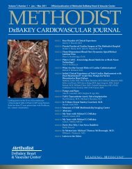
Journal of THE METHodisT dEbakEy HEarT cEnTEr VoluME 2, nuMbEr 1, 2006<br />
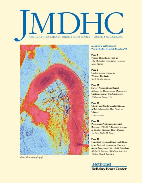
Photo illustration of a graft<br />
A quarterly publication of<br />
<strong>The</strong> <strong>Methodist</strong> <strong>Hospital</strong>, Houston, TX<br />
Page 4<br />
Venous Thrombosis Trials at<br />
<strong>The</strong> <strong>Methodist</strong> <strong>Hospital</strong> in Houston<br />
James Muntz<br />
Page 8<br />
Cardiovascular Disease in<br />
Women: <strong>The</strong> <strong>Facts</strong><br />
<strong>Karla</strong> M. <strong>Ku</strong>rrelmeyer<br />
Page 13<br />
Surgery Versus Alcohol Septal<br />
Ablation for Hypertrophic Obstructive<br />
Cardiomyopathy: <strong>The</strong> Controversy<br />
William H. Spencer, III<br />
Page 18<br />
Obesity and Cardiovascular Disease:<br />
A Bad Relationship That Needs to<br />
Change<br />
Peter H. Jones<br />
Page 22<br />
Peroxisome Proliferator-Activated<br />
Receptors (PPAR): A Potential Strategy<br />
to Combat Lipotoxic Heart Disease<br />
Qi Tian, Philip M. Barger<br />
Page 25<br />
Combined Open and Stent Graft Repair<br />
of an Arch and Descending Thoracic<br />
Aortic Aneurysm: <strong>The</strong> Hybrid Procedure<br />
Michael J. Reardon, Wei Zhou, Jon-Cecil<br />
Walkes, Alan B. Lumsden
We Welcome your<br />
questions a nd comments<br />
inquiries and letters to the editor can be<br />
directed to jmdhc@tmh.tmc.edu.<br />
William l. Winters, Jr., Md<br />
Editor-in-chief<br />
Journal of the <strong>Methodist</strong><br />
DeBakey Heart Center<br />
Journal of the<br />
me thodist deBake y<br />
he art center<br />
Volume 2, numBer 1<br />
William L. Winters, Jr., M.D.<br />
Editor-in-Chief<br />
Sheshe Giddens<br />
Managing Editor<br />
John K. Dietrich<br />
Assistant Editor<br />
Advisory Board:<br />
Christie Ballantyne, M.D.<br />
Stan Duchman, M.D.<br />
Raphael Espada, M.D.<br />
Robert Hust, M.D.<br />
<strong>Karla</strong> <strong>Ku</strong>rrelmeyer, M.D.<br />
Mike Reardon, M.D.<br />
JMDHC provides an update from<br />
<strong>Methodist</strong> DeBakey Heart Center<br />
specialists about leading edge research,<br />
diagnosis and treatment.<br />
U.S.News & World Report ranks the<br />
<strong>Methodist</strong> DeBakey Heart Center’s<br />
cardiology, cardiothoracic and<br />
vascular surgery programs number<br />
16 in the nation.<br />
JMDHC is written for physicians and<br />
should be relied upon for medical<br />
education purposes only. It does<br />
not provide a complete overview of<br />
the topics covered and should not<br />
replace the independent judgment of<br />
a physician about the appropriateness<br />
or risks of a procedure or treatment<br />
for a given patient.<br />
© 2006 <strong>The</strong> <strong>Methodist</strong> <strong>Hospital</strong><br />
Houston, Texas<br />
<strong>Methodist</strong> DeBakey<br />
Heart Center<br />
6565 Fannin<br />
Houston, Texas 77030<br />
Telephone: 713-DEBAKEY<br />
debakeyheartcenter.com
TO T E AC H I S TO L E A R N<br />
W i l l i a m L . W i n t e r s<br />
F r o m M e t h o d i s t D e B a k e y H e a r t C e n t e r a n d B a y l o r C o l l e g e o f M e d i c i n e , H o u s t o n , Te x a s<br />
How often have you heard someone<br />
say, “I learn more from preparing a<br />
lesson than my class will ever learn<br />
from listening to me deliver it”? It’s no<br />
secret that sharing personal experiences<br />
with others who have had similar experiences<br />
reinforces the message shared.<br />
Teaching and learning may be delivered<br />
in a variety of formats, especially in the<br />
field of medicine.<br />
I recently attended a superb conference<br />
structured around the discipline<br />
of echocardiography. As a disclaimer, I<br />
do admit to some bias toward echocardiography,<br />
having been seduced by the<br />
potential by-product of cardiac ultrasound<br />
some 40 years ago and then<br />
befriended by Drs. Inge Edler and<br />
Helmuth Hertz, whose fairly simple<br />
idea has blossomed into an extraordinary<br />
science.<br />
<strong>The</strong> message delivered at this conference<br />
was well prepared and presented.<br />
Some information was new, some old<br />
and revisited with new perspective, but<br />
when delivered expertly it is always<br />
worth hearing again. What has stuck<br />
with me after leaving that conference<br />
has been the message from the many<br />
case reports — including a brief clinical<br />
review and reports of the laboratory,<br />
ECG, X-ray and echocardiography<br />
findings — that were presented each<br />
morning.<br />
Echocardiography provided the<br />
opportunity to visualize the problem,<br />
discuss the pathophysiology, make<br />
differential diagnoses and highlight the<br />
important features. Each report was<br />
followed by a brief discussion directed<br />
by the case presenter and the moderator.<br />
<strong>The</strong> experience was absolutely spellbinding<br />
and guaranteed to have everyone<br />
awake and eager for the first lecturer.<br />
Upon my return, I came across an<br />
editorial by Dr. C. Richard Conti,<br />
editor-in-chief of Clinical Cardiology. 1<br />
For those of you who enjoy reading<br />
editorials, I believe his are among<br />
the most interesting and informative<br />
around. His topic is case-based teaching<br />
and learning. He and I are of an<br />
era when presentations of clinical cases<br />
by students to mentors, followed by<br />
brief discussions by all involved, was a<br />
major modus operandi for teaching clinical<br />
sciences. That put the onus on all<br />
parties to be prepared. Over the years,<br />
as the sheer volume and complexity of<br />
medical information has increased, not<br />
to mention the array of diagnostic capabilities<br />
and therapeutic options, it is my<br />
perception that didactic lectures are now<br />
the standard when planning educational<br />
programs — albeit with fascinating<br />
technical computer tricks to keep attention<br />
focused.<br />
In years past, medical grand rounds<br />
at <strong>The</strong> <strong>Methodist</strong> <strong>Hospital</strong> was a major<br />
weekly event where physicians vied for<br />
the opportunity to participate. Slide<br />
lectures were important, but the really<br />
entertaining and educational programs<br />
were those in which clinical cases were<br />
presented and discussed. I don’t believe<br />
that has occurred for the past 20 years.<br />
Roughly 35 years ago, in an attempt<br />
to stimulate an educational collegial<br />
spark among those interested in cardiovascular<br />
education, a small group of us<br />
from the American Heart Association’s<br />
Physicians’ Education Committee<br />
formed the Houston Society of<br />
Cardiology. <strong>The</strong> format required participation<br />
on a rotating basis of physicians<br />
interested in cardiovascular medicine.<br />
Physicians, students and trainees from<br />
each major hospital in the city, most<br />
of which were in the Texas Medical<br />
Center at that time, were all invited<br />
to participate. Attendance at those<br />
monthly meetings often exceeded 100<br />
individuals — far exceeding our expectations.<br />
Most presentations involved<br />
case reports, and it proved to be one<br />
of the most exciting and interesting<br />
educational enterprises I’ve ever been<br />
associated with. Prominent physicians<br />
joined in, among them cardiologists,<br />
cardiovascular surgeons, radiologists<br />
and pathologists. Rather than lectures,<br />
the cardiovascular surgeons presented<br />
fascinating surgical problems, the radiologists<br />
some unbelievable X-ray problems,<br />
and the pathologists those beguiling<br />
things we often overlooked as clinicians.<br />
On one occasion, Dr. Miguel Quiñones<br />
and I presented a town-meeting-type<br />
program around the discipline of echocardiography.<br />
We each stood at one side<br />
of a large screen and presented echocardiographic<br />
cases to each other with<br />
audience participation. It was a howling<br />
success, being entertaining as well as<br />
educational.<br />
<strong>The</strong>n a funny thing began to happen.<br />
After a number of years, respected guest<br />
lecturers began to appear in place of case<br />
reports. <strong>The</strong> requirement for program<br />
presentation by participating hospitals<br />
was dropped, and soon attendance at<br />
the meetings declined to the point where<br />
the American Heart Association lost<br />
interest and ultimately declined further<br />
support — ironic, given that the idea<br />
was originally conceived in the AHA<br />
Physicians’ Education Committee. I<br />
have always believed that a major culprit<br />
in the Houston Cardiology Society’s<br />
demise was the loss of local physician<br />
participation from the major hospital<br />
groups and the case-based teaching and<br />
discussion.<br />
As I read the editorial by Dr. Conti,<br />
I was reminded how echocardiography<br />
is the heart of cardiovascular imaging.<br />
Continued on page 28<br />
JMDHC | II (1) 2006 1
let te Rs to tHe e dItoR<br />
Send letters to the editor to<br />
William L. Winters, M.D.,<br />
jmdhc@tmh.tmc.edu or mail<br />
a disk or CDROM and a print<br />
out to JMDHC, 8060 El Rio<br />
St., Houston, TX 77054. Letters<br />
discussing a recent JMDHC article<br />
should not exceed 600 words in<br />
length and limited to one figure<br />
or table and five references. <strong>The</strong>y<br />
should be double-spaced and a<br />
word count should be provided.<br />
<strong>The</strong> names, academic degrees, and<br />
primary institutional affiliations of<br />
all authors as well as the address,<br />
telephone and fax numbers, and email<br />
address for the corresponding<br />
author must be included in the text<br />
of the letter in order to be considered<br />
for publication. Letters will be<br />
published at the discretion of the<br />
editor-in-chief and are subject to<br />
editing for style and space requirements.<br />
L E T T E R T O T H E E d I T O R<br />
Consultant Roles<br />
Dear Dr. Winters:<br />
I had occasion recently to read your<br />
essay on “Etiquette Guidelines for<br />
Consultants? Are <strong>The</strong>re Any?”<br />
JMDHC 1(4) 2005. I enjoyed what<br />
you had to say. A colleague at the<br />
University of Miami Miller School of<br />
Medicine sent it to me after I sent him<br />
an expanded version of something he<br />
requested, which I prepared many years<br />
ago for GI fellows in training at UM.<br />
Having retired December 12, 2001, I<br />
had almost forgotten that I had.<br />
<strong>The</strong> teaching of important nontechnical<br />
(in the traditional sense) skills<br />
designed to improve doctor-doctor and<br />
doctor-patient communication, confidence,<br />
and trust should begin early in<br />
training.<br />
I hope you enjoy reading what I have<br />
prepared on the topic of becoming an<br />
effective consultant, whether performing<br />
a cognitive or technical service.<br />
Best wishes,<br />
Arvey I. Rogers, MD, FACP<br />
Professor Emeritus (Retired)<br />
Internal Medicine/Gastroenterology<br />
University of Miami Miller<br />
School of Medicine<br />
IntRoduCtIon<br />
• You will be asked frequently to<br />
provide assistance/expertise (consult)<br />
in the management of patients with<br />
suspected or established disorders of<br />
the digestive system.<br />
• <strong>The</strong> assistance may be of an urgent or<br />
elective nature.<br />
• <strong>The</strong> request for your expertise may<br />
come in written or verbal form.<br />
• You possess expertise which the<br />
consulting physician does not.<br />
• How you provide your expertise<br />
reflects on you specifically and the<br />
service you represent generally.<br />
• While there are many ways to func-<br />
tion at a high level as a consultant, a<br />
proposed set of guidelines follows:<br />
GuIdelInes<br />
1. Respond promptly to the consultation<br />
request.<br />
2. Communicate verbally with the<br />
consulting physician for several<br />
reasons:<br />
a. Establish a personal relationship.<br />
b. Assess the urgency of the consult.<br />
c. Determine the specific reason for<br />
the consult, i.e., To perform a procedure?<br />
To answer a question? To<br />
follow up on a consult provided<br />
previously by a colleague?<br />
d. Assess the level of knowledge of the<br />
consulting physician.<br />
e. Gather additional data to assist you<br />
in planning the consult, i.e., what is<br />
needed, in what setting, with what<br />
preparation, etc.<br />
f. Inform the consulting physician<br />
when you will be seeing the patient.<br />
3. Establish a consultant relationship<br />
with the patient, explaining<br />
your role; that you will be working<br />
closely with the patient’s primary<br />
physician who will be informed of<br />
your findings and recommendations;<br />
and that you will follow up as<br />
necessary.<br />
4. Provide answers and recommendations<br />
(verbally and in writing)<br />
expeditiously and be certain they are<br />
understood by the consulting physician.<br />
5. Teach with humility.<br />
6. Provide appropriate current or<br />
classic references (affix a copy of an<br />
article when appropriate).<br />
7. Be certain that the consulting physician<br />
wishes you to remain involved<br />
in the care of the patient; avoid<br />
assuming control or “taking over”<br />
the case.<br />
8. Write legibly!<br />
9. “Sign off” the case with the knowledge<br />
of the consulting physician.<br />
10. Be certain that your attending or<br />
responsible fellow (more senior to<br />
you) knows about the consult, your<br />
2 II (1) 2006 | JMDHC
ecommendations, and (when possible)<br />
actually reads what you have<br />
written.<br />
11. Follow up to learn the patient’s<br />
outcome!<br />
Pe RfoRmInG a PRoCeduRe —<br />
unIque GuIdelInes<br />
Many of the consultations you receive<br />
will be for the purpose performing<br />
a diagnostic/therapeutic procedure,<br />
urgently or electively. <strong>The</strong> following<br />
suggestions are offered to maximize<br />
your role as a consultant under these<br />
specific circumstances:<br />
1. If received “after hours,” assess the<br />
urgency of the request.<br />
2. Assess the level of urgency and<br />
whether or not to “come in” depend<br />
upon the level of knowledge/competency<br />
of the consulting physician,<br />
your assessment of same, and your<br />
assessment as well as to the level of<br />
anxiety experienced by the “consulting<br />
physician.”<br />
3. When in doubt, speak to the most<br />
senior member of the patient care<br />
team.<br />
4. When in doubt, meet the team at<br />
the bedside.<br />
5. In doing so, you accomplish the<br />
following:<br />
a. Make your own assessment of the<br />
situation<br />
b. Transmit knowledge and confidence<br />
to the patient and the team.<br />
c. Determine whether a procedure or<br />
needed or not and, if needed, when.<br />
d. You represent your department in<br />
the right light.<br />
6. Whenever a procedure is required,<br />
whether urgently or electively, and<br />
whether for diagnostic and/or therapeutic<br />
purposes, do the following:<br />
a. Be certain the consulting physician<br />
understands what you plan to do.<br />
b. Be certain s/he and the patient or<br />
a surrogate understand the limitations/risks<br />
of the procedure.<br />
c. Invite the consulting physician to be<br />
present at the procedure.<br />
7. Be certain that the consultant physi-<br />
JMDHC | II (1) 2006<br />
L E T T E R T O T H E E d I T O R<br />
cians (your colleagues) involved<br />
in performing the procedure are<br />
sufficiently skilled to handle the<br />
fundamentals as well as anticipated<br />
adverse occurrences. Involve your<br />
attending!<br />
8. Discuss your findings and implications<br />
for management with the<br />
consulting physician.<br />
9. In a progress note, summarize your<br />
findings/recommendations/plans for<br />
follow-up.<br />
10. If biopsies are obtained or other<br />
tissue removed, accept responsibility<br />
for obtaining the results, discussing<br />
them and their implications for<br />
further management; write a brief<br />
note.<br />
11. Follow up to assess the patient’s<br />
outcome!<br />
e R R at u m<br />
In the Letter to the Editor, “Percutaneous Versus Surgical Treatment of<br />
Hypertrophic Obstructive Cardiomyopathy: the Pendulum Continues to<br />
Swing” by Tsung O. Cheng, M.D., published in the Journal of the <strong>Methodist</strong><br />
DeBakey Heart Center 2005;1(4): 2 , the second sentence of the first paragraph<br />
should read “As Buergler and associates pointed out in their paper, since<br />
Sigwart’s original report of PTSMA in 1995, 4 several publications confirming<br />
the efficacy and safety of the procedure have appeared all around the world,<br />
including China. 5 ”
V E N O U S T H R O M B O S I S T R I A L S AT<br />
T H E M E T H O d I S T H O S P I TA L I N H O U S TO N<br />
J a m e s M u n t z<br />
F r o m M e t h o d i s t D e B a k e y H e a r t C e n t e r, H o u s t o n , Te x a s<br />
IntRoduCtIon<br />
Venous thromboembolism continues to be a significant problem in hospitalized patients. 1 according to Geerts et al.,<br />
“the rationale for thromboprophylaxis is based on the high prevalence of venous thromboembolism among hospitalized<br />
patients, the clinically silent nature of the disease in the majority of patients, and the morbidity, costs and<br />
potential mortality associated with unprevented thrombi.” 2,3 approximately two million americans suffer from deep<br />
vein thrombosis (dVt) each year, 4 but because most of these clots are silent, the true incidence is actually unknown.<br />
In addition to the acute thrombotic event with its inherent morbidity and time missed from work, the late sequelae<br />
from even properly treated thrombi can present as venous stasis and leg ulcers.<br />
thromboembolic disease is responsible for approximately 200,000 deaths annually in the united states alone. 5<br />
the elderly are at highest risk, with a one-year mortality approaching 21%. 6 although excellent drugs are currently<br />
available to prevent and treat dVt, an investigation into new drugs with new targets and designer end points of anticoagulation<br />
seemed clinically relevant and timely.<br />
the following paper will discuss the different antithrombotic/anticoagulant agents that have been used in clinical<br />
trials at the methodist <strong>Hospital</strong> in Houston, texas. several of the trials have been successfully completed and<br />
featured in medical publications such as the New England Journal of Medicine and <strong>The</strong> Lancet.<br />
n e W a n t I t H R o m B o t I C<br />
s t R at e G I e s<br />
According to a review by Hirsh and<br />
Weitz, “anticoagulant strategies to block<br />
thrombogenesis have focused on inhibiting<br />
thrombin, preventing thrombin<br />
generation, or blocking the initiation<br />
of coagulation” (Figure 1). 7 <strong>The</strong> drugs<br />
studied at <strong>The</strong> <strong>Methodist</strong> <strong>Hospital</strong> will<br />
be discussed in chronological order,<br />
with the first new class of anticoagulant<br />
drugs being one of the Factor Xa inhibitors,<br />
pentasaccharide.<br />
f o n d a Pa R I n u X<br />
Fondaparinux is the first of a new class of<br />
antithrombotics: the synthetic inhibitors<br />
of Factor Xa (SIXa). Originally termed<br />
ORG31540/SR90107A, fondaparinux<br />
is a unique and novel pentasaccharide<br />
produced by total chemical synthesis<br />
and designed specifically to bind its<br />
target, the protein antithrombin, with<br />
very high affinity. Fondaparinux (trade<br />
name Arixtra) is obtained exclusively<br />
by chemical synthesis from basic building<br />
blocks synthesized from glucose,<br />
glucosamine and cellobiose. It has guar-<br />
anteed batch-to-batch consistency that<br />
eliminates the risk of contamination by<br />
pathogenic agents. 8<br />
<strong>Methodist</strong> physicians participated in<br />
the drug’s Phase II and Phase III trials,<br />
with ours being the largest enrolling site<br />
worldwide in the Phase II Pentathlon<br />
trial. Phase IIb studies, which included<br />
more than 900 patients undergoing<br />
total hip replacement surgery and 400<br />
undergoing knee replacement surgery,<br />
demonstrated statistically significant<br />
dose-dependent reductions in the risk<br />
of DVT with this agent. 9<br />
We also participated in a multi-center,<br />
double-blinded, dose-ranging Phase II<br />
study to determine optimal dosing in<br />
933 patients undergoing primary total<br />
hip replacement surgery. 10 Figures 2 and<br />
3 outline the efficacy and safety results<br />
from this trial. 11 Following this were<br />
four Phase III trials-randomizing more<br />
than 7,000 patients on five continentswhich<br />
demonstrated that once-daily<br />
fondaparinux in a 2.5 mg subcutaneous<br />
dose reduced the risk of overall<br />
DVT by 50% compared with the established<br />
standard of care (enoxaparin) in<br />
patients undergoing major orthopedic<br />
surgery. 12 <strong>The</strong>se trials provided significant<br />
support of fondaparinux and were<br />
the basis of eventual FDA approval of<br />
the drug in orthopedic populations.<br />
X I m e l a G at R a n<br />
A prodrug of the active site-directed<br />
thrombin inhibitor melagatran, ximelagatran<br />
is absorbed from the small<br />
intestine and was the next drug to be<br />
studied in orthopedic populations at <strong>The</strong><br />
<strong>Methodist</strong> <strong>Hospital</strong>. 13 Ximelagatran<br />
has a plasma half-life of three to four<br />
hours and was administered in a twicedaily<br />
dose. This drug was considered a<br />
savior in the anticoagulation world as it<br />
has no food or drug interactions and,<br />
due to its very predictable response, did<br />
not appear to require any anticoagulant<br />
monitoring.<br />
To evaluate the utility of this drug<br />
in North American orthopedic populations,<br />
we participated in the Platinum<br />
hip and knee trials. <strong>The</strong> Platinum<br />
Hip Trial randomized 1,557 patients<br />
with adequate venography undergoing<br />
total hip replacement to receive oral<br />
II (1) 2006 | JMDHC
��������<br />
�����������<br />
����������<br />
�����������<br />
�����������������<br />
figure 1. Steps in blood coagulation. Initiation of coagulation is triggered by the factor<br />
VIIa/tissue factor complex (VIIa/TF), which activates factor IX (IX) and factor X (X).<br />
Activated factor IX (IXa) propagates coagulation by activating factor X in a reaction that<br />
utilizes activated factor VIII (VIIIa) as a cofactor. Activated factor X (Xa), with activated<br />
factor V (Va) as a cofactor, converts prothrombin (II) to thrombin (IIa). Thrombin then<br />
converts fibrinogen to fibrin. Tissue factor pathway inhibitor (TFPI) and nematode<br />
anticoagulant peptide (NAPc2) target VIIa/TF, whereas synthetic pentasaccharide and<br />
DX-9065a inactivate Xa. Hirudin, bivalirudin, argatroban, and H376/95 inactivate IIa.<br />
ximelagatran (24 mg twice daily) or<br />
enoxaparin (30 mg SQ twice daily)<br />
for seven to 12 days. 14 <strong>The</strong> results of<br />
the trial showed that enoxaparin was<br />
superior to ximelagatran when started<br />
postoperatively in the 24-mg, twicedaily<br />
dosage. Further trials altered the<br />
dosages of ximelagatran to 36 mg twice<br />
a day with an additional subcutaneous<br />
dose given on the day of surgery, which<br />
then demonstrated superiority over<br />
the enoxaparin regimen. 15 However,<br />
release of the drug in the U.S. market<br />
has been delayed due to FDA safety<br />
concerns regarding liver enzyme elevations<br />
that occurred in longer-term atrial<br />
fibrillation trials.<br />
I d R a Pa R I n u X<br />
A more highly sulfated derivative of<br />
fondaparinux, idraparinux is an inves-<br />
JMDHC | II (1) 2006<br />
sites of action of new anticoagulants<br />
����������<br />
�<br />
�����������<br />
�������<br />
��<br />
��<br />
���<br />
��<br />
�������<br />
�����<br />
������<br />
���<br />
��<br />
����<br />
����<br />
����<br />
���������������<br />
��������<br />
�������<br />
�����������<br />
����������<br />
�������<br />
tigational drug with a half-life of 150<br />
hours. This promising and sophisticated<br />
new anticoagulant, to be studied<br />
in acute DVT and pulmonary embolism<br />
patients, was presented to us in<br />
the Van Gogh trials by Sanofi Aventis.<br />
In a Phase II trial performed elsewhere,<br />
idraparinux therapy was compared with<br />
warfarin therapy in 659 patients with<br />
acute proximal DVT. 16 After five to<br />
seven days of initial treatment with<br />
enoxaparin, patients were randomized<br />
to receive once-weekly subcutaneous<br />
doses of idraparinux (2.5, 5.0, 7.5 or<br />
10 mg.) or warfarin for 12 weeks. <strong>The</strong><br />
primary end point, changes in compression<br />
ultrasound or new findings on<br />
perfusion lung scans, was similar in all<br />
of the idraparinux groups and no different<br />
in the warfarin group. 17<br />
Based on the above results and the<br />
fact that there was less bleeding in the<br />
lowest-dosage idraparinux group than<br />
in the warfarin group, the 2.5 mg, onceweekly<br />
dose of idraparinux was chosen<br />
for the Phase III trials. <strong>The</strong> Van Gogh<br />
DVT and pulmonary embolism trials<br />
are now completed, with the data to be<br />
reviewed and published.<br />
s o l u B l e<br />
t H R o m B o m o d u l I n<br />
<strong>The</strong> <strong>Methodist</strong> <strong>Hospital</strong> was next<br />
recruited by the Hamilton, Ontario<br />
group to participate in the soluble<br />
thrombomodulin trial. Figure 1 reveals<br />
where thrombomodulin affects propagation<br />
of thrombin. <strong>The</strong> initiation of<br />
coagulation is triggered by the tissue<br />
factor/factor VIIa complex that activates<br />
factor IX and factor X. Activated factor<br />
IX propagates coagulation by activating<br />
factor X in a reaction utilizing factor<br />
VIII as a cofactor. Activated protein C<br />
then blocks the propagation of coagulation<br />
by inactivating factors Va and<br />
VIIIa at the thrombin/thrombomodulin<br />
site. Protein C and thrombomodulin<br />
also target this step: According to Weitz<br />
et al., “soluble thrombomodulin binds<br />
thrombin and induces a conformational<br />
change in the active site of the enzyme<br />
that converts it into a potent activator of<br />
protein C.” 17<br />
In an open-label, dose-escalating<br />
study, soluble thrombomodulin positively<br />
affected coagulation abnormalities<br />
in patients with disseminated intravascular<br />
coagulation. 18 After these results<br />
were demonstrated, the Canadian group<br />
organized a Phase II dose-ranging study<br />
in patients undergoing elective total hip<br />
replacement surgery. 19 Patients were<br />
given thrombomodulin subcutaneously<br />
(0.3 or 0.45 mg/kg) two to four<br />
hours after surgery, and patients receiving<br />
the lower thrombomodulin dosage<br />
received another dose five days later.<br />
<strong>The</strong> primary end point, a composite<br />
of venographically detected and symptomatic<br />
venous thromboembolism,<br />
occurred in only 4.3% of those patients<br />
receiving the lower dose and in none<br />
of the 99 patients in the higher-dose
���������������������<br />
��������������������������<br />
��<br />
��<br />
��<br />
�<br />
�<br />
��<br />
��<br />
��<br />
��<br />
��<br />
�<br />
�<br />
����<br />
�������<br />
�����<br />
�������<br />
�����<br />
���<br />
������<br />
�����<br />
figure 2. Efficacy results from fondaparinux Phase II dose-finding study in patients<br />
with total hip replacement. Data are expressed as percent of treated patients with<br />
venous thromboembolism. Enrollment in the 6 mg and 8 mg groups was discontinued<br />
early after an increase in bleeding incidence was noted.<br />
������<br />
�����<br />
���<br />
����<br />
�����<br />
������������<br />
����<br />
�����<br />
������������<br />
����<br />
����<br />
�������<br />
����<br />
����<br />
����������<br />
�����<br />
figure 3. Safety results from fondaparinux Phase II dose-finding study in patients<br />
with total hip replacement. Data are expressed as percent of treated patients with<br />
major bleeding. Enrollment in the 6 and 8 mg groups was discontinued early because<br />
of this complication.<br />
���<br />
����<br />
����<br />
����<br />
����<br />
����������<br />
�����<br />
group. 17 Phase III trials will next be<br />
required to compare ART-123 (soluble<br />
thrombomodulin) to either enoxaparin<br />
or fondaparinux.<br />
C o n C l u s I o n<br />
Recently developed, highly selective,<br />
novel therapeutic agents such as those<br />
explored in our trials can optimize antithrombotic<br />
efficacy and improve the<br />
prevention of hemorrhagic complications.<br />
It is hoped that <strong>The</strong> <strong>Methodist</strong><br />
<strong>Hospital</strong>’s participation in several landmark<br />
trials — with particular attention<br />
to patient care and safety — will lead<br />
to the development of new and possibly<br />
better anticoagulants that ultimately<br />
have a positive benefit for the future of<br />
medicine.<br />
R e f e R e n C e s<br />
1. Bergmann JF, Elkharrat D. Prevention of<br />
venous thromboembolic risk in non-surgical<br />
patients. Haemostasis. 1996;26 Suppl<br />
2:16-23.<br />
2. Geerts WH, Heit JA, Clagett GP, Pineo<br />
GF, Colwell CW, Anderson FA Jr, et al.<br />
Prevention of venous thromboembolism.<br />
Chest. 2001 Jan;119(1 Suppl):132S-175S.<br />
3. Prandoni P, Lensing AW, Cogo A, Cuppini<br />
S, Villalta S, Carta M, et al. <strong>The</strong> longterm<br />
clinical course of acute deep venous<br />
thrombosis. Ann Intern Med. 1996 Jul<br />
1;125(1):1-7.<br />
4. Hirsh J, Hoak J. Management of deep<br />
vein thrombosis and pulmonary embolism.<br />
A statement for health care professionals.<br />
Council on Thrombosis (in consultation with<br />
the Council on Cardiovascular Radiology),<br />
American Heart Association. Circulation.<br />
1996 Jun 15; 93(12):2212-45.<br />
5. Ferris EJ. George W. Holmes Lecture.<br />
Deep vein thrombosis and pulmonary<br />
embolism: correlative evaluation and therapeutic<br />
implications. Am J Roentgenol. 1992<br />
Dec;159(6):1149-55.<br />
6. Kniffin WD Jr, Baron JA, Barrett J,<br />
Birkmeyer JD, Anderson FA Jr. <strong>The</strong> epidemiology<br />
of diagnosed pulmonary embolism<br />
and deep vein thrombosis in the elderly.<br />
Arch Int Med. 1994 Apr 25;154(8):861-6.<br />
7. Weitz JI, Hirsh J. New anticoagulant drugs.<br />
Chest. 2001 Jan;119(1Suppl):95S-107S.<br />
6 II (1) 2006 | JMDHC<br />
���
8. Casu B. Structure and biological activity of<br />
heparin. Adv Carbohydr Chem Biochem.<br />
1985;43:51-134.<br />
9. Herbert JM, Petitou M, Lormeau JC,<br />
Cariou R, Necciari J, Magnani HN, et al.<br />
SR 90107A/Org 31540, a novel anti-factor<br />
Xa antithrombotic agent. Cardiovasc Drug<br />
Rev. 1997;15:1-26.<br />
10. Turpie AG, Gallus AS, Hoek JA. A<br />
synthetic pentasaccharide for the prevention<br />
of deep-vein thrombosis after total hip<br />
replacement. N Engl J Med. 2001 Mar<br />
1;344(9):619-25.<br />
11. Muntz JE. Fondaparinux for Prophylaxis.<br />
Techniques in Orthopaedics. 2004<br />
Dec;19(4):278-82.<br />
12. Turpie AG, Bauer KA, Eriksson BI, Lassen<br />
MR. Fondaparinux vs enoxaparin for the<br />
prevention of venous thromboembolism in<br />
major orthopedic surgery: a meta-analysis<br />
of 4 randomized double-blind studies.<br />
Arch Intern Med. 2002 Sep 9;162(16):<br />
1833-40.<br />
13. Gustafsson D, Nystrom J, Carlsson S, Bredberg<br />
U, Eriksson U, Gyzander E, et al.<br />
<strong>The</strong> direct thrombin inhibitor melagatran<br />
and its oral prodrug H 376/95: intestinal<br />
absorption properties, biochemical and<br />
pharmacodynamic effects. Throm Res. 2001<br />
Feb 1;101(3):171-81.<br />
14. Colwell CW, Berkowitz SD, Davidson BL,<br />
Lotke PA, Ginsberg JS, Lieberman JR, et<br />
al. Randomized, double-blind, comparison<br />
of ximelagatran, an oral direct thrombin<br />
inhibitor, and enoxaparin to prevent venous<br />
thromboembolism after total hip arthroplasty<br />
[abstract]. Blood. 2001;98:2952.<br />
15. Francis CW, Berkowitz SD, Comp PC,<br />
Lieberman JR, Ginsberg JS, Paiement G,<br />
et al. Comparison of ximelagatran with<br />
warfarin for the prevention of venous<br />
thromboembolism after total knee<br />
replacement. N Engl J Med. 2003 Oct<br />
30;349(18):1703-12.<br />
16. Büller HR, Cohen AT, Lensing AW, Prins<br />
MH, Monreal M, Schulman S, et al.; <strong>The</strong><br />
PERSIST Investigators. A novel long-acting<br />
synthetic Xa inhibitor (SanOrg34006) to<br />
replace warfarin for secondary prevention in<br />
deep vein thrombosis: a Phase II evaluation.<br />
J Thromb Haemost. 2004 Jan;2:47-53.<br />
Erratum: J Thromb Haemost. 2004<br />
Mar;2(3):540.<br />
JMDHC | II (1) 2006<br />
17. Weitz JI, Hirsh J, Samama MM. New<br />
anticoagulant drugs: the Seventh ACCP<br />
Conference on Antithrombotic and Thrombolytic<br />
<strong>The</strong>rapy. Chest. 2004 Sep;126(3<br />
Suppl):265S-286S.<br />
18. Maruyama I. Recombinant thrombomodulin<br />
and activated protein C in the<br />
treatment of disseminated intravascular<br />
coagulation. Thromb Haemost. 1999<br />
Aug;82(2):718-21.<br />
19. Kearon C, Comp P, Douketis J, Royds R,<br />
Yamada K, Gent M. Dose-response study<br />
of recombinant human soluble thrombomodulin<br />
(ART-123) in the prevention of<br />
venous thromboembolism after total hip<br />
replacement. J Thromb Haemost. 2005<br />
May;3(5):962-8.
C A R d I OVA S C U L A R d I S E A S E I N W O M E N : T H E FAC T S<br />
K a r l a M . K u r r e l m e y e r<br />
F r o m M e t h o d i s t D e B a k e y H e a r t C e n t e r, H o u s t o n , Te x a s<br />
IntRoduCtIon<br />
Cardiovascular disease (CVd) is the leading cause of death in women. one of every 2.4 women will die of CVd,<br />
and it claims almost 500,000 female lives annually. 1 since women are more likely than men to have sudden death<br />
as their initial presenting symptom, early prevention measures are even more important in women to prevent this<br />
catastrophic outcome. 2<br />
Ironically, more than 50% of women who were surveyed did not know CVd was the leading killer of women, and<br />
only 5% identified heart disease as their greatest health problem. 3 this unawareness likely has contributed to the<br />
increase of cardiovascular deaths among women, whereas the number of cardiovascular deaths among men has<br />
slowly decreased since 1984. 4<br />
P R e V e n t I o n o f C V d<br />
I n W o m e n<br />
<strong>The</strong> risk factors for heart disease in<br />
women are similar to men, though<br />
the weighting differs. For example,<br />
smoking, hypertension, LDL cholesterol<br />
and family history increase risk<br />
in women as much as they do in men. 5<br />
Diabetes, however, is a stronger risk<br />
factor in women, as are elevated triglycerides<br />
and low HDL levels. 5,7 Diabetic<br />
women are five times more likely<br />
to develop CVD than non-diabetic<br />
women, whereas diabetic men are only<br />
two times more likely to develop CVD<br />
than non-diabetic men. 6<br />
Recommendation Class,<br />
by Risk level<br />
<strong>The</strong> most prevalent modifiable risk<br />
factor in American women is excess<br />
weight, and approximately 62% of<br />
American women are either overweight<br />
or obese. 8 Prospective data from the<br />
Nurses’ Health Study clearly demonstrates<br />
that the relative risk of death<br />
from CVD increases with body mass<br />
index (BMI – defined as weight in kilograms<br />
divided by the square of the<br />
height in meters) once the BMI exceeds<br />
19, the upper limit of normal; those<br />
with BMIs greater than 29, defined as<br />
obesity, are at greatest risk. 9<br />
<strong>The</strong> second most prevalent modifiable<br />
risk factor in women is elevated<br />
Guidelines outline best practices for treating CVd in women<br />
CVD treatment/prevention options for various risk levels<br />
High • Aspirin therapy<br />
• ACE inhibitor therapy<br />
• Glycemic control<br />
• Treatments for low/<br />
intermediate risk<br />
Intermediate • BP control<br />
• Lipid control<br />
• Treatments for low risk<br />
Low/Optimal • Smoking cessation<br />
• Physical activity<br />
• Weight reduction<br />
table 1. Current ACC/AHA guidelines for treating CVD in women<br />
total and LDL cholesterol. 8 Diet and<br />
aerobic exercise have been shown to<br />
lower total and LDL cholesterol and<br />
raise HDL cholesterol in women if they<br />
are performed together, although either<br />
in isolation failed to reach statistical<br />
significance. 10 Unfortunately, the third<br />
most prevalent risk factor in women is<br />
physical inactivity. In 2002, 43% of<br />
American women reported that they<br />
were physically inactive. 8 <strong>The</strong> good<br />
news is their cholesterol abnormalities<br />
can be treated with medication. In the<br />
large statin trials, women responded<br />
similarly to drug therapy as men and<br />
benefited from a similar reduction in<br />
I IIa IIb III<br />
• depression<br />
evaluation and<br />
treatment<br />
• Omega-3<br />
fatty acid<br />
supplementation<br />
• Folic acid<br />
supplementation<br />
• Hormone therapy<br />
• Antioxidant<br />
supplementation<br />
• Aspirin therapy N/A • Hormone therapy<br />
• Antioxidant<br />
supplementation<br />
N/A N/A • Aspirin therapy<br />
• Hormone therapy<br />
• Antioxidant<br />
supplementation<br />
Source: Mosca et al. Circulation: 2/10/04.<br />
II (1) 2006 | JMDHC
major coronary events. 11 <strong>The</strong> bad news<br />
is they will not benefit from the myriad<br />
other effects of aerobic exercise.<br />
<strong>The</strong> current ACC/AHA guidelines<br />
for treating CVD in women are listed<br />
in Table 1. 38 Women have been stratified<br />
into four groups based on their risk<br />
for developing CVD: high risk means<br />
> 20% chance of developing CVD in<br />
10 years, intermediate risk means a 10-<br />
20% chance of developing CVD in<br />
10 years, and low/optimal risk means<br />
< 10% chance of developing CVD in<br />
10 years. For each group there are four<br />
different levels of recommendations:<br />
Class I describes interventions deemed<br />
safe and effective; Class IIa describes<br />
treatments favored by clinical evidence;<br />
Class IIb describes therapies for which<br />
evidence is less well established; and<br />
Class III describes interventions that are<br />
not useful and may be harmful. 38 Table<br />
2 demonstrates how to estimate 10-year<br />
risk for developing CVD in women. 39<br />
d I a G n o s I n G C o R o n a R Y<br />
a R t e R Y d I s e a s e I n<br />
W o m e n<br />
Diagnosing coronary artery disease<br />
(CAD) in women is difficult for several<br />
reasons. First, chest pain in women does<br />
not accurately predict the presence of<br />
CAD, making risk stratification based<br />
on this symptom alone practically<br />
useless. However, refining the diagnoses<br />
of chest pain into definite angina,<br />
probable angina, or nonspecific chest<br />
pain may improve the predictive value<br />
of symptoms. 18 Like men, nearly 90%<br />
of women with myocardial infarction<br />
(MI) in the Myocardial Infarction Trial<br />
and Intervention Project presented with<br />
chest pain. However, women with MI<br />
were also more likely than men to<br />
present with upper abdominal pain,<br />
dyspnea, nausea, and fatigue. 12-14<br />
<strong>The</strong>refore, both chest pain and atypical<br />
symptoms of angina should be pursued<br />
in women based on the appropriate<br />
clinical context and the underlying<br />
probability of disease. Second, for a<br />
given age and risk factor profile, the<br />
prevalence of CAD in women when<br />
JMDHC | II (1) 2006<br />
compared to men is significantly less. 18<br />
Women develop CAD on average 10<br />
years later than men and therefore have<br />
frequently developed other diseases,<br />
which affect their ability to exercise. 17<br />
<strong>The</strong>se factors decrease the accuracy of<br />
all available noninvasive tests, which<br />
may partially explain why women are<br />
less likely than men to be referred<br />
for diagnostic testing. 15,16 Some earlier<br />
studies demonstrated that women<br />
with positive test results are less likely<br />
to undergo coronary angiography<br />
and revascularization procedures and<br />
therefore are twice as likely as men<br />
to experience a cardiac event during<br />
follow-up. 19 However, these biases<br />
appear to be disappearing as physician<br />
awareness increases.<br />
<strong>The</strong> following gender-specific issues<br />
should be considered when choosing<br />
a noninvasive stress test to evaluate<br />
chest pain in women. <strong>The</strong> sensitivity<br />
and specificity of electrocardiography<br />
(ECG) stress testing is much lower<br />
in women than men. 20 This not only<br />
reflects the lower prevalence of CAD in<br />
women < 50 years old, higher prevalence<br />
of single-vessel disease in women 50-75<br />
years and lower exercise capacity of older<br />
women as previously stated, but also<br />
the ST-response used to evaluate ECG<br />
results is less accurate in women since<br />
it varies with the menstrual cycle and<br />
estrogen replacement therapy. Mitral<br />
valve prolapse, syndrome X, and coronary<br />
artery spasm are more prevalent in<br />
women and can cause an abnormal ST<br />
response in the absence of CAD. 21,22<br />
<strong>The</strong> sensitivity and specificity<br />
of pharmacologic or exercise testing<br />
is enhanced by adding imaging techniques.<br />
Myocardial perfusion imaging<br />
with gated SPECT has improved sensitivity<br />
over conventional treadmill<br />
testing, however breast tissue artifacts<br />
may lead to a false-positive test result<br />
(decreased specificity). 23,24 Breast tissue<br />
artifacts may be reduced and specificity<br />
increased with the use of newer highenergy<br />
agents such as technetium-99m<br />
sestamibi rather than thallium-201. 25<br />
Application of computerized breast<br />
attenuation correction algorithms may<br />
also lead to enhanced specificity.<br />
Likewise, echocardiography has<br />
improved sensitivity and specificity<br />
over conventional treadmill testing and<br />
is considered the most cost-effective<br />
approach to diagnosing CAD in<br />
women. 26 However, test accuracy is<br />
highly dependent on the image quality<br />
and the technician’s and interpreter’s<br />
skills. Inadequate imaging occurs in<br />
approximately 10% of cases secondary<br />
to obesity, severe lung disease, chest wall<br />
deformities and inexperience. Using<br />
newer techniques such as harmonic<br />
imaging and echocardiography contrast<br />
agents improves image quality. 27<br />
P R o G n o s I s a f t e R<br />
R e Va s C u l a R I Z at I o n<br />
Several studies have demonstrated a<br />
worse prognosis for women than men<br />
with ST elevation myocardial infarction,<br />
which may reflect increased<br />
severity of illness at presentation,<br />
increased age, more comorbidities in<br />
women when compared to men, and<br />
lower referral rates for coronary angiography<br />
and revascularization procedures.<br />
For example, the Framingham Study<br />
demonstrated that women were more<br />
likely than men to die in the hospital<br />
and within one year after infarction. 17<br />
<strong>The</strong> International Study of Infarct<br />
Survival (ISIS-1) examined the effects<br />
of atenolol post-MI and demonstrated<br />
greater one-week mortality for women<br />
compared with men. 36 ISIS-4 examined<br />
the effects of captopril after thrombolysis<br />
for acute MI and also suggested an<br />
increased short and long-term mortality<br />
in women. 35 Finally, the Global<br />
Utilization of Streptokinase and Tissue<br />
Plasminogen Activator for Occluded<br />
Arteries trial demonstrated higher 30day<br />
mortality in women and higher<br />
rates of intracerebral hemorrhage, possibly<br />
due to smaller body size and lack of<br />
thrombolytic dose adjustments. 33<br />
Meta-analysis of 10 randomized trials<br />
of primary angioplasty versus thrombolytic<br />
therapy demonstrated that primary<br />
angioplasty results in lower rates of
death and MI at 30 days in women<br />
presenting with acute ST elevation<br />
MI. 34 <strong>The</strong> CADILLAC trial examined<br />
the benefit of stent and abciximab in<br />
patients presenting within 12 hours of<br />
ST elevation MI and found a higher<br />
one-year mortality in women, possibly<br />
due to a longer time from symptom<br />
onset to percutaneous coronary inter-<br />
estimating the 10-Year Risk of CVd in Women<br />
Step 1. age<br />
Age, y 20-34 35-39 40-44 45-49 50-54 55-59 60-64 65-69 70-74 75-79<br />
Points -7 -3 0 3 6 8 10 12 14 16<br />
Point<br />
Total<br />
10-y<br />
Risk, %<br />
HdL,<br />
mg/dL<br />
TC, mg/dL<br />
Age, y<br />
20-39<br />
Step 2. total Cholesterol<br />
Age, y<br />
40-49<br />
Age, y<br />
50-59<br />
vention (PCI). Women who received<br />
a stent demonstrated no difference in<br />
mortality when compared to angioplasty<br />
alone, however they had less<br />
major adverse cardiac events at one<br />
year. 31<br />
<strong>The</strong> outcome of women and men<br />
with unstable angina and non-Q-wave<br />
MI (NQWMI) was found to be similar<br />
Age, y<br />
60-69<br />
< 160 0 0 0 0 0<br />
160-199 4 3 2 1 1<br />
200-239 8 6 4 2 1<br />
240-279 11 8 5 3 2<br />
> 280 13 10 7 4 2<br />
Age, y<br />
20-39<br />
Step 3. smoking status<br />
Age, y<br />
40-49<br />
Age, y<br />
50-59<br />
Age, y<br />
60-69<br />
Nonsmoker 0 0 0 0 0<br />
Smoker 9 7 4 2 1<br />
Age, y<br />
70-79<br />
Age, y<br />
70-79<br />
Step 4. Hdl Step 5. Blood Pressure<br />
> 60 50-59 40-49 < 40<br />
Systolic BP,<br />
mm Hg<br />
Untreated Treated<br />
Points -1 0 1 2 < 120 0 0<br />
Step 6. add up the Points<br />
120-149 1 3<br />
130-139 2 4<br />
140-159 3 5<br />
> 160 4 6<br />
< 9 9 10 11 12 13 14 15 16 17 18 19 20 21 22 23 24 > 25<br />
< 1 1 1 1 1 2 2 3 4 5 6 8 11 14 17 22 27 > 30<br />
table 2. Steps 1-6 demonstrate how to estimate the 10-year risk for CVD in women.<br />
in the Thrombolysis in Myocardial<br />
Ischemia (TIMI) IIIB Registry. 28<br />
TACTICS: TIMI 18 demonstrated<br />
that, like men, women with NQWMI<br />
receiving both aspirin and tirofiban<br />
benefited from early invasive treatment.<br />
29 Likewise, women and men<br />
have similar prognoses after successful<br />
high-risk PCI. EPIC, EPILOGUE and<br />
10 II (1) 2006 | JMDHC
EPISTENT demonstrated that the clinical<br />
benefit of abciximab in high-risk<br />
PCI is independent of gender, although<br />
women experience more minor bleeding.<br />
30 <strong>The</strong> Taxus-IV trial demonstrated<br />
that women benefit equally from the<br />
Taxus drug-eluting stent. 37<br />
<strong>The</strong> Coronary Artery Surgery Study<br />
demonstrated increased operative<br />
mortality for women, which may be<br />
secondary to differences in functional<br />
class, age, size of coronary arteries and<br />
greater likelihood of emergent surgery.<br />
However, coronary artery bypass surgery<br />
provides excellent relief of symptoms<br />
and comparable long-term survival in<br />
women. 18,32<br />
C o n C l u s I o n<br />
With respect to coronary artery disease,<br />
women are not “small men.” Differences<br />
in prevention, diagnosis, prognosis,<br />
and response to treatment have been<br />
identified. <strong>The</strong> reasons for these differences<br />
and the optimal management of<br />
coronary artery disease in women will<br />
hopefully be elucidated as more and<br />
more women are included in large,<br />
multicenter, randomized cardiovascular<br />
research trials.<br />
R e f e R e n C e s<br />
1. National Vital Statistics Report, Vol. 50.<br />
Hyattsville (MD): Centers for Disease<br />
Control and Prevention, National Center<br />
for Health Statistics; 2002 Sept 16. Report<br />
No. 15.<br />
2. American Heart Association. Heart Disease<br />
and Stroke Statistics — 2004 Update. Dallas<br />
(Texas): American Heart Association.<br />
2003:10.<br />
3. Excel Omnibus Study #R825/R925;<br />
National Vital Statistics Report, Vol. 50.<br />
Hyattsville (MD): Centers for Disease<br />
Control and Prevention, National Center<br />
for Health Statistics; 2002 Sept 16.<br />
Report No.15. Commissioned by Society of<br />
Women’s Health Research.<br />
4. Cooper R, Cutler J, Desvigne-Nickens P,<br />
Fortmann SP, Friedman L, Havlik R, et al.<br />
Trends and Disparities in Coronary Heart<br />
Disease, Stroke, and Other Cardiovascular<br />
Diseases in the United States: Findings of<br />
the National Conference on Cardiovascular<br />
Disease Prevention Circulation. 2000 Dec<br />
19;102(25):3137-47.<br />
5. Mosca L, Manson JE, Sutherland SE,<br />
Langer RD, Manolio T, Barrett-Connor<br />
E. Cardiovascular disease in women:<br />
a statement for healthcare professionals<br />
from the American Heart Association.<br />
Writing Group. Circulation. 1997 Oct<br />
7;96(7):2468-82.<br />
6. Kannel WB, McGee DL. Diabetes and<br />
cardiovascular disease: <strong>The</strong> Framingham<br />
Study. JAMA. 1979;241:2035-8.<br />
7. Cui Y, Blumenthal RS, Flaws JA, Whiteman<br />
MK, Langenberg P, Bachorik PS, et<br />
al. Non-High-Density Lipoprotein Cholesterol<br />
Level as a Predictor of Cardiovascular<br />
Disease Mortality. Arch Intern Med. 2001<br />
Jun 11;161(11):1413-9.<br />
8. American Heart Association. Heart Disease<br />
and Stroke Statistics — 2004 Update. Statistical<br />
Fact Sheet — Physical Inactivity.<br />
Dallas (Texas): American Heart Association.<br />
2003:10.<br />
9. Manson JE, Willett WC, Stampfer<br />
MJ, Colditz GA, Hunter DJ, Hankinson<br />
SE, et al. Body weight and mortality<br />
among women. N Engl J Med. 1995 Sept<br />
14;333(11):677-85.<br />
10. Stefanick ML, Mackey S, Sheehan M,<br />
Ellsworth N, Haskell WL, Wood PD. Effects<br />
of diet and exercise in men and postmenopausal<br />
women with low levels of HDL cholesterol<br />
and high levels of LDL cholesterol.<br />
N Engl J Med. 1998 Jul 2;339(1):12-20.<br />
11. LaRosa JC, He J, Vupputuri S. Effect of<br />
statins on risk of coronary disease: a metaanalysis<br />
of randomized controlled trials.<br />
JAMA. 1999 Dec 22-29;282(24):2340-6.<br />
12. Philpott S, Boynton PM, Feder G, Hemingway<br />
H. Gender differences in descriptions<br />
of angina symptoms and health problems<br />
immediately prior to angiography: the<br />
ACRE study. Appropriateness of Coronary<br />
Revascularisation study. Soc Sci Med. 2001<br />
May;52(10):1565-75.<br />
13. Neuzil KM, Reed GW, Mitchel EF<br />
Jr, Griffin MR. Influenza-associated<br />
morbidity and mortality in young and<br />
middle-aged women. JAMA. 1999 Mar<br />
10;281(10):901-7.<br />
14. Penque S, Halm M, Smith M, Deutsch<br />
J, Van Roekel M, McLaughlin L, et al.<br />
Women and coronary disease: relationship<br />
between descriptors of signs and symptoms<br />
and diagnostic and treatment course. Am J<br />
Crit Care. 1998 May;7(3):175-82.<br />
15. Ayanian JZ, Epstein AM. Differences in<br />
the use of procedures between women and<br />
men hospitalized for coronary heart disease.<br />
N Engl J Med. 1991 Jul 25;325(4):221-<br />
5.<br />
16. Steingart RM, Packer M, Hamm P, Coglianese<br />
ME, Gersh B, Geltman EM, et<br />
al. Sex differences in the management of<br />
coronary artery disease. Survival and Ventricular<br />
Enlargement Investigators. N Engl<br />
J Med. 1991 Jul 25;325(4):226-30.<br />
17. Kannel WB, Sorlie P, McNamara PM.<br />
Prognosis after initial myocardial infarction:<br />
the Framingham study. Am J Cardiol.<br />
1979 Jul;44(1):53-9.<br />
18. Chaitman BR, Bourassa MG, Davis<br />
K, Rogers WJ, Tyras DH, Berger R, et<br />
al. Angiographic prevalence of high-risk<br />
coronary artery disease in patient subsets<br />
(CASS). Circulation. 1981;64:360-7.<br />
19. Shaw LJ, Miller DD, Romeis JC, Kargl D,<br />
Younis LT, Chaitman BR. Gender Differences<br />
in the Noninvasive Evaluation and<br />
Management of Patients with Suspected<br />
Coronary Artery Disease. Ann Intern Med.<br />
1994 Apr;120(7):559-66.<br />
20. Gibbons RJ, Balady GJ, Bricker JT, Chaitman<br />
BR, Fletcher GF, Froelicher VF, et al.<br />
ACC/AHA 2002 guideline update for exercise<br />
testing: a report of the American College<br />
of Cardiology/American Heart Association<br />
Task Force on Practice Guidelines (Committee<br />
on Exercise Testing). Maryland:<br />
American College of Cardiology; 2002.<br />
Available at: www.acc.org/clinical/guidelines/exercise/dirindex.htm.<br />
21. Barolsky SM, Gilbert CA, Faruqui A,<br />
Nutter DO, Schlant RC. Differences in<br />
electrocardiographic response to exercise of<br />
women and men. A non-Bayesian factor.<br />
Circulation. 1979 Nov;60(5):1021-7.<br />
22. Rupp M, Grill HP, Granato JE. Acute<br />
Myocardial infarction (AMI) in the<br />
young: Gender specific etiologies and treatment.<br />
(Abstract). J Am Coll Cardiol.<br />
1992;19:82A.<br />
23. Hung J, Chaitman BR, Lam J, Lesperance<br />
J, Dupras G, Fines P, et al. Noninvasive<br />
diagnostic test choices for the evaluation<br />
JMDHC | II (1) 2006 11
of coronary artery disease in women: a<br />
multivariate comparison of cardiac<br />
fluoroscopy, exercise electrocardiography,<br />
and exercise thallium myocardial perfusions<br />
scintigraphy. J Am Coll Cardiol. 1984<br />
Jul;4(1):8-16.<br />
24. Goodgold HM, Rehder JG, Samuels LD,<br />
Chaitman BR. Improved interpretation<br />
of exercise TI-201 myocardial perfusions<br />
scintigraphy in women: characterization<br />
of breast attenuation artifacts. Radiology.<br />
1987 Nov;165(2):361-6.<br />
25. Taillefer R, DePuey EG, Udelson JE, Beller<br />
GA, Latour Y, Reeves F. Comparative<br />
Diagnostic Accuracy of TI-201 and Tc-99m<br />
Sestamibi SPECT Imaging (Perfusion and<br />
ECG-Gated SPECT) in Detecting Coronary<br />
Artery Disease in Women. J Am Coll<br />
Cardiol. 1997 Jan;29(1):69-77.<br />
26. Marwick TH, Anderson T, Williams MJ,<br />
Haluska B, Melin JA, Pashkow F, et al.<br />
Exercise echocardiography is an accurate<br />
and cost-efficient technique for detection<br />
of coronary artery disease in women. J Am<br />
Coll Cardiol. 1995 Aug;26(2):335-41.<br />
27. Cohen JL, Cheirif J, Segar DS, Gillam<br />
LD, Gottdiener JS, Hausnerova E, et al.<br />
Improved left ventricular endocardial<br />
border delineation and opacification with<br />
OPTISON (FS069), a new echocardiographic<br />
contrast agent. Results of a phase III<br />
Multicenter Trial. J Am Coll Cardiol. 1998<br />
Sep;32(3):746-52.<br />
28. Hochman JS, McCabe CH, Stone PH,<br />
Becker RC, Cannon CP, DeFeo-Fraulini<br />
T, et al. Outcome and profile of women<br />
and men presenting with acute coronary<br />
syndrome: a report for TIMI IIIB. TIMI<br />
Investigators. Thrombolysis in Myocardial<br />
Infarction. J Am Coll Cardiol. 1997<br />
Jul;30(1):141-8.<br />
29. Glaser R, Herrmann HC, Murphy SA,<br />
Demopoulos LA, DiBattiste PM, Cannon<br />
CP, et al. Benefit of an early invasive<br />
management strategy in women with<br />
acute coronary syndromes. JAMA. 2002<br />
Dec;288(24):3124-9.<br />
30. Cho L, Topol EJ, Balog C, Foody JM,<br />
Booth JE, Cabot C, et al. Clinical benefit<br />
of glycoprotein IIb/IIIa blockade with<br />
Abciximab is independent of gender:<br />
pooled analysis from EPIC, EPILOG and<br />
EPISTENT trials. Evaluation of 7E3 for<br />
the Prevention of Ischemic Complications.<br />
Evaluation in Percutaneous Transluminal<br />
Coronary Angioplasty to Improve Long-<br />
Term Outcome with Abciximab GP<br />
IIb/IIIa blockade. Evaluation of Platelet<br />
IIb/IIIa Inhibitor for Stent. J Am Coll<br />
Cardiol. 2000 Aug;36(2):381-6.<br />
31. Lansky AJ, Pietras C, Costa RA, Tsuchiya<br />
Y, Brodie BR, Cox DA, et al. Gender differences<br />
in outcomes after primary angioplasty<br />
versus primary stenting with and without<br />
abciximab for acute myocardial infarction:<br />
results of the Controlled Abciximab and<br />
Device Investigation to Lower Late Angioplasty<br />
Complications (CADILLAC) trial.<br />
Circulation. 2005 Apr; 111(13):1611-8.<br />
32. Abramov D, Tamariz MG, Sever JY, Christakis<br />
GT, Bhatnagar G, Heenan AL, et al.<br />
<strong>The</strong> influence of gender on the outcome of<br />
coronary artery bypass surgery. Ann Thorac<br />
Surg. 2000 Sep;70(3):800-5.<br />
33. Weaver WD, White HD, Wilcox RG,<br />
Aylward PE, Morris D, Guerci A, et al.<br />
Comparisons of characteristics and outcomes<br />
among women and men with acute myocardial<br />
infarction treated with thrombolytic<br />
therapy. GUSTO-I Investigators. JAMA.<br />
1996 Mar 13;275(10):777-82.<br />
34. Weaver WD, Simes RJ, Betriu A,<br />
Grines CL, Zijlstra F, Garcia E, et al.<br />
Comparison of primary coronary angioplasty<br />
and intravenous thrombolytic<br />
therapy for acute myocardial infarction:<br />
a quantitative review. JAMA. 1997 Dec<br />
17;278(23):2093-8.<br />
35. ISIS-4: a randomised factorial trial<br />
assessing early oral captopril, oral mononitrate,<br />
and intravenous magnesium sulfate<br />
in 58,050 patients with suspected acute<br />
myocardial infarction. ISIS-4 (Fourth<br />
International Study of Infarct Survival)<br />
Collaborative Group. Lancet. 1995 Mar<br />
18;345(8951):669-85.<br />
36. Randomised trial of intravenous atenolol<br />
among 16,027 cases of suspected acute<br />
myocardial infarction: ISIS-1. First<br />
International Study of Infarct Survival<br />
Collaborative Group. Lancet. 1986 Jul<br />
12;2(8498):57-66.<br />
37. Lansky AJ, Costa RA, Mooney M, Midei<br />
MG, Lui HK, Strickland W, et al. Genderbased<br />
outcomes after paclitaxel-eluting stent<br />
implantation in patients with coronary<br />
artery disease. J Am Coll Cardiol. 2005 Apr<br />
19:45(8):1180-5.<br />
38. Mosca L, Appel LJ, Benjamin EJ, Berra<br />
K, Chandra-Strobos N, Fabunmi RP, et<br />
al. Evidence-based guidelines for cardiovascular<br />
disease prevention in women.<br />
Circulation. 2004 Feb 10;109(5):672-93.<br />
Epub 2004 Feb 4.<br />
39. Expert Panel on Detection, Evaluation and<br />
Treatment of High Blood Cholesterol in<br />
Adults. Executive Summary of the Third<br />
Report of the National Cholesterol Education<br />
Program (NCEP) Expert Panel on<br />
Detection, Evaluation and Treatment of<br />
High Blood Cholesterol in Adults (Adult<br />
Treatment Panel III). JAMA. 2001 May<br />
16;285(19):2486-97.<br />
Recently, the National Heart, Lung, and<br />
Blood Institute (NHLBI), the American<br />
Heart Association (AHA), the Sister to<br />
Sister: Everyone Has a Heart Foundation<br />
and the American College of Cardiology<br />
(ACC) have partnered to increase public<br />
awareness. <strong>The</strong> Red Dress — promoted<br />
by the NHLBI’s “<strong>The</strong> Heart Truth”<br />
campaign and the recognized<br />
symbol of heart disease awareness in<br />
women — cautions women to heed the<br />
threat of heart disease and take care<br />
of themselves. <strong>The</strong> AHA’s “Go Red for<br />
Women” campaign designated a National<br />
Wear Red Day to increase awareness<br />
and help stimulate donations for research<br />
and education. <strong>The</strong> Sister to Sister<br />
Foundation, dedicated to bringing free<br />
screening to women, designated February<br />
17 as National Woman’s Heart Day and<br />
offered free screenings and counseling in<br />
Dallas, Philadelphia, Miami, San Diego<br />
and Chicago.<br />
12 II (1) 2006 | JMDHC
S U R G E RY V E R S U S A L C O H O L S E P TA L A B L AT I O N F O R<br />
H Y P E R T R O P H I C O B S T R U C T I V E C A R d I O M YO PAT H Y:<br />
T H E C O N T R OV E R SY<br />
W i l l i a m H . S p e n c e r , I I I<br />
F r o m M e d i c a l U n i v e r s i t y o f S o u t h C a r o l i n a , C h a r l e s t o n , S o u t h C a r o l i n a<br />
I n t R o d u C t I o n<br />
Hypertrophic cardiomyopathy is a common genetic illness affecting approximately one in 500 of the general population.<br />
the disease may occur spontaneously or be inherited in an autosomal dominant pattern. 1 Initially, it was felt that<br />
most patients with this illness had severe symptoms of heart failure, angina and syncope and were at high risk for<br />
sudden death. However, population studies have revealed that many patients are asymptomatic and do not have the<br />
high risk of sudden death originally found in symptomatic patients referred to tertiary medical centers.<br />
Roughly one-third of those with hypertrophic cardiomyopathy have obstructed left ventricular outflow, and the<br />
severity of the obstruction is measured by the left ventricular outflow tract gradient. therapies that reduce the<br />
pressure gradient have been shown to improve symptoms and outcomes of patients with hypertrophic obstructive<br />
cardiomyopathy (HoCm). 2 many live with minimal symptoms while taking drugs such as beta blockers or calcium<br />
channel blockers. dual-chamber cardiac pacemakers, while originally thought to effectively reduce the left ventricular<br />
outflow tract gradient, have in reality had very limited applicability in treating HoCm. 1 for severely symptomatic<br />
patients, cardiac surgery has been a long-standing therapy. Recently, however, alcohol septal ablation (asa) has<br />
emerged as an alternative to surgery. 3<br />
s u R G e R Y V e R s u s a s a<br />
Ventricular septal myectomy is an<br />
established treatment for symptomatic<br />
HOCM, 4 and more than 2,000<br />
patients have received this surgery in the<br />
last 40 years. Long-term follow-up has<br />
revealed that surgery effectively reduces<br />
the left ventricular outflow tract gradient,<br />
improving symptoms and exercise<br />
tolerance. Refined surgical techniques<br />
have produced better outcomes, and a<br />
relatively low (1-2%) surgical mortality<br />
is possible in experienced centers.<br />
Recent reports show better long-term<br />
survival in patients with HOCM who<br />
undergo surgery. 4<br />
By contrast, ASA is a catheter-based<br />
procedure that instills pure alcohol into<br />
the hypertrophied ventricular septum via<br />
a septal perforator artery. 3 This results<br />
in a therapeutic myocardial infarction.<br />
<strong>The</strong> technique has been refined since<br />
its emergence a decade ago, and the<br />
procedural mortality even in the initial<br />
experience has been quite low. Ablation<br />
therapy has been shown to effectively<br />
lower the left ventricular outflow tract<br />
gradient and improve symptoms and<br />
exercise tolerance. 5 Longer-term followup<br />
of ASA shows that mortality from<br />
sudden cardiac death, which was originally<br />
feared to be high, actually is less<br />
than what might be expected in an<br />
untreated population of severely symptomatic<br />
patients. 5 Studies also show an<br />
improvement in left ventricular diastolic<br />
function and mitral regurgitation as<br />
well as long-term regression of left<br />
ventricular hypertrophy. 6 Unlike myectomy<br />
surgery, ASA can not be applied<br />
to all patients with HOCM. Those<br />
with abnormalities of the cardiac valves<br />
or papillary muscles must be treated<br />
with myectomy surgery combined with<br />
other techniques such as valve repair or<br />
replacement.<br />
t H e d e B at e o V e R a s a<br />
<strong>The</strong> advent of ASA as an alternate<br />
treatment for severely symptomatic<br />
HOCM patients has created controversy. 7<br />
Some say that the induced therapeutic<br />
myocardial infarction could result in<br />
an “arrhythmogenic scar.” Some also<br />
claim that this “arrhythmogenic scar”<br />
will result in an increased incidence of<br />
sudden death, ventricular dysfunction<br />
and/or heart failure during long-term<br />
follow-up. Because large numbers of<br />
patients have undergone ASA worldwide,<br />
many believe that the indications for<br />
therapy in terms of disease severity<br />
and symptoms have been lowered<br />
considerably below those for surgery.<br />
Finally, there have been reports that<br />
short-term results with ASA are inferior<br />
to surgery because they produced less<br />
gradient reduction and more mitral<br />
regurgitation on follow-up, and that a<br />
number of ASA-treated patients have<br />
required a second procedure because<br />
results from the first were inadequate.<br />
s t u d Y R e s u lt s f R o m t H e<br />
m e t H o d I s t H o s P I ta l<br />
Several studies comparing the results<br />
of ASA and surgical myectomy have<br />
been published, and all have been non<br />
randomized, uncontrolled and observational.<br />
<strong>The</strong> results of ASA performed<br />
at <strong>The</strong> <strong>Methodist</strong> <strong>Hospital</strong> in Houston,<br />
JMDHC | II (1) 2006 1
Texas were compared with those of<br />
myectomy surgery performed at the<br />
Mayo Clinic in Rochester, Minnesota. 8<br />
Both techniques produced a similar<br />
reduction in left ventricular outflow<br />
tract gradient and improvement in<br />
patient symptomatology, and both<br />
resulted in similar statistically significant<br />
improvement in exercise tolerance<br />
as measured by a treadmill exercise test.<br />
<strong>The</strong>re was no procedural mortality in<br />
either group. However, the incidence of<br />
complete heart block requiring a pacemaker<br />
was considerably higher in the<br />
ASA group because this was an early<br />
cohort, whereas the incidence of postoperative<br />
atrial fibrillation and aortic<br />
regurgitation was higher in the surgery<br />
group.<br />
Several other non randomized<br />
studies of both procedures found<br />
similar results in terms of reductions of<br />
the left ventricular outflow tract gradient<br />
and improved symptoms. <strong>The</strong>se<br />
studies also have shown some relatively<br />
minor benefits favoring surgery versus<br />
ASA. 9,10 While there have been calls for<br />
a randomized trial of these two therapies,<br />
such a study appears to be unlikely<br />
due to: 1) the ethical and moral issues<br />
involved in randomizing patients to<br />
open-heart surgery, 2) the low volume<br />
of patients available at any one site, and<br />
3) the lack of sites having comparable<br />
expertise in both therapies to make a<br />
valid comparison.<br />
C o n C l u s I o n<br />
Both ventricular septal myectomy<br />
surgery and ASA have proven to<br />
be effective therapies for treating<br />
symptomatic patients with hypertrophic<br />
obstructive cardiomyopathy, although<br />
further delineation of the roles of both<br />
procedures is needed. In the future,<br />
longer-term results of ASA will clarify<br />
its effect on the long-term outcome<br />
of patients with HOCM. Because of<br />
the perceived simplicity of the catheter<br />
technique, however, there is a danger<br />
that ASA will be widely used by<br />
operators with minimal experience<br />
and in practices that lack broad-based<br />
institutional support in terms of treating<br />
hypertrophic cardiomyopathy.<br />
For now, it is recommended that<br />
both ASA and myectomy surgery<br />
be limited to tertiary centers<br />
with relatively high volumes and<br />
expertise in treating hypertrophic<br />
cardiomyopathy — including the<br />
clinical, genetic, electrophysiologic<br />
and echocardiographic aspects of the<br />
disease.<br />
R e f e R e n C e s<br />
1. Maron BJ, McKenna W, Danielson GK,<br />
Kappenberger LJ, <strong>Ku</strong>hn HJ, Seidman CE,<br />
et al. American College of Cardiology/<br />
European Society of Cardiology clinical<br />
expert consensus document on hypertrophic<br />
cardiomyopathy. A report of the American<br />
College of Cardiology Foundation Task Force<br />
on Clinical Expert Consensus Documents<br />
and the European Society of Cardiology<br />
Committee for Practice Guidelines. J Am<br />
Coll Cardiol. 2003 Nov;42(9):1687-713.<br />
2. Maron MS, Olivotto I, Betocchi S, Casey<br />
SA, Lesser JR, Losi MA, et al. Effect<br />
of left ventricular outflow tract obstruction<br />
on clinical outcome in hypertrophic<br />
cardiomyopathy. N Engl J Med. 2003<br />
Jan;348(4):295-303.<br />
3. Sigwart U. Non-surgical myocardial<br />
reduction for hypertrophic<br />
obstructive cardiomyopathy. Lancet. 1995<br />
Jul;346(8969):211-4.<br />
4. Ommen SR, Maron BJ, Olivotto I, Maron<br />
MS, Cecchi F, Betocchi S, et al. Longterm<br />
effects of surgical septal myectomy on<br />
survival in patients with obstructive hypertrophic<br />
cardiomyopathy. J Am Coll Cardiol.<br />
2005;46(3):470-6.<br />
5. Fernandes VL, Nagueh SF, Wang W, Roberts<br />
R, Spencer WH 3rd. A prospective followup<br />
of alcohol septal ablation for symptomatic<br />
hypertrophic obstructive cardiomyopathy—<br />
the Baylor experience (1996-2002). Clin<br />
Cardiol. 2005 Mar;28(3):124-30.<br />
6. Mazur W, Nagueh SF, Lakkis NM,<br />
Middleton KJ, Killip D, Roberts R, et al.<br />
Regression of left ventricular hypertrophy<br />
after non-surgical septal reduction therapy<br />
for hypertrophic obstructive cardiomyopathy.<br />
Circulation. 2001;103:1492-6.<br />
7. Maron BJ, Surgery for Hypertrophic Obstruc-<br />
tive Cardiomyopathy: Alive and Quite Well.<br />
Circulation. 2005;111:2016-18.<br />
8. Nagueh SF, Ommen SR, Lakkis NM, Killip<br />
D, Zoghbi WA Schaff HV, et al. Comparison<br />
of ethanol septal reduction therapy with<br />
surgical myectomy for the treatment of<br />
hypertrophic obstructive cardiomyopathy. J<br />
Am Coll Cardiol. 2001;38:1701-6.<br />
9. Qin JX, Shiota T, Lever HM, Kapadia<br />
SR, Sitges M, Rubin DN, et al. Outcome<br />
of patients with hypertrophic obstructive<br />
cardiomyopathy after percutaneous transluminal<br />
septal myocardial ablation and septal<br />
myectomy surgery. J Am Coll Cardiol. 2001<br />
Dec;38(7):1994-2000.<br />
10. Firoozi S, Elliott PM, Sharma S, Murday<br />
A, Brecker SJ, Hamid MS, et al. Septal<br />
myotomy-myectomy and transcoronary septal<br />
alcohol ablation in hypertrophic obstructive<br />
cardiomyopathy. A comparison of clinical,<br />
haemodynamic and exercise outcomes. Eur<br />
Heart J. 2002 Oct;23(20):1617-24.<br />
1 II (1) 2006 | JMDHC
How often is a new academic department born?<br />
Rarely. Would such an event create a ripple of interest?<br />
Absolutely. Is the department likely to survive and<br />
thrive? That depends primarily on the new chairman<br />
and available resources…which brings me to the reason<br />
for this essay.<br />
As part of its strategic plan, <strong>The</strong> <strong>Methodist</strong> <strong>Hospital</strong><br />
administration in a bold stroke has created its own<br />
department of Cardiology —<br />
separate from the department of<br />
Medicine, where it resides in most<br />
instances. A national search was<br />
dutifully conducted for a chairman<br />
with the vision and courage to raise<br />
such a department to prominence<br />
from scratch. (And, I daresay,<br />
someone with thick skin and a<br />
sated ego, which seem to me to be<br />
prerequisites for a fledgling department<br />
chairman.) Such an individual<br />
was found “in house” in the person<br />
of dr. Miguel A. Quiñones, who<br />
has worked as a cardiologist at<br />
<strong>Methodist</strong> since his return from mili-<br />
d E B A k E Y H E A R T C E N T E R U P d A T E<br />
P O R T R A I T O F A C H A I R M A N<br />
by echocardiographic techniques. In addition to<br />
his educator activities as professor of medicine at<br />
Baylor, dr. Quiñones has been central to various<br />
educational programs at the American College of<br />
Cardiology (ACC), where he chaired the Learning<br />
Center Committee and still serves on the Strategic<br />
Education directions Committee. In 2004 he was<br />
elected to the ACC Board of Trustees. He has<br />
participated on committees related<br />
to education and cardiac imaging<br />
for the American Heart Association<br />
Council of Clinical Cardiology, has<br />
served on the board of the American<br />
Society of Echocardiography (ASE)<br />
in addition to a variety of ASE<br />
educational writing committees, and<br />
his professional activities have led to<br />
appointments on many cardiovascular<br />
journal editorial boards.<br />
Closer to home, dr. Quiñones has<br />
served as the director of Continuing<br />
Medical Education at Baylor and<br />
as acting chairman of Baylor’s<br />
division of Cardiology. He presided<br />
tary service in 1977.<br />
His background seems ideal for<br />
Dr. Miguel A. Quiñones<br />
as medical director of the <strong>Methodist</strong><br />
deBakey Heart Center and as the<br />
the challenge. A native of Puerto Rico, dr. Quiñones center’s interim chief. In September 2005 he assumed<br />
graduated from the University of Puerto Rico School of the chair of <strong>Methodist</strong>’s department of Cardiology.<br />
Medicine with an Alpha Omega Alpha award. A summer He is blessed with a loving and supportive family and<br />
clerkship at Columbia University <strong>Hospital</strong> in New York for years has had the respect and admiration of his<br />
City led to a position in their internal medicine resi- colleagues. <strong>The</strong> thickness of his skin remains to be<br />
dency program. In 1971 he came under the influence seen, but when asked how he would like the success<br />
of dr. J.k. Alexander at the VA hospital here in Houston of his chairmanship to be judged in years to come,<br />
during his cardiology fellowship training at Baylor<br />
his response was one from a sated ego: “I want<br />
College of Medicine. dr. Alexander became his primary the department to be recognized for its individual<br />
mentor during his cardiology training, shaping his<br />
members — not its chairman.”<br />
education while instilling the seeds of inquiry and scien- As for resources to manage and develop a new<br />
tific research. In 1975, dr. Quiñones went on to serve academic department, the hospital — with its new<br />
as chief of cardiology at the Moncrief Army <strong>Hospital</strong> in affiliate partner, Weill Medical College of Cornell<br />
Fort Jackson, South Carolina.<br />
University — has already provided a strong founda-<br />
Returning to Houston and Baylor in 1977, dr.<br />
tion and the promise of continued support. <strong>Methodist</strong><br />
Quiñones joined <strong>The</strong> <strong>Methodist</strong> <strong>Hospital</strong> staff as<br />
is in a strong financial position to address its strategic<br />
director of the echocardiography laboratory, which plan for patient care, research and education, and the<br />
had been established in 1969. This marked the begin- new department of Cardiology, with its rich heritage<br />
ning of a long, illustrious research career in the field in the cardiovascular field, will be a major player in<br />
of echocardiography — complementing his growth those plans. <strong>The</strong> Journal of the <strong>Methodist</strong> deBakey<br />
as an educator, mentor to many young physicians<br />
Heart Center wishes dr. Quiñones Godspeed in his<br />
and superb clinician. His CV is replete with seminal quest.<br />
publications addressing cardiac function defined<br />
— William L. Winters, Jr., M.D., Editor-in-Chief<br />
JMDHC | II (1) 2006 1
d E B A k E Y H E A R T C E N T E R U P d A T E<br />
C O N T I N U I N G M E d I C A L E d U C AT I O N<br />
VulneRaBle Plaque summIt 2006<br />
methodist deBakey Heart Center<br />
saturday, June 10, 2006<br />
8 a.m. – 3 p.m.<br />
the Houstonian Hotel<br />
Houston, texas<br />
Is there a way to detect a heart attack before it<br />
happens? Researchers across the globe are hoping so,<br />
and they’re looking at vulnerable plaque for possible<br />
answers. Vulnerable plaque — the culprit lesion<br />
responsible for most heart attacks — is one of the most<br />
pressing and debated topics in cardiology, and MdHC<br />
is illuminating the debate by hosting the first Vulnerable<br />
Plaque Summit.<br />
directed by MdHC cardiologists Juan F. Granada<br />
and Albert E. Raizner, the summit brings together the<br />
country’s top researchers, clinicians and thought<br />
leaders to present the most up-to-date information in<br />
the field of vulnerable plaque research. With more than<br />
20 consecutive presentations, this fast-paced, information-packed<br />
program will address key issues from all<br />
perspectives, including the benefits and risks of early<br />
detection in asymptomatic patients and plenty of firsthand<br />
experience in detection technologies and<br />
therapies.<br />
Scientists and clinicians will share their research,<br />
experiences and theories on the correlation between<br />
vulnerable plaque and future coronary events,<br />
identifying at-risk patients, invasive identification and<br />
characterization of non-obstructive lesions, the role of<br />
blood cells in assessing risk, the role of systemic and<br />
local interventional therapies, and the future of<br />
vulnerable plaque research.<br />
<strong>The</strong> goal, says Granada, is to present the most<br />
current data from the “cream of the crop” in vulnerable<br />
plaque research.<br />
“Most meetings are biased, with like-minded people<br />
in the same field presenting corroborating information,”<br />
Granada explains. “This summit is designed to<br />
assemble experts from different universities, industries<br />
and specialties so that all facets of the topic are<br />
represented, even opposing views. This first meeting is<br />
intentionally broad to present an overview, whereas<br />
future meetings will focus on those areas that have<br />
seen the most advances in vulnerable plaque research.”<br />
Granada, Raizner and fellow MdHC investigator<br />
Greg kaluza are developing an animal model that<br />
simulates human vulnerable plaque, with the ultimate<br />
goal of understanding how, why and — most<br />
importantly — when a heart attack occurs.<br />
This program will be jointly sponsored by Weill<br />
Medical College of Cornell University and <strong>Methodist</strong><br />
deBakey Heart Center.<br />
faculty<br />
• John Ambrose • Marco Costa<br />
• Zahi A Fayad • Antonio M. Gotto, Jr.<br />
• Juan F. Granada • Greg kaluza<br />
• Neal kleiman • Martin B. Leon<br />
• Eli Lev • Mohammed Madjid<br />
• James Muller • Harry Phillips<br />
• Albert Raizner • Robert Schwartz<br />
• Guillermo Tearney • Renu Virmani<br />
• Ron Waksman • James Willerson<br />
• Marvin Woodall<br />
To register for this activity, please call<br />
713-441-4CME (4263).<br />
16 II (1) 2006 | JMDHC
mdHC ReseaRCH at aCC sCIentIfIC<br />
sessIons 2006<br />
Research and expertise from <strong>Methodist</strong> deBakey<br />
Heart Center (MdHC) cardiologists was highlighted<br />
in more than 25 talks at the 2006 American College<br />
of Cardiology (ACC) Scientific Sessions in Atlanta in<br />
March.<br />
Topics ranged from advanced cardiac diagnostic<br />
imaging to novel therapies for heart failure to new<br />
treatments for conditions such as arterial stenosis,<br />
atrial fibrillation and valvular heart disease, among<br />
others.<br />
“This conference was an exceptional forum to share<br />
new ideas in research with our peers across the nation<br />
and around the world,” said Miguel A. Quiñones, M.d.,<br />
chair of the department of cardiology at <strong>The</strong> <strong>Methodist</strong><br />
<strong>Hospital</strong> and medical director of the <strong>Methodist</strong> deBakey<br />
Heart Center. “<strong>The</strong> spirit of collaboration, ingenuity<br />
and excellence is central as we prepare our research<br />
for presentation at the American College of Cardiology<br />
Scientific Sessions.”<br />
d E B A k E Y H E A R T C E N T E R U P d A T E<br />
M d H C R E S E A R C H<br />
Four sessions at the prestigious conference were cochaired<br />
by MdHC cardiologists, dr. William Zoghbi,<br />
medical director of the echocardiography laboratory<br />
at <strong>Methodist</strong>, dr. Guillermo Torre-Amione, medical<br />
director of the heart transplant service at <strong>Methodist</strong>,<br />
dr. Neal kleiman, medical director of the cardiac<br />
catheterization laboratory at <strong>Methodist</strong>, and dr. John<br />
Mahmarian, medical director of nuclear cardiology at<br />
<strong>Methodist</strong>.<br />
MdHC researchers and cardiologists collaborate with<br />
<strong>The</strong> <strong>Methodist</strong> <strong>Hospital</strong> Research Institute in pioneering<br />
research in areas such as advanced heart failure/<br />
transplant, atherosclerosis, preventative cardiology,<br />
cardiovascular sciences, cardiovascular surgery,<br />
echocardiography, genetics, proteomics, interventional<br />
cardiology, nuclear cardiology and vascular surgery.<br />
Presentations given by MdHC physicians at this year’s<br />
ACC represent six primary areas of research: advanced<br />
heart failure, ischemic heart disease, arrhythmia<br />
therapy, valvular heart disease, vascular disease and<br />
atherosclerosis, and metabolic syndrome and diabetes.<br />
o n G o I n G C l I n I C a l<br />
t R a I l s<br />
<strong>The</strong> <strong>Methodist</strong> deBakey Heart<br />
Center is dedicated to improving<br />
clinical outcomes through evidence<br />
based-based medicine. Visit<br />
www.debakeyheartcenter.com<br />
to access information regarding<br />
ongoing clinical trails related to<br />
cardiovascular disease.<br />
JMDHC | II (1) 2006 1
O B E S I T Y A N d C A R d I OVA S C U L A R d I S E A S E :<br />
A B A d R E L AT I O N S H I P T H AT N E E d S TO C H A N G E<br />
P e t e r H . J o n e s<br />
F r o m M e t h o d i s t D e B a k e y H e a r t C e n t e r, H o u s t o n , Te x a s<br />
IntRoduCtIon<br />
obesity is strongly associated with an increased risk of cardiovascular disease, and conventional wisdom reasons<br />
that reducing weight would favorably reduce that risk. 1 an abundance of evidence associates excess adipose tissue,<br />
particularly in the visceral compartment, with insulin resistance, a risk for developing diabetes, increased blood<br />
pressure and lipoprotein disorders (low high-density lipoprotein, cholesterol and high triglycerides). there also is<br />
evidence that short-term weight loss can improve insulin sensitivity, reduce blood pressure and improve lipoprotein<br />
levels. 2 unfortunately, there are no long-term randomized clinical trials that demonstrate a reduction in cardiovascular<br />
endpoints with sustained weight loss. an obvious explanation is the lack of effective methods to induce and sustain<br />
weight loss over many years in most people.<br />
the complex biologic and genetic relationship of hunger and energy balance is finally receiving much-needed<br />
scientific attention, and new research is focusing on lifestyle habits, such as the composition of food intake and<br />
exercise, and drugs that modulate hunger/satiety and energy expenditure. even so, the dramatic global rise in obesity<br />
threatens the progress thus far in reducing cardiovascular disease (CVd) risk factors. this article examines the<br />
epidemic of obesity, its effect on traditional CVd risk factors and the effect of weight loss on controlling CVd risk.<br />
o B e s I t Y a n d<br />
C a R d I o Va s C u l a R d I s e a s e<br />
Observational studies have established<br />
that the risk of total and cardiovascular<br />
mortality increases progressively as<br />
the body mass index (BMI) rises over<br />
25 kg/m 2 . Over the past decade, the<br />
prevalence of obesity has increased<br />
to where more than 30% of U.S.<br />
adults have a BMI >30 kg/m 2 . 3 <strong>The</strong><br />
prevalence is influenced by age, gender<br />
and ethnicity, with a graded increase<br />
as one ages and a higher prevalence in<br />
women and Hispanics.<br />
Obesity influences numerous disease<br />
states that impact longevity and quality<br />
of life, such as type 2 diabetes, hypertension,<br />
coronary heart disease (CHD),<br />
stroke, obstructive sleep apnea, nonalcoholic<br />
fatty liver disease, osteoarthritis<br />
and cancer. Recent analysis from the<br />
National Health and Nutrition<br />
Examination Survey (NHANES) I, II<br />
and III that followed patients from<br />
1971 to 1994 show that Caucasian men<br />
and women at age 20 with a BMI >45<br />
have 8-13 years of lost life compared to<br />
similar-aged subjects with a BMI of 24. 4<br />
Likewise, African-American men and<br />
women with a BMI >45 at age 20 have<br />
5-20 years of lost life.<br />
This increase in obesity prevalence<br />
has not escaped our children, and this<br />
alarming trend is threatening their<br />
life expectancy. 5 <strong>The</strong> cause is probably<br />
not the result of genetic alterations.<br />
Although genetics play a major role in<br />
one’s propensity for obesity, the change<br />
in lifestyle habits — larger portion sizes,<br />
readily available high-calorie foods,<br />
sedentary lifestyle — over the past few<br />
decades has been a major contributor. 6<br />
<strong>The</strong> health benefits of weight loss have<br />
been confined to various low-caloric<br />
diets that produced at least a 5-10%<br />
reduction in body weight, which in turn<br />
showed significant reductions in systolic<br />
and diastolic blood pressure, glucose,<br />
LDL cholesterol and triglycerides and<br />
increases in HDL cholesterol. A recent<br />
meta-analysis of clinical outcomes<br />
from bariatric surgery has confirmed<br />
the impressive improvement in obesityrelated<br />
comorbidities with substantial<br />
weight loss. 7 With an average loss of<br />
60% of excess weight following surgery,<br />
77% of type 2 diabetics had normal<br />
glucoses, 70% of dyslipidemic patients<br />
improved, 62% of hypertensives had<br />
resolution, and 86% of obstructive<br />
sleep apnea subjects resolved their<br />
symptoms. Although there have been<br />
no hypocaloric diet or anorectic drug<br />
randomized clinical trials performed to<br />
evaluate hard cardiovascular endpoints,<br />
it is presumed that the improvement<br />
in surrogate cardiovascular risk factors<br />
(blood pressure, glucose control,<br />
lipoproteins) with short-term weight loss<br />
would translate to long-term benefits.<br />
With this in mind, the National Heart<br />
Lung and Blood Institute (NHLBI)<br />
recommends that weight-loss treatment<br />
be offered to patients with a BMI >30 or<br />
between 27-29.9 with two or more risk<br />
factors for vascular disease, one of which<br />
can be high waist circumference. 8 <strong>The</strong><br />
goal of a weight-loss treatment should<br />
be 10% of baseline weight within six<br />
months followed by a maintenance<br />
program and further weight loss if<br />
feasible. <strong>The</strong> NIH classification of<br />
overweight and obese — with the risk<br />
for type 2 diabetes, hypertension and<br />
1 II (1) 2006 | JMDHC
cardiovascular disease associated with<br />
waist circumference — is shown in<br />
Table 1.<br />
o B e s I t Y a n d I n s u l I n<br />
R e s I s ta n C e<br />
As the prevalence of obesity has risen<br />
over the last decade, so too has the<br />
incidence of type 2 diabetes. <strong>The</strong> vast<br />
majority of these cases are insulin<br />
resistant, meaning that the liver,<br />
skeletal muscle and adipose tissue are<br />
less responsive to insulin-medicated<br />
actions such as glucose uptake and<br />
lipogenesis. Although the exact cause of<br />
insulin resistance is not known, adipose<br />
tissue, particularly in the visceral<br />
location, is thought to play a permissive<br />
role. Adipocytes are known to secrete<br />
many products that can negatively<br />
affect insulin action — products such<br />
as nonesterified fatty acids (NEFA),<br />
tumor necrosis factor (TNF-A), resistin,<br />
interleukin-6e — and they fail to release<br />
a beneficial hormone, adiponectin. 9<br />
<strong>The</strong> compensatory hyperinsulinemia<br />
of insulin resistance, combined with<br />
increased free fatty acids, leads to hepatic<br />
steatosis and hepatic overproduction<br />
of triglycerides as well as increased<br />
renal sodium absorption that can<br />
result in hypertension. <strong>The</strong> clustering<br />
of these clinical findings has been<br />
termed the “metabolic syndrome” and<br />
has been defined by several consensus<br />
organizations. <strong>The</strong> National Cholesterol<br />
Education Program Adult Treatment<br />
Panel (ATP) III requires three out<br />
of five for a diagnosis of metabolic<br />
syndrome (Table 2). 10 To highlight the<br />
importance of abdominal obesity, the<br />
International Diabetes Federation now<br />
uses increased waist circumference plus<br />
two of several other criteria to define<br />
metabolic syndrome. 11 At least 25%<br />
of the U.S. adult population meets the<br />
ATP III metabolic syndrome criteria,<br />
and this increases with age and in<br />
certain ethnic groups. 12 Recent studies<br />
confirm that individuals with or without<br />
CHD who have metabolic syndrome<br />
are at increased risk of cardiovascular<br />
disease. 13,14<br />
Risk for type 2 diabetes, Hypertension and CVd<br />
table 1. Classification of overweight and obesity<br />
Research has shown that people with<br />
metabolic syndrome who lose weight<br />
(approximately 7% of baseline) and<br />
exercise regularly (at least 150 minutes/<br />
week) reduce the progression of diabetes<br />
by 41%. 15<br />
It appears clear that central or<br />
abdominal obesity is not only a culprit<br />
in causing insulin resistance but<br />
that reducing weight can reverse the<br />
components of metabolic syndrome.<br />
t H e e f f e C t s o f<br />
W e I G H t l o s s<br />
<strong>The</strong> <strong>Methodist</strong> <strong>Hospital</strong> Wellness<br />
Services has provided a rapid weight-<br />
loss program for patients with BMI<br />
>30 for the past decade. It uses a very<br />
low-calorie (VLCD, 800 calories), lowcarbohydrate,<br />
liquid protein plan that<br />
results in consistent weight loss of 2-5<br />
pounds/week. Research conducted at<br />
the <strong>Methodist</strong> DeBakey Heart Center<br />
found this drastic caloric deficit diet to<br />
improve the components of metabolic<br />
syndrome within weeks, well before the<br />
patients lose substantial weight (Table<br />
3). 16 Accompanying the reduction in<br />
glucose, triglycerides and blood pressure<br />
was a lowering of plasma insulin and<br />
an improvement in the homeostatic<br />
model assessment of insulin resistance.<br />
JMDHC | II (1) 2006 1<br />
BmI<br />
obesity<br />
Class<br />
Waist Circumference<br />
normal High<br />
Overweight 25.0-29.9 Increased High<br />
Obesity 30.0 -34.9 I High Very High<br />
35.0-39.9 II Very High Very High<br />
Extreme >40 III Extremely High Extremely High<br />
High waist circumference: men >40 inches; women >35 inches<br />
National Institutes of Health. Obes Res. 1998;6(suppl 2):51S. 1998;6(suppl 2):51S–209S.<br />
atP III Criteria for the metabolic syndrome;<br />
diagnose with any 3 of the following:<br />
Risk factor defining level<br />
Abdominal Obesity<br />
(waist circumference)<br />
Men >102 cm (>40 inches)<br />
Women >88 cm (>35 inches)<br />
Triglycerides >150mg/dL<br />
HdL-C<br />
Men 85 mm Hg<br />
Fasting Glucose >110 mg/dL (>100 mg/dL)<br />
Expert Panel on Detection, Evaluation, and Treatment of High Blood Cholesterol in Adults. JAMA. 2001;285:2486.<br />
table 2. ATP III criteria for the metabolic syndrome
Based on previous assumptions that<br />
certain peptides are central to causing<br />
insulin resistance, we measured serum<br />
levels in metabolic syndrome patients<br />
on the liquid protein diet and found no<br />
reduction in TNF-A or NEFA nor an<br />
increase in adiponectin in the first four<br />
weeks (Table 4). 17 <strong>The</strong>se results suggest<br />
that while there are strong correlations<br />
between the peptides/hormones<br />
produced by adipocytes and insulin<br />
resistance, the observed improvement<br />
in glucose tolerance is not dependent<br />
on correcting them nor on losing<br />
substantial fat mass. Continued work<br />
from our group on fat obtained from<br />
subjects before and after 4-6 weeks of<br />
VLCD participation has focused on<br />
the genetic expression of an array of<br />
important steps in energy metabolism.<br />
Preliminary results suggest a marked<br />
suppression of stearoyl coenzyme A<br />
desaturase 1 (SCD1), which would<br />
cause fat to be shunted from storage to<br />
energy metabolism. 18 It is possible that<br />
the early benefit on insulin sensitivity<br />
from marked caloric restriction is<br />
through suppression of SCD1 rather<br />
than increases in adiponectin, and that<br />
sustained improvement in metabolic<br />
syndrome components are from the<br />
increase in adiponectin that occurs with<br />
substantial weight loss.<br />
C o n C l u s I o n<br />
As obesity approaches epidemic status<br />
in the United States, there are important<br />
implications on cardiovascular disease<br />
risk. <strong>The</strong> American Heart Association<br />
has addressed the issue in a conference<br />
symposium and states that weight<br />
loss and increased physical activity<br />
are the cornerstones for preventing<br />
obesity-related CHD risk factors. 2<br />
Recent studies with rapid weight-loss<br />
diets suggest that early improvements<br />
in insulin resistance and components<br />
of metabolic syndrome are mediated<br />
through mechanisms that regulate<br />
energy metabolism, and that long-term<br />
maintenance of insulin sensitivity is<br />
probably controlled by reduced visceral<br />
fat and improved adipose tissue function<br />
Variable Initial 4 Weeks P value<br />
Weight 261 244 6.5% < 0.001<br />
BMI 40.7 38.2 < 0.001<br />
Systolic BP 140 mm Hg 129 mm Hg < 0.001<br />
diastolic BP 85 mm Hg 75 mm Hg < 0.001<br />
Glucose 115 98 15% < 0.001<br />
Triglycerides 232 137 40% < 0.001<br />
Cholesterol 208 171 18% < 0.001<br />
table 3. Weight loss and the metabolic syndrome at 4 weeks<br />
through release of adiponectin.<br />
R e f e R e n C e s<br />
1. Sowers JR. Obesity as a cardiovascular risk<br />
factor. Am J Med. 2003;115(Suppl 8A):<br />
S37-41.<br />
2. Klein S, Burke LE, Bray GA, Blair S,<br />
Allison DB, Pi-Sunyer X, et al. Clinical<br />
implications of obesity with specific<br />
focus on cardiovascular disease: A statement<br />
for professionals from the American<br />
Heart Association Council on Nutrition,<br />
Physical activity and Metabolism.<br />
Circulation. 2004;110:2952-2967.<br />
Diab. Met. Obes. 2002;4:407<br />
Variable Before after P value<br />
BMI 39 36 < 0.001<br />
Weight 257 239 < 0.001<br />
SBP 135 129 0.01<br />
dBP 85 80 < 0.001<br />
Glucose 108 92 0.01<br />
TG 280 127 < 0.001<br />
HdL-C 44 41 < 0.001<br />
LdL-C 118 98 0.001<br />
NEFA 0.5 0.6 NS<br />
hs-CRP 5.0 4.3 0.02<br />
Insulin 168 59 < 0.001<br />
HOMA-IR 7.7 2.3 < 0.001<br />
Leptin 2964 1727 < 0.001<br />
Adiponectin 7.5 7.1 NS<br />
TNF-a 3.3 3.3 NS<br />
table 4. VLCD effects in metabolic syndrome at 4 weeks<br />
3. Hedley AA, Ogden CL, Johnson CL, Carroll<br />
MD, Curtin LR, Flegal KM. Prevalence of<br />
overweight and obesity among US children,<br />
adolescents and adults, 1999-2002. JAMA.<br />
2004;291:2847-2850.<br />
4. Fontaine KR, Redden DT, Wang C, Westfall<br />
AO, Allison DB. Years of life lost due to<br />
obesity. JAMA. 2003:289:187-193.<br />
5. Osharsky SJ, Passaro DJ, Hershow RC,<br />
Layden J, Carnes BA, Brody J, et al. A<br />
potential decline in life expectancy in the<br />
United States in the 21st century. N Engl J<br />
Med. 2005;352:1138-1145.<br />
6. Manson JE, Skerrett PJ, Greenland P,<br />
20 II (1) 2006 | JMDHC
Van Itallie TB. <strong>The</strong> escalating pandemics<br />
of obesity and sedentary lifestyle. Arch<br />
Intern Med. 2004;164:249-258.<br />
7. Buchwald H, Avido Y, Braunwald E,<br />
Jensen MD, Pories W, Fahrbach K, et al.<br />
Bariatric surgery; A systematic review and<br />
meta-analysis. JAMA. 2004;292:1724-<br />
1737.<br />
8. National Institutes of Health. Clinical<br />
guidelines on the Identification, Evaluation<br />
and Treatment of Overweight and Obesity<br />
in Adults—<strong>The</strong> Evidence Report. Obes Res.<br />
1998 Sept; 6 (Suppl 2):S51-209.<br />
9. Ruan H, Lodish HF. Regulation of insulin<br />
sensitivity by adipose tissue-derived hormones<br />
and inflammatory fytokines. Curr Opin<br />
Lipidol. 2004 Jun; 15(3):297-302.<br />
10. Expert Panel on Detection, Evaluation<br />
and Treatment of High Blood Cholesterol<br />
in Adults. Executive summary of the third<br />
report of the National Cholesterol Education<br />
Program (NCEP) expert panel on detection,<br />
evaluation and treatment of high blood<br />
cholesterol in adults (Adult Treatment<br />
Panel III). JAMA. 2001;285:2486-2497.<br />
11. International Diabetes Federation [homepage<br />
on the Internet]. Belgium; c2003<br />
[updated 2005 Apr 14; accessed 2005 Jul].<br />
<strong>The</strong> IDF consensus worldwide definition<br />
of the metabolic syndrome. Available from<br />
www.idf.org.<br />
12. Ford ES, Giles WH, Dietz WH. Prevalence<br />
of the metabolic syndrome among US<br />
adults: Findings from the third National<br />
Health and Nutrition Examination Survey.<br />
JAMA. 2002;287:356-359.<br />
13. Lakka HM, Laaksonen DE, Lakka TA,<br />
Niskanen LK, <strong>Ku</strong>mpusalo E, Tuomilehto J,<br />
et al. <strong>The</strong> metabolic syndrome and total and<br />
cardiovascular disease mortality in middleaged<br />
men. JAMA. 2002;288:2709-2716.<br />
14. Levantesi G, Macchia A, Marfisi RM,<br />
Franzosi MG, Maggioni AP, Nicolosi GL,<br />
et al. Metabolic syndrome and risk of cardiovascular<br />
events after myocardial infarction.<br />
J Am Coll Cardiol. 2005; 46(2):277-283.<br />
15. Orchard TJ, Temprosa M, Goldberg R,<br />
Haffner S, Ratner R, Marcovina S, Fowler<br />
S, for the Diabetes Prevention Program<br />
Research Group (2005). <strong>The</strong> effect of<br />
metformin and intense lifestyle intervention<br />
on the metabolic syndrome: <strong>The</strong> Diabetes<br />
Prevention Program Randomized Trial.<br />
Ann Intern Med. 2005;142:611-619.<br />
16. Case CC, Jones PH, Nelson K, O’Brian<br />
Smith E, Ballantyne CM. Impact of weight<br />
loss on the metabolic syndrome. Diab<br />
Obesity Metab. 2002;4:407-414.<br />
17. Xydakis AM, Case CC, Jones PH,<br />
Hoogeveen RC, Liu M-Y, Smith EO, et al.<br />
Adiponectin, inflammation, and the expression<br />
of the metabolic syndrome in obese<br />
individuals: the impact of rapid weight loss<br />
through caloric restriction. J Clin Endocrinol<br />
Metab. 2004;89(6):2697-2703.<br />
18. Cohen P, Friedman JM. Leptin and<br />
the control of metabolism: role for stearoyl-CoA<br />
desaturase 1 (SCD-1). J Nutr.<br />
2004;134(Suppl):S2455-2463.<br />
JMDHC | II (1) 2006 21
P E R OX I S O M E P R O L I F E R ATO R - AC T I VAT E d<br />
R E C E P TO R S ( P PA R ) : A P OT E N T I A L S T R AT E GY TO<br />
C O M B AT L I P OTOX I C H E A R T d I S E A S E<br />
Q i T i a n , P h i l i p M . B a r g e r<br />
F r o m W i n t e r s C e n t e r f o r H e a r t F a i l u r e R e s e a r c h , B a y l o r C o l l e g e o f M e d i c i n e<br />
a n d Te x a s H e a r t I n s t i t u t e , S t L u k e’s E p i s c o p a l H o s p i t a l , H o u s t o n , Te x a s<br />
IntRoduCtIon<br />
obesity has been increasing dramatically in developed countries, which portends a higher incidence of cardiovascular<br />
risk factors such as insulin resistance, type 2 diabetes and hypertension. accumulating evidence shows that<br />
the occurrence of heart disease in obesity is associated with the systemic dysregulation of lipid metabolism and not<br />
necessarily with hypertension and coronary artery disease. the impaired lipid metabolism may lead to cardiac lipid<br />
accumulation or “lipotoxic heart disease.” 1 this is also encountered in diabetic cardiomyopathy and in late stages of<br />
heart failure when global cardiac energy production is impaired. the accumulation of excess lipids has been known to<br />
cause cell dysfunction and/or cell death in non-adipose tissues such as the pancreas, although it is unknown whether<br />
a direct link between lipid accumulation and cell death occurs in the heart. nevertheless, prevention or antagonism of<br />
cardiac lipid accumulation may serve as an adjunctive therapy for a variety of heart disorders that are accompanied<br />
by metabolic derangement.<br />
m e C H a n I s m s o f<br />
l I P o t o X I C H e a R t d I s e a s e<br />
Normally, fatty acid (FA) supply is<br />
tightly coupled to need in non-adipose<br />
cells, leaving little or no unoxidized FAs<br />
in these cells. In pathological states such<br />
as obesity or overnutrition, circulating<br />
FA levels increase and thus oversupply<br />
FAs to non-adipose cells. After entering<br />
cells, FAs are esterified to fatty acyl-<br />
CoAs and then transported across the<br />
mitochondrial membrane to undergo<br />
b-oxidation, producing acetyl CoA that<br />
enters the tricarboxylic acid cycle and<br />
yields ATP. If FA overload is imposed<br />
and/or FA utilization is suppressed or<br />
impaired, the fatty acyl-CoAs can accumulate<br />
in the cytoplasm. <strong>The</strong>se excess<br />
fatty acyl-CoAs will be diverted to<br />
synthesize triglycerides (TG), which<br />
are relatively inert and keep fatty acyl-<br />
CoAs away from pathways leading to<br />
apoptosis. When the content of intracellular<br />
fatty acyl-CoAs exceeds the<br />
TG storage capacity, which is limited<br />
in cardiac myocytes, fatty acyl-CoAs<br />
enter nonoxidative pathways such as<br />
ceramide synthesis. Ceramide has long<br />
been implicated in FA-induced apoptosis<br />
in non-adipose cells and could<br />
predictably result in loss of cardiac function.<br />
<strong>The</strong> accumulated acyl-CoAs also<br />
disrupt the insulin-signaling cascade<br />
that normally causes movement of<br />
glucose transporter 4 to the cell surface,<br />
resulting in impaired glucose uptake<br />
and cellular insulin resistance. <strong>The</strong> fates<br />
of fatty acyl-CoAs in cardiomyocytes<br />
are summarized in Figure 1.<br />
K e Y R o l e o f P Pa R s<br />
I n C a R d I a C l I P I d<br />
m e ta B o l I s m<br />
It has been well established that FA<br />
metabolism is transcriptionally<br />
regulated by peroxisome proliferatoractivated<br />
receptors (PPARs), members<br />
of the ligand-activated nuclear<br />
receptor superfamily. 2 PPAR isoforms<br />
heterodimerize with the retinoid-<br />
X receptor (RXR) for binding to a<br />
conserved specific DNA sequence in<br />
the promoters of PPAR target genes<br />
and then activate transcription. To<br />
date, three PPAR isoforms — a, b/δ<br />
and γ have been identified with<br />
specific tissue distribution; PPARa<br />
and b/δ are abundantly expressed in<br />
cardiac myocytes. Activation of PPAR<br />
transcriptional complexes occurs via<br />
binding of ligands, including both<br />
naturally occurring long-chain FAs<br />
and synthetic compounds such as the<br />
fibrate class of hypolipidemic drugs.<br />
In the heart, activation of PPARa and<br />
b/δ increases the expression of genes<br />
involved in cellular FA uptake as well<br />
as mitochondrial and peroxisomal boxidation,<br />
3 thus creating a positive<br />
feedback loop to handle the normal<br />
flow of intracellular FAs.<br />
<strong>The</strong> important role of PPARa and<br />
b/δ in cardiac physiology has been illustrated<br />
in genetically engineered mice.<br />
Heart-restricted PPARb/δ knockout<br />
mice exhibit a considerable reduction<br />
of FA oxidative capacity and undergo<br />
progressive accumulation of neutral<br />
lipids in the heart. 4 Similarly, systemic<br />
PPARa knockout mice have lower<br />
constitutive cardiac expression of FA<br />
oxidative enzymes and exhibit agedependent<br />
histological abnormalities<br />
such as contraction band necrosis and<br />
myocardial fibrosis when compared<br />
with wild-type controls. 5 In the fasting<br />
state, stored triglycerides in adipose<br />
tissue are hydrolyzed to release FAs that<br />
are taken up by the liver, heart or other<br />
tissues to yield ATP. However, this<br />
22 II (1) 2006 | JMDHC
fasting-induced increase of FA utilization<br />
is completely abolished in PPARa<br />
knockout mice and is accompanied<br />
by myocardial lipid accumulation. 6<br />
Whether PPARγ plays a physiologic<br />
role in the heart is still a matter for<br />
debate, but PPARγ activity in skeletal<br />
muscle and adipose tissue may impact<br />
the function of myocardial PPARa and<br />
b/δ through the effects of reducing<br />
circulating FA levels.<br />
I n V o lV e m e n t o f P Pa R s I n<br />
C a R d I a C l I P o t o X I C I t Y<br />
As shown in Figure 1, cardiac lipotoxicity<br />
is thought to result from two<br />
different situations: oversupply of FA<br />
to the heart or impairment of FA utilization.<br />
<strong>The</strong> first situation is frequently<br />
encountered in obesity, hyperlipidemia,<br />
high-fat diet and diabetes mellitus,<br />
while impaired FA utilization is mainly<br />
seen in aging, hypertrophied or failing<br />
hearts. Through proton magnetic<br />
resonance spectroscopy, Petersen et<br />
al. showed that the elderly exhibit<br />
increased fat accumulation in muscle<br />
due to reduced mitochondrial oxidative<br />
activity. 7 Moreover, healthy, young,<br />
lean but insulin-resistant offspring of<br />
patients with type 2 diabetes have 80%<br />
higher intramyocellular lipid content<br />
when compared to insulin-sensitive<br />
controls. <strong>The</strong>se studies beg the question<br />
as to whether the incidence of<br />
lipotoxic disease involves an impaired<br />
PPAR regulatory pathway due to its<br />
critical role in lipid metabolism. Indeed,<br />
evidence shows that down-regulation of<br />
FA oxidative enzymes and their transcription<br />
factor, PPARa, is the main<br />
reason for intracellular lipid accumulation<br />
in hearts from obese rats and results<br />
in marked apoptosis and contractile<br />
dysfunction. 8,9 During cardiac hypertrophy<br />
and heart failure, reduced FA<br />
oxidation (FAO) rates result in lipid<br />
accumulation. This metabolic derangement<br />
is mediated, at least in part,<br />
through the down-regulation of the<br />
PPARa signaling pathway as the expression<br />
of PPARa, RXRa, and the PPAR<br />
interacting protein PGC-1 is reduced<br />
���<br />
����������<br />
���������<br />
����������<br />
��<br />
�������������<br />
����������������������<br />
���� ��������<br />
��������������<br />
figure 1. Schematic mechanisms of lipotoxic heart disease. In the normal state, most<br />
fatty acids (FAs) that are imported to the heart enter the mitochondrial b-oxidation<br />
pathway. Oversupply of fatty acids or impaired fatty acid oxidative capacity leads<br />
to the accumulation of fatty acyl-CoAs in cardiac myocytes. A surplus of nonesterified<br />
FAs may increase the expression of FA oxidative enzymes (represented by gold<br />
stars) and thus normalize the intracellular FA level by serving as PPAR agonists.<br />
Surplus fatty acyl-CoAs can also be used to synthesize triglycerides. When the<br />
surplus intracellular FAs exceed the limit of these compensatory mechanisms, they<br />
will enter nonoxidative pathways such as ceramide synthesis and may contribute to<br />
lipotoxicity (indicated by red lines). <strong>The</strong>refore, reducing the supply of long-chain FAs<br />
may keep the content of intracellular acyl-CoAs within the limits of oxidative capacity<br />
(A). Alternatively, increasing FA oxidation will divert the accumulated acyl-CoAs from<br />
nonoxidative pathways (B) and thus ameliorate lipotoxicity.<br />
Abbreviations: ACO=acyl-CoA oxidase; ACS=acyl-CoA synthetase; CPT I=carnitine<br />
palmitoyltransferase I; FATP=fatty acid transportation protein; FAT/CD36=fatty acid<br />
translocase CD36; PPAR=peroxisome proliferator-activated receptors; RXR=retinoid-X<br />
receptor; TCA cycle=tricarboxylic acid cycle; TG=triglycerides.<br />
to fetal levels during cardiac hypertrophy.<br />
10 Collectively, disturbance of the<br />
normal PPAR signaling pathway is a<br />
hallmark signature for lipotoxic heart<br />
disease. Although the role of PPARb/<br />
δ and γ in lipotoxic heart disease has<br />
not been completely investigated, their<br />
importance in FA metabolism presumes<br />
that both also play key roles in the<br />
pathogenesis or treatment of cardiac<br />
lipotoxicity.<br />
u s e o f P Pa R a G o n I s t s I n<br />
C a R d I a C l I P o t o X I C I t Y<br />
Figure 1 suggests there are two potential<br />
strategies to prevent or ameliorate<br />
cardiac lipotoxicity: reduce the supply<br />
of FAs to the heart and/or improve<br />
FAO in the heart. PPAR agonists<br />
might be ideal candidates to combat<br />
cardiac lipotoxicity by either increasing<br />
myocardial FAO capacity or reducing<br />
plasma FAs available to the heart through<br />
their actions in the liver, muscle and<br />
adipose tissue. In Zucker diabetic fatty<br />
rats, treatment with the PPARγ agonist,<br />
troglitazone, lowers myocardial TG and<br />
prevents apoptosis of cardiomyocytes<br />
and loss of cardiac function. 9 In other<br />
experimental models, administration of<br />
JMDHC | II (1) 2006 2<br />
���<br />
��������<br />
�����<br />
��������<br />
�����������<br />
�����������������<br />
�<br />
��<br />
�<br />
���������<br />
��<br />
����<br />
����������������������<br />
������������<br />
����<br />
���������<br />
������������<br />
�������
PPARa agonists diminishes myocardial<br />
TG accumulation, inhibits production<br />
of inflammatory factors such as tumor<br />
necrosis factor, attenuates cardiac<br />
fibrosis and improves cardiac function. 11<br />
<strong>The</strong>oretically, PPARa activation could<br />
also induce the expression of FA<br />
importing proteins and thus increase FA<br />
uptake from the circulation. <strong>The</strong>refore,<br />
the question of whether PPARa agonists<br />
induce or normalize an imbalance<br />
between the uptake and utilization of<br />
FAs requires further investigation.<br />
<strong>The</strong>re are no reports to date of deteriorating<br />
cardiac function in humans<br />
treated with fibrates — synthetic<br />
PPARa agonists — despite widespread<br />
use to treat hyperlipidemia in patients<br />
with known cardiac disease, suggesting<br />
that PPARa agonists may provide a net<br />
benefit to the cardiovascular system.<br />
C o n C l u s I o n<br />
PPARs transcriptionally regulate<br />
every aspect of fatty acid metabolism.<br />
<strong>The</strong>refore, development of efficient<br />
and targeted PPAR agonists may serve<br />
as a novel strategy to combat heart<br />
disease complicated by myocyte lipid<br />
imbalance.<br />
R e f e R e n C e s<br />
1. Unger RH. Lipotoxic diseases. Annu Rev<br />
Med. 2002;53:319-36.<br />
2. Barger PM, Kelly DP. PPAR signaling<br />
in the control of cardiac energy metabolism.<br />
Trends Cardiovasc Med. 2000<br />
Aug;10(6):238-45.<br />
3. Tian Q, Barger PM. Do PPARs play a role<br />
in cardiac hypertrophy and heart failure?<br />
Drug Discovery Today: Disease Mechanism.<br />
2005;2(1):109-14.<br />
4. Cheng L, Ding G, Qin Q, Huang Y, Lewis<br />
W, He N, et al. Cardiomyocyte-restricted<br />
peroxisome proliferator-activated receptordelta<br />
deletion perturbs myocardial fatty acid<br />
oxidation and leads to cardiomyopathy. Nat<br />
Med. 2004 Nov;10(11):1245-50.<br />
5. Watanabe K, Fujii H, Takahashi T,<br />
Kodama M, Aizawa Y, Ohta Y, et al.<br />
Constitutive regulation of cardiac fatty<br />
acid metabolism through peroxisome proliferator-activated<br />
receptor alpha associated<br />
with age-dependent cardiac toxicity. J Biol<br />
Chem. 2000 Jul;275(29):22293-9.<br />
6. Leone TC, Weinheimer CJ, Kelly DP. A<br />
critical role for the peroxisome proliferatoractivated<br />
receptor alpha (PPARalpha) in<br />
the cellular fasting response: the PPARalpha-null<br />
mouse as a model of fatty acid<br />
oxidation disorders. Proc Natl Acad Sci U<br />
S A. 1999 Jun;96(13):7473-8.<br />
7. Petersen KF, Dufour S, Befroy D, Garcia<br />
R, Shulman GI. Impaired mitochondrial<br />
activity in the insulin-resistant offspring of<br />
patients with type 2 diabetes. N Engl J Med.<br />
2004 Feb;350(7):664-71.<br />
8. Young ME, Guthrie PH, Razeghi P,<br />
Leighton B, Abbasi S, Patil S, et al.<br />
Impaired long-chain fatty acid oxidation<br />
and contractile dysfunction in the<br />
obese Zucker rat heart. Diabetes. 2002<br />
Aug;51(8):2587-95.<br />
9. Zhou YT, Grayburn P, Karim A, Shimabukuro<br />
M, Higa M, Baetens D, et al.<br />
Lipotoxic heart disease in obese rats: implications<br />
for human obesity. Proc Natl Acad<br />
Sci U S A. 2000 Feb;97(4):1784-9.<br />
10. Sack MN, Disch DL, Rockman HA, Kelly<br />
DP. A role for Sp and nuclear receptor transcription<br />
factors in a cardiac hypertrophic<br />
growth program. Proc Natl Acad Sci USA.<br />
1997 Jun;94(12):6438-43.<br />
11. Aasum E, Belke DD, Severson DL,<br />
Riemersma RA, Cooper M, Andreassen M,<br />
et al. Cardiac function and metabolism in<br />
type 2 diabetic mice after treatment with<br />
BM 17.0744, a novel PPAR-alpha activator.<br />
Am J Physiol Heart Circ Physiol. 2002<br />
Sept;283(3):H949-57.<br />
2 II (1) 2006 | JMDHC
C O M B I N E d O P E N A N d S T E N T G R A F T R E PA I R O F A N A R C H<br />
A N d d E S C E N d I N G T H O R AC I C AO R T I C A N E U RYS M :<br />
T H E H Y B R I d P R O C E d U R E<br />
M i c h a e l J . R e a r d o n , W e i Z h o u , J o n - C e c i l W a l k e s , A l a n B . L u m s d e n<br />
F r o m M e t h o d i s t D e B a k e y H e a r t C e n t e r, H o u s t o n , Te x a s<br />
IntRoduCtIon<br />
surgeons at the methodist <strong>Hospital</strong> and the methodist deBakey Heart Center have a long history of contributions<br />
to the treatment of thoracic aortic aneurysms. In the 1950s, dr. michael e. deBakey pioneered the use of artificial<br />
grafts for aortic replacement and drs. deBakey and denton Cooley introduced the use of cardiopulmonary bypass<br />
to repair aneurysms of the ascending aorta and aortic arch. dr. stanley Crawford then introduced surgical repair of<br />
the descending and thoracoabdominal aneurysm. Half a century later, the methodist deBakey Heart Center has now<br />
performed more descending and thoracoabdominal aortic aneurysm repairs than any other institution in the world.<br />
despite these advances, treatment of aneurysms involving both the ascending aorta/aortic arch segment and the<br />
descending thoracic aorta have continued to pose a formidable surgical challenge: aortic arch repair traditionally<br />
requires circulatory arrest and profound hypothermia via median sternotomy followed by a later descending thoracic<br />
aorta repair through a left thoracotomy.<br />
Recently, a patient who had undergone previous ascending thoracic aortic aneurysm repair for dissection<br />
presented with a large arch and descending thoracic aortic aneurysm and a high risk of standard surgical repair. a<br />
unique, minimally-invasive hybrid approach of open and stent graft repair with aortic arch debranching was successfully<br />
employed, again advancing the field of thoracic aortic surgery at the methodist deBakey Heart Center.<br />
C a s e H I s t o R Y<br />
Mr. TF is an 81-year-old male with a<br />
history of previous graft replacement<br />
of his ascending thoracic aorta for<br />
dissection in 1996. His current medical<br />
history included hypertension, COPD<br />
with a 120-pack-year history of smoking,<br />
history of a CVA and parkinsonism.<br />
He presented with chest and back<br />
pain and a chest X-ray showing a wide<br />
mediastinum (Figure 1). Arteriography<br />
evaluation revealed a large aneurysm<br />
beginning at his distal ascending aortic<br />
graft and extending through his aortic<br />
arch into the distal descending thoracic<br />
aorta (Figure 2). Evaluation by the<br />
consulting medical services placed<br />
his risk of standard surgical repair at<br />
30-50%. Further, the patient refused<br />
consideration of a standard open, twostage<br />
repair because of prior difficulty<br />
in recovering from his initial dissection<br />
surgery.<br />
A hybrid procedure of minimallyinvasive<br />
debranching of the aortic arch<br />
and stent graft repair of the arch and<br />
C L I N C I A L B R I E F<br />
figure 1. Chest X-ray showing wide mediastinum<br />
JMDHC | II (1) 2006 2
figure 2. Arteriogram showing arch and<br />
descending aortic aneurysm<br />
descending thoracic aortic aneurysm<br />
was offered as a therapeutic approach.<br />
t R a d I t I o n a l t e C H n I q u e<br />
<strong>The</strong> ascending aorta and aortic arch<br />
are typically approached via median<br />
sternotomy, and the descending<br />
thoracic aorta and thoracoabdominal<br />
aorta are approached through a left<br />
thoracotomy. Aneurysms involving<br />
both of these segments typically require<br />
figure 3. Surgical incisions<br />
figure 4. Left subclavian embolization<br />
a two-stage surgical approach. In<br />
addition, surgical repair of the aortic<br />
arch is usually performed under deep<br />
hypothermia with circulatory arrest<br />
for neurologic protection. Standard<br />
repair of these problems represents<br />
a formidable undertaking even in<br />
relatively healthy patients. Furthermore,<br />
in most series, about 50% of patients<br />
who undergo Stage 1 refuse to undergo<br />
the second-stage surgical repair because<br />
of difficulties they encountered in<br />
recovering from their initial surgery.<br />
Since our patient had multiple medical<br />
comorbidities, the risk would be very<br />
large. Our challenge was to design a<br />
one-stage, minimally-invasive approach<br />
acceptable to the patient. Surgical<br />
planning would include provisions<br />
for debranching and subsequent<br />
revascularization of the aortic arch,<br />
provision of adequate “landing zones”<br />
or zones of attachment proximally and<br />
distally for the stent grafts, and access<br />
for introducing the stent grafts.<br />
s u R G I C a l t e C H n I q u e<br />
Extra anatomic reconstruction<br />
of the aortic arch and debranching<br />
or disconnection of the arch vessels<br />
was provided via small right anterior<br />
thoracotomy, left and right neck<br />
incisions for carotid artery access, and<br />
left supraclavicular incision for left<br />
subclavian artery access (Figure 3). A<br />
small right fourth interspace anterior<br />
thoracotomy allowed exposure of the<br />
previous graft and dissection of a<br />
proximal portion of that graft. A sidebiting<br />
clamp was placed on the graft,<br />
and a 14 mm Dacron graft was sewn<br />
into the side of his old ascending aortic<br />
graft.<br />
Previously, this graft had a 10 mm<br />
graft sewn into its side, forming an<br />
“L” going superiorly where it came off<br />
the ascending graft. <strong>The</strong> 14 mm graft<br />
allowed access to the ascending aorta,<br />
and the 10 mm graft was brought up<br />
through the thoracic outlet and anastomosed<br />
in the side into the right carotid<br />
artery. An 8 mm Dacron graft was<br />
then taken from this graft at the level<br />
of the carotid over to the left carotid<br />
artery, passing in a “U” fashion below<br />
the sternal notch. Another 8 mm graft<br />
was then taken from the graft to the left<br />
carotid artery and anastomosed end to<br />
side into the left subclavian artery.<br />
<strong>The</strong> innominate artery was ligated<br />
and divided as was the left carotid<br />
artery, disconnecting these two vessels<br />
from the aortic arch. <strong>The</strong> left subclavian<br />
artery then had coils placed proximally,<br />
occluding the takeoff left subclavian<br />
26 II (1) 2006 | JMDHC
figure 5. Graft deployment figure 6. Final graft placement<br />
artery at its origin but allowing retrograde<br />
flow from our bypass grafts into<br />
the left vertebral artery (Figure 4). This<br />
resulted in an extra anatomic bypass of<br />
the aortic arch with disconnection of all<br />
the arch vessels. By placing the 14 mm<br />
graft low on the ascending graft, there<br />
was approximately 7 cm of ascending<br />
graft distal to this that could be used<br />
as a proximal landing zone for our stent<br />
graft. Anatomically, the patient’s aneurysm<br />
stopped at the distal descending<br />
figure 7. End result<br />
aorta, leaving a 5 cm segment proximal<br />
to the celiac axis appropriate for a distal<br />
landing zone.<br />
<strong>The</strong> 14 mm graft was then brought<br />
through a second, small stab incision<br />
in the right anterior chest through the<br />
eighth interspace. This allowed the angle<br />
of the graft approaching the ascending<br />
graft to be somewhat parallel. <strong>The</strong><br />
introduction system for the stent graft<br />
was then placed through the 14 mm<br />
graft, and stents were deployed start-<br />
ing with an overlap at the preexisting<br />
ascending graft and extending down to<br />
the distal aorta (Figure 5). Completion<br />
of an aortogram shows exclusion of the<br />
entire arch and descending aneurysm<br />
with good revascularization of the arch<br />
vessels (Figure 6).<br />
R e C o V e R Y<br />
<strong>The</strong> patient recovered from this<br />
procedure without neurologic sequelae<br />
and with good cardiac function. His<br />
wounds healed very quickly, and he<br />
experienced little impairment of activity<br />
because of his minimally-invasive<br />
incisions (Figure 7).<br />
C o n C l u s I o n<br />
Aneurysms that involve both the arch<br />
and descending aorta can be approached<br />
in a one-stage hybrid procedure with<br />
debranching of the aortic arch and<br />
stent graft repair of the aneurysms. We<br />
believe this provides a simpler approach<br />
for our patients, with less risk and<br />
enhanced recovery.<br />
JMDHC | II (1) 2006 2
Continued from page 1<br />
(You are free to take issue — if you<br />
dare.) <strong>The</strong>re is no better technique<br />
to complement and augment the<br />
cardiovascular physical diagnosis.<br />
It provides a wonderful unifying<br />
discipline upon which to conduct casebased<br />
educational programs. It is safe,<br />
noninvasive and relatively inexpensive.<br />
It epitomizes the old adage, “A picture<br />
is worth a thousand words.” If it<br />
becomes sufficiently compact, it may<br />
one day become an extension of the<br />
stethoscope.<br />
What I hope for most is<br />
that the various applications of<br />
echocardiography will provide a<br />
foundation to return to more casebased<br />
teaching by virtue of its ability<br />
to define structure and function of the<br />
heart and blood vessels, something no<br />
other discipline has been able to do. If<br />
that should happen, then the fun and<br />
usefulness of a vanishing art (the art<br />
of auscultation) may be resurrected<br />
by combining the auditory and visual<br />
senses at the bedside.<br />
R e f e R e n C e s<br />
1. Conti, CR. Case-based teaching and<br />
learning. Clin Cardiol. 2006;29:1-2.<br />
2 II (1) 2006 | JMDHC
6565 Fannin<br />
Houston, Texas 77030<br />
713-DeBakey<br />
www.debakeyheartcenter.com<br />
A B o u t t H e M e t H o D i s t<br />
D e B A k e y H e A r t C e n t e r<br />
<strong>The</strong> <strong>Methodist</strong> debakey Heart center<br />
continues the groundbreaking work begun<br />
by famed heart care pioneer, dr. Michael E.<br />
debakey and his associates, who developed<br />
many of today’s life-saving techniques, tools<br />
and procedures at <strong>The</strong> <strong>Methodist</strong> <strong>Hospital</strong>.<br />
located in Houston, Texas, the <strong>Methodist</strong><br />
debakey Heart center combines research,<br />
prevention, diagnostic care, surgery and<br />
rehabilitation services in a coordinated<br />
multi-disciplinary program with one focus:<br />
delivering compassionate, effective care and<br />
treatment to patients suffering from heart<br />
disease.<br />
nonprofit<br />
organization<br />
u.s. Postage<br />
Paid<br />
houston, tX<br />
Permit no. 6311