DeBAKEy CARDIOvASCuLAR JOuRNAL - Methodist Hospital
DeBAKEy CARDIOvASCuLAR JOuRNAL - Methodist Hospital
DeBAKEy CARDIOvASCuLAR JOuRNAL - Methodist Hospital

You also want an ePaper? Increase the reach of your titles
YUMPU automatically turns print PDFs into web optimized ePapers that Google loves.
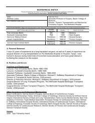
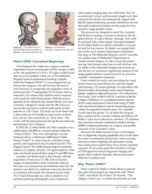
Figure 2. “The Visible Human”<br />
from the1995 National Library of<br />
Medicine, rendered with Fovia, Inc.<br />
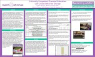
Figure 3. Archimedes<br />
Circulatory System from 287 B.C.<br />
Plato’s CAVE: Conceptual Beginnings<br />
I first designed the single-user surgeon computeraugmented<br />
virtual environment (CAvE) concept in 2006<br />
as the sole proprietor of a 20 ft x 15 ft glass-walled kiosk<br />
that was to be installed within one of The <strong>Methodist</strong><br />
<strong>Hospital</strong> System’s professional buildings. Initially<br />
called the Surgeons EDGE TM , it was designed for cardiovascular<br />
surgeons who did not have the time or<br />
tools necessary to manipulate and integrate a series of<br />
patient-specific CT angiography (CTA) studies into an<br />
interactive 3-D volume that could be used to measure<br />
aortic grafts for individual patients. With the encouragement<br />
of Drs. Richard Geis, Samuel Henly, and Alan<br />
Lumsden, I designed a kiosk near the OR where cardiovascular<br />
physicians could fit aortic grafts to their<br />
patients on a 24/7 basis. The kiosk was to be fitted with<br />
a state-of-the-art 3-D TeraRecon large-screen workstation,<br />
and my role evolved into an “aorta tailor” who<br />
works with the physicians and sets up the procedure for<br />
measurement and validation.<br />
In 2007, Drs. E. Brian Butler and Alan Lumsden were<br />
collaborating with IBM on a research project called the<br />
“virtual Patient.” They were attempting to use the<br />
National Library of Medicine’s MRI-based visible<br />
Human Project ® as a base map for a detailed, imageguided,<br />
auto-segmented atlas of patient-specific CTAs<br />
(Figures 2 and 3). The visible Human Project involved the<br />
creation of complete, detailed, 3-D representations of the<br />
male and female human bodies using CT and MRI slices.<br />
Lumsden and Butler built on this concept with the<br />
acquisition of transverse CT, MR, and cryosection<br />
images of representative male and female cadavers.<br />
The male was sectioned at one-millimeter intervals, the<br />
female at one-third of a millimeter intervals; this level<br />
of resolution still exceeds the standard of care today.<br />
The virtual Patient data set, with its detailed crosssectional<br />
pathology photographs, was coregistered<br />
with medical imaging data sets of the body. The 3-D<br />
reconstructed virtual cardiovascular images were then<br />
automatically labeled and subsequently tagged with<br />
SQuID (superconducting quantum interference device)<br />
detectable anatomical markers for the proposed intraoperative<br />
image-guided system.<br />
This process was designed to assist Drs. Lumsden<br />
and Butler in creating a vascular roadmap for the targeted<br />
delivery of a gene therapy “package” that could<br />
be activated with a time-release viral agent designed<br />
by Dr. Butler. Before it could be concluded, it was put<br />
on hold for two reasons: Dr. Butler was named chairman<br />
of the newly formed Department of Radiation<br />
Oncology at The <strong>Methodist</strong> <strong>Hospital</strong>, and Dr. Lumsden<br />
was named chair of <strong>Methodist</strong>’s Department of<br />
Cardiovascular Surgery. In order to keep the project<br />
moving, both doctors asked me to work full-time with<br />
Dr. Butler at The <strong>Methodist</strong> <strong>Hospital</strong> and to look outside<br />
the box for nonclinical engineering solutions to the<br />
image-guided delivery system based on my previous<br />
scientific visualization experience.<br />
I have worked for many years providing the visual<br />
system hardware and software for real-time fighter<br />
pilot training, 3-D seismic/geologic oil exploration, and<br />
pharmaceutical drug design using supercomputing<br />
tightly coupled to high-performance 3-D workstations.<br />
Previously, I had worked with Dr. Lumsden at Baylor<br />
College of Medicine on a peripheral arterial disease<br />
(PAD) multi-institutional clinical trial using 3T MRI<br />
with specialized bilateral coils for measuring plaque<br />
within the superficial femoral artery. Dr. Lumsden<br />
would repeat the phrase “it’s all about the imaging”<br />
like a mantra for the vascular residents and fellows. Dr.<br />
Butler’s vision for an interactive, real-time, 3-D radiation<br />
dose cloud for intensity modulated radiation therapy<br />
(IMRT) treatment planning was a logical and parallel<br />
extension of Dr. Lumsden’s vision.<br />
However, Dr. Butler needed to have a full integration<br />
of diagnostic imaging capabilities that would allow<br />
for clearer visualization and provide a more accurate,<br />
interactive tumor targeting and delineation system<br />
that could protect normal tissue from adverse radiation<br />
exposure. It was at this point that I decided to design<br />
an FDA-approved clinical diagnostic imaging integration<br />
that would be clinically relevant and make a<br />
difference in patient care.<br />
Why “Plato’s CAVE?”<br />
When I asked Dr. Butler to think about a name for<br />
this pilot clinical project, he responded with “Plato’s<br />
CAvE” from Book vII of Plato’s The Republic, “The<br />
Allegory of the Cave,” in which the prisoners see and<br />
28 vII (1) 2011 | MDCvJ