DeBAKEy CARDIOvASCuLAR JOuRNAL - Methodist Hospital
DeBAKEy CARDIOvASCuLAR JOuRNAL - Methodist Hospital
DeBAKEy CARDIOvASCuLAR JOuRNAL - Methodist Hospital

You also want an ePaper? Increase the reach of your titles
YUMPU automatically turns print PDFs into web optimized ePapers that Google loves.
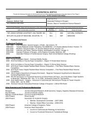
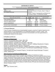
Figure 4. Plato, in The Republic, Book VII, 514a-520a. 1 Figure 5. “The Anatomy Lecture of Dr. Nicolaes Tulp” by Rembrandt,<br />
1632.<br />
hear shadows and echoes cast by objects that they<br />
cannot see clearly (Figure 4 and 5). 1<br />
It was the perfect code name for exploring the art and<br />
science of “seeing” the invisible.<br />
Dr. Butler’s research over the past decade has been<br />
focused on image-guided radiation and gene therapies<br />
for cancer. In the past year, he and I have broadened the<br />
focus to include visualization-guided clinical interventions<br />
that are within the domain of surgical oncology,<br />
including reconstructive surgery and transplantation.<br />
This expanded focus was made possible by the development<br />
of an N-dimensional (space, time, color, and<br />
multimodality images) image-guided intervention<br />
system, more correctly a visualization system, that has<br />
been operational in our laboratory since April 2009.<br />
Plato’s CAVE: The Advanced Clinical<br />
Visualization Process<br />
The current standard of care for medical imaging is<br />
to interpret slices of 2-D shades-of-gray images in an<br />
attempt to translate them into a physician’s “personal<br />
virtual 3-D anatomical and disease world,” then interpreting<br />
the problem and rendering a written diagnosis<br />
— granted, an expert professional diagnosis by a welltrained<br />
imaging specialist, but still an interpretation of<br />
a 2-D dataset nonetheless. Plato’s CAvE raises the bar<br />
with an advanced clinical visualization process that<br />
creates 3-D images derived from a patient’s DICOM<br />
image studies that have been pulled from <strong>Methodist</strong>’s<br />
Picture Archiving and Communication System (PACS),<br />
from other institutional PACS via electronic transfer, or<br />
from CDs that patients bring with them. Regardless of<br />
the image source, the 3-D visualizations are ready for<br />
projection and viewing on a variety of screen formats<br />
within minutes. The region of interest is measurable,<br />
can be manipulated in three dimensions, and can be<br />
presented stereoscopically like the movie Avatar. This<br />
approach makes full use of the strengths of each imaging<br />
modality by displaying and/or fusing all of the<br />
images available for the patient (CT, MRI, PET and,<br />
soon, HR ultrasound) in a sequence of the events that<br />
are relevant to the treatment team. The scene is adaptable<br />
to all surgical interventions, from reconstruction<br />
through transplantation, and capable of bringing quality<br />
improvements to the diagnosis and treatment<br />
planning for many medical conditions.<br />
Our technology is totally complementary to all<br />
standard-of-care imaging modalities currently in use<br />
and some that are in development, i.e., physiodynamic<br />
processes, beating heart, mitral valves opening and<br />
closing. There is a high level of clinical imaging and<br />
medical device industry interest in experimenting with<br />
or enhancing Plato’s CAvE with natural user interactions.<br />
Our surgeons have discovered, once they work<br />
with this system, that they cannot easily justify returning<br />
to the current standard of viewing images for their<br />
patients. We have designed and integrated the CAvE<br />
to function as a node/portal on a physician grid or<br />
network. It is not an island: the visualization can be<br />
distributed to the scientist’s or clinician’s desktop<br />
because it is server-based, thin-client enabled, and<br />
HIPAA compliant.<br />
The system offers enormous opportunity for educating<br />
primary care and referring physicians, patients and<br />
their families, medical students, residents and staff surgeons<br />
by narrowing the degree of uncertainty that each<br />
brings to the interaction (Figure 6 and 7). It also gives<br />
the medical team the ability to make a more informed<br />
decision regarding the best intervention or treatment<br />
of the disease. In some cases, the visualization has<br />
revealed previously unrecognized ancillary anatomical<br />
findings or additional disease complications.<br />
Development, testing, validation and translation of<br />
this technology into practice will shape the metrics<br />
MDCvJ | vII (1) 2011 29