DeBAKEy CARDIOvASCuLAR JOuRNAL - Methodist Hospital
DeBAKEy CARDIOvASCuLAR JOuRNAL - Methodist Hospital
DeBAKEy CARDIOvASCuLAR JOuRNAL - Methodist Hospital

You also want an ePaper? Increase the reach of your titles
YUMPU automatically turns print PDFs into web optimized ePapers that Google loves.
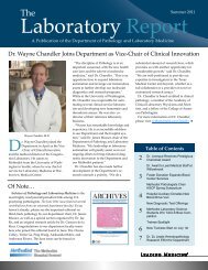
MuseuM of tMH MultiModality iMaging Center<br />
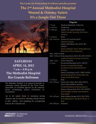
Diastolic Mitral Valve Regurgitation. A 41-year-old man was referred to <strong>Methodist</strong> with suspected bacterial endocarditis. 2-D<br />
echocardiography was performed and a large aortic valve vegetation was identified along with severe aortic regurgitation. Panel A depicts<br />
a mildly enlarged left ventricle (5.9 cm diameter) with open mitral leaflets near end-diastole. Color Doppler demonstrates simultaneous aortic<br />
regurgitation (AR) and mitral regurgitation (MR). Panel B depicts a continuous wave Doppler signal across the mitral valve. Regurgitant mitral<br />
flow begins 80ms before the onset of systole. In general, diastolic MR is diagnostic of highly elevated left ventricular end-diastolic pressure.<br />
For this patient, the presence of diastolic MR confirmed that the aortic regurgitation was severe and likely acute. In this era of increasingly<br />
sophisticated imaging technologies, it is important to continue to recognize these classic Doppler findings.<br />
Image courtesy of Stephen H. Little, M.D.<br />
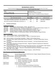
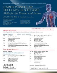
Cardiac Computed Tomography<br />
Angiogram. Cardiac computed<br />
tomography angiogram (CTA) of a 48-yearold<br />
male with atypical chest pain. A small<br />
muscular ventricular septal defect (arrow)<br />
with left to right shunting as shown by the<br />
passage of iodinated contrast from left<br />
ventricle (LV) to right ventricle (RV).<br />
Image courtesy of Su Min Chang, M.D.<br />
MDCvJ | vII (1) 2011 55