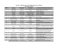
Nature Immunology
Nature Immunology
Nature Immunology

Create successful ePaper yourself
Turn your PDF publications into a flip-book with our unique Google optimized e-Paper software.
© 2009 <strong>Nature</strong> America, Inc. All rights reserved.<br />
METHODS<br />
Mice. C57BL/6 mice were from Harlan; 2D2 TCR–transgenic (006912), UBC-<br />
GFP (004353) and Ifng –/– (002287) mice were from the Jackson Laboratory.<br />
OT-II TCR–transgenic mice (provided by J. Kirberg) were bred onto backgrounds<br />
of various Cd45 alleles in the animal facility of the Institute for<br />
Research in Biomedicine. Ccr6 –/– mice have been described 31 , Cxcr3 –/– mice<br />
were provided by C. Gerard, and Ccr7 –/– mice were provided by M. Lipp. Mice<br />
were treated in accordance with guidelines of the Swiss Federal Veterinary<br />
Office and experiments were approved by the Dipartimento della Sanità<br />
eSocialità.<br />
EAE model. Groups of female C57BL/6 mice 8–10 weeks of age were<br />
immunized subcutaneously on day 0 with 100 mg MOG(35–55) (MEVG<br />
WYRSPFSRVVHLYRNGK; Servei de Proteòmica, Pompeu Fabra University,<br />
Barcelona) emulsified in CFA (Difco). Pertussis toxin in 100 ml salinewas<br />
injected intravenously twice, on days 0 and 2 or days 1 and 3. Disease severity<br />
was assigned scores on the following scale: 0, no disease; 1, tail weakness; 2,<br />
paraparesis; 3, paraplegia; 4, paraplegia with forelimb weakness or paralysis; 5,<br />
moribund or dead. In some EAE experiments, naive T cells (1 10 5 )from<br />
GFP + 2D2-transgenic mice or 2D2 CCR6-knockout mice or total CD4 +<br />
T cells (10 10 6 ) from wild-type, CXCR3-knockout, IFN-g-knockout or<br />
CCR7-knockout mice were transferred intravenously into mice 16 h before<br />
immunization. In some experiments, a mixture of GFP + 2D2-transgenic T cells<br />
(2.5 10 6 ) and 2D2 CCR6-knockout T cells (at a ratio of 1:1) were<br />
transferred intravenously into mice 16 h before immunization. Alternatively,<br />
effector TH-17 cells were generated in vitro from GFP + 2D2-transgenic T cells<br />
(2.5 10 6 ) and 2D2 CCR6-knockout T cells (at a ratio of 1:1) and were<br />
transferred intravenously in wild-type mice at various times after EAE induction.<br />
For the preparation of CNS lymphocytes, mice were perfused through the<br />
left cardiac ventricle with cold PBS. The forebrain and cerebellum were<br />
dissected, were cut into pieces and were digested for 45 min at 37 1C with<br />
collagenase D (1 mg/ml; Roche Diagnostics) and DNaseI (1 mg/ml; Sigma).<br />
CD45 + cells were isolated by passage of the tissue through a cell strainer<br />
(70 mm), followed by incubation with beads coated with antibody to CD45<br />
(anti-CD45; 130-052-301; Milteny). After passage through the column, CD45 +<br />
cells were washed and resuspended in culture medium for further analysis.<br />
Flow cytometry analysis and in vitro stimulation. For analysis of mouse<br />
phenotypes, the following monoclonal antibodies were used: anti-L-selectin<br />
(MEL14), anti-CD44 (IM7), anti-CD4 (RM4-5), anti-CD8a (53-6.7), anti-CD3<br />
(145-2C11), anti-CD28 (37.51), anti-B220 (RA3-6B2), anti-CD19 (1D3), anti-<br />
CD11b (M1/70), anti-CD45.1 (A20), anti-CD45.2 (104), anti-CD127 (A7R34)<br />
and anti-IFN-g (XMG1.2; all from eBiosciences); and anti-IL-17A (TC11-<br />
18H10) and anti-CCR6 (140706; both from BD Biosciences). For intracellular<br />
cytokine staining, cells were stimulated for 4 h with phorbol 12-myristate<br />
13-acetate (100 nM; Sigma) and ionomycin (1 mg/ml; Sigma), with the final 2 h<br />
of culture in the presence of brefeldin A (10 mg/ml; Sigma). Labeled antibodies<br />
were used after cells were fixed in 4% (wt/vol) paraformaldehyde and made<br />
permeable with 0.5% (wt/vol) saponin (Sigma-Aldrich). Six-color staining of<br />
the cell surface was done with the appropriate combinations of antibodies<br />
conjugated to fluorescein isothiocyanate, phycoerythrin, peridinine chlorophyll<br />
protein complex, phycoerythrin-indotricarbocyanine, allophycocyanin, allophycocyanin-indotricarbocyanine<br />
or biotin, and with streptavidin labeled with<br />
phycoerythrin-indotricarbocyanine or allophycocyanin-indotricarbocyanine<br />
(BD Biosciences). Samples were acquired on a FACSCanto (BD Biosciences)<br />
and were analyzed with FlowJo software (TreeStar).<br />
For studies of human cell phenotypes, peripheral blood and cerebrospinal<br />
fluid were obtained from eight patients (after informed consent was provided)<br />
who presented with a first demyelinating event suggestive of multiple sclerosis<br />
and underwent venipuncture and lumbar puncture for diagnostic purposes.<br />
The study was approved by the Ethical Committee and Board of the Department<br />
of Neurosciences, Ophthalmology and Genetics, University of Genoa.<br />
Peripheral blood mononuclear cells were isolated with Ficoll and cerebrospinal<br />
fluid leukocytes were collected after centrifugation, then these cells were<br />
incubated for 30 min with the following monoclonal antibodies: fluorescein<br />
isothiocyanate–conjugated anti-CD4 (RPA-T4), phycoerythrin-conjugated<br />
anti-CCR6 (11A9), phycoerythrin-indodicarbocyanine–conjugated anti-CD25<br />
ARTICLES<br />
(M-A251) and allophycocyanin-conjugated anti-CD45RO (UCHL1; all from<br />
BD Biosciences). At the end, cells were washed with PBS, were resuspended,<br />
were counterstained with propidium iodide (1 mg/ml; Sigma Aldrich) and were<br />
analyzed by flow cytometry of the propidium iodide–negative population. A<br />
FACSCanto (BD Biosciences) was used for all flow cytometry and data were<br />
analyzed with FACSDiva & FloJo software.<br />
For antigen-specific restimulation of mouse T cells, 5 10 6 splenocytes or<br />
2.5 10 6 lymph node cells were cultured for 3 d in presence of MOG(35–55).<br />
Cells purified from the brain were cultured for 3 d in presence of fixed<br />
splenocytes loaded with MOG(35–55). For fixation, spleens were removed from<br />
naive C57BL/6 mice and erythrocytes were lysed. Splenocytes were incubated<br />
for 90 min at 37 1C with MOG(35–55) (50 mg/ml), then were washed in 1%<br />
(vol/vol) FCS in PBS and were fixed for 30 s at 20 1C in 0.05% (vol/vol)<br />
glutaraldehyde (Merck). An equal volume of a solution of glycine (0.2 M;<br />
Sigma-Aldrich) was added for 30 s and then cells were washed and used for<br />
coculture. Cytokines in culture supernatants were measured by enzyme-linked<br />
immunosorbent assay according to the manufacturer’s protocols (BD Biosciences).<br />
Data were analyzed with the SoftMax program.<br />
Immunohistology and immunofluorescence. Mice were perfused through the<br />
left cardiac ventricle with 1% (vol/vol) formaldehyde (Grogg Chemie) in PBS.<br />
Brains and spinal cords were removed, were embedded in Tissue-Tek optimum<br />
cutting temperature compound (Haslab) and were ‘snap-frozen’ in a bath of<br />
dry ice and isopentane (Grogg Chemie). Cryostat sections 6 mm inthickness<br />
were air-dried overnight, were fixed in acetone and were stained for immunohistology<br />
with a three-step immunoperoxidase staining kit according to the<br />
manufacturer’s protocol (Vectastain; Reactolab). For immunofluorescence<br />
staining, sections were blocked for 20 min with skim milk and then were<br />
incubated for 1 h each with primary and secondary antibody diluted in skim<br />
milk, with washing steps of Tris-buffered saline between incubations. After a<br />
final wash in Tris-buffered saline, sections were mounted in Mowiol solution<br />
(Calbiochem). Antibodies used were as follows: fluorescein isothiocyanate–<br />
conjugated anti-CD45 (30F11; BD Biosciences), anti-CCL20 (AB9829; Abcam),<br />
anti-laminin (Z0097; Dako), anti-PECAM-1 (Mec13.3; BD Biosciences) and<br />
anti–‘pan cytokeratin’ (C2562; Sigma).<br />
LifeSpan Biosciences analyzed CCL20 expression in healthy and diseased<br />
human tissues. Formalin-fixed, paraffin-embedded tissues were stained with<br />
rabbit polyclonal anti-CCL20 (15 mg/ml; AB9829; Abcam). The detection<br />
system consisted of anti-rabbit secondary antibody (BA-1000; Vector) and an<br />
ABC-AP kit (avidin–biotinylated enzyme complex–alkaline phosphatase;<br />
AK-5000; Vector) with a Red substrate kit (SK-5100; Vector), which was used<br />
to produce a fuchsia-colored deposit. Only tissues that were positive for<br />
staining of CD31 and vimentin were used. The negative control consisted<br />
of immunohistochemistry of adjacent sections in the absence of primary<br />
antibody. Slides were imaged with a DVC 1310C digital camera coupled to a<br />
Nikon microscope.<br />
Intravital microscopy. Active EAE was induced by immunization with<br />
MOG(35–55) in CFA. Mice with a score of 0.5 (limp tail) to 1 (hind leg<br />
weakness) were used for intravital fluorescence videomicroscopy experiments.<br />
The spinal cord window was created as described in the Supplementary<br />
Methods. Normal body temperature was maintained throughout the entire<br />
experiment. Epi-illumination techniques were used for intravital fluorescence<br />
videomicroscopy with an IVM500 microscope (Mikron Instruments) coupled<br />
to a 50-Watt mercury lamp (HBO 50 microscope illuminator; Zeiss) and<br />
combined with blue filter blocks (exciter, 455DF70; dichroic, 515DRLP;<br />
emitter, 515ALP) and green filter blocks (exciter, 525DF45; dichroic, 560DRLP;<br />
emitter, 565ALP). Observations were made with 4 , 10 and 20 longdistance<br />
working objectives (Zeiss). All experiments were recorded by means of<br />
a low-light silicon-intensified target video camera with an optional image<br />
intensifier for weak fluorescence (Dage-MTI of Michigan City). Data were<br />
transferred to a digital video system for later offline analysis of the interaction<br />
of cells with CNS microvessels. The injection of 1% (vol/vol) tetramethylrhodamine<br />
isothiocyanate–conjugated dextran in 0.9% (wt/vol) NaCl allowed<br />
visualization of the spinal cord microvasculature. In parallel, mouse T cells were<br />
stained for 45 min at 37 1C with2.5mM CellTracker green (Molecular Probes)<br />
and were injected into the carotid artery (4 10 6 T cells in three<br />
NATURE IMMUNOLOGY VOLUME 10 NUMBER 5 MAY 2009 521