Nature Immunology
Nature Immunology
Nature Immunology

Create successful ePaper yourself
Turn your PDF publications into a flip-book with our unique Google optimized e-Paper software.
© 2009 <strong>Nature</strong> America, Inc. All rights reserved.<br />
ARTICLES<br />
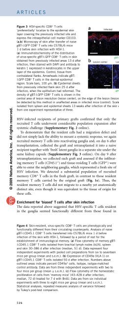
Figure 3 HSV-specific CD8 + Tcells<br />
‘preferentially’ localize to the epidermal skin<br />
layer covering the previously infected site and<br />
express the intraepithelial cell marker CD103.<br />
(a,b) Microscopy of skin after transfer of naive<br />
gBT-I.GFP CD8 + T cells into C57BL/6 mice<br />
1 d before skin infection with HSV-1.<br />
(a) Immunohistochemistry of the distribution<br />
of virus-specific gBT-I.GFP CD8 + T cells in skin<br />
obtained from previously infected areas 13 d after<br />
infection, then stained with DAPI and antibody to<br />
keratin 1 expressed in keratinocytes in the upper<br />
layer of the epidermis. Control, tissue from<br />
contralateral flanks. Arrowheads indicate gBT-<br />
I.GFP CD8 + T cells in the dermal-epidermal<br />
region. Scale bars, 100 mm. (b) Epidermal sheets<br />
from previously infected flank skin 25 d after<br />
infection, when the epithelium had reformed. The<br />
density of gBT-I.GFP CD8 + T cells is shown in the<br />
central area of lesion resolution (lesion center), on the edge of the lesion (lesion periphery) and in an unaffected area (control). Virus-specific T cells could not<br />
be detected by this method in unaffected areas in infected mice (control). Scale bars, 50 mm. (c) CD103 expression by memory gBT-I.CD45.1 CD8 + T cells<br />
isolated from spleen and epidermal sheets 13 weeks after infection of the skin with HSV. Numbers above lines (right) indicate percent CD103 + Infected<br />
Control<br />
Spleen<br />
1.5<br />
b Lesion center<br />
gBT-l (GFP) Keratin 1 DAPl<br />
Lesion periphery Control<br />
Vα2 CD103<br />
Epidermis<br />
98.1<br />
Vα2 CD103<br />
gBT-l (GFP) DAPl<br />
cells. Data are<br />
from one experiment representative of three.<br />
HSV-infected recipients of primary grafts confirmed that only the<br />
recruited T cells underwent considerable population expansion after<br />
systemic challenge (Supplementary Fig. 2 online).<br />
To demonstrate that the resident cells had a migration defect and<br />
did not simply lack the ability to mount a systemic response, we again<br />
recruited memory T cells into reactivating ganglia and, on day 6 after<br />
transplantation, collected the graft and retransplanted it into a naive<br />
recipient together with ‘fresh’ latent ganglia in a separate site under the<br />
same kidney capsule (Supplementary Fig. 3 online). On day 9 after<br />
retransplantation, we collected each graft and assessed if the infiltrating<br />
memory T cells (CD45.1 + ) and tissue-residing T cells (GFP + ) were<br />
able to enter the neighboring ganglia, which represented a fresh site of<br />
HSV infection. We detected a substantial population of recruited<br />
memory CD8 + T cells in the fresh graft, in contrast to those resident<br />
memory T cells carried by the original graft (Fig. 1e). Thus, the<br />
resident memory T cells did not migrate to a nearby yet anatomically<br />
distinct site, even though it was equivalent to the tissue of origin for<br />
these cells.<br />
Enrichment for ‘biased’ T cells after skin infection<br />
The data reported above suggested that HSV-specific T cells resident<br />
in the ganglia seemed functionally different from those found in<br />
Figure 4 Skin-resident, virus-specific CD8 + T cells are phenotypically and<br />
functionally different from their circulating counterparts. Analysis of naive<br />
gBT-I.CD45.1 CD8 + T cells transferred into C57BL/6 mice 1 d before<br />
infection of the skin with HSV-1, followed by a period of rest for the<br />
establishment of immunological memory. (a) Flow cytometry of memory gBT-<br />
I.CD45.1 CD8 + T cells isolated from brachial lymph nodes (bLN), spleen<br />
and skin 30–386 d after infection (median, 51 d). Data represent four<br />
independent experiments with pooled cell preparations from six to seventeen<br />
mice per group (mean and s.e.m.). (b) Expression of CD49a (VLA-1) on<br />
gBT-I.CD45.1 CD8 + T cells isolated 50 d after infection. Numbers above<br />
outlined areas indicate percent CD49a + cells. Isotype, isotype-matched<br />
control antibody. Data are from three independent experiments with two to<br />
four mice per group (mean ± s.e.m.). (c) Flow cytometry of the homeostatic<br />
proliferation of cells from ‘memory mice’ (43–428 d after infection;<br />
median, 72 d) treated for 7 d with BrdU. Data are from six individual<br />
experiments with three to eight mice per group (mean and s.e.m.).<br />
Statistical analyses, repeated measures analysis of variance followed<br />
by Tukey’s post-test comparison.<br />
a c<br />
CD45.1<br />
CD45.1<br />
the circulation. We reasoned that an analogous population of tissueresident<br />
memory T cells would also be found in other peripheral sites<br />
of infection, notably the skin. Evidence for this took the form of<br />
‘preferential’ retention of virus-specific T cells in flank skin involved in<br />
overt HSV disease relative to that in the uninvolved contralateral<br />
flanks. There was ‘preferential’ enrichment for gBT-I T cells in<br />
flank skin ipsilateral to the origin of infection at all times assessed<br />
(Fig. 2a,b). In contrast, there was no significant difference in the<br />
number of endogenous CD8 + T cells in opposing flanks after clearance<br />
of replicating virus, which occurs by day 8 after infection 18 (Fig. 2c). It<br />
should be noted that gBT-I cells represented a relatively small<br />
proportion of all CD8 + T cells in the skin, especially at these later<br />
times (day 30 or beyond; Fig. 2d). The accumulation of HSV-specific<br />
T cells was not restricted to transgenic cells, because repeat experiments<br />
with tetramer staining to detect HSV-specific T cells showed<br />
similar biases (Supplementary Fig. 4 online). Retention of T cells in<br />
the skin had a nonspecific component, as demonstrated by the use of<br />
attenuated recombinant viruses (HSV strains rgB and rgB-L8A) 25<br />
(Supplementary Fig. 5 online). The number of long-term gB-specific<br />
T cells was the same in flanks infected with gB-sufficient rgB virus and<br />
control gB-deficient rgB-L8A in dual-inoculated mice. Even scarification<br />
alone led to equivalent infiltration into rgB-infected and control<br />
a<br />
CD62L hi gBT-l (%)<br />
b<br />
Isotype CD49a<br />
100 P < 0.01<br />
75<br />
50<br />
25<br />
0<br />
bLN Spleen Skin<br />
bLN<br />
35.2 ± 16.2<br />
CD45.1<br />
P < 0.001 P < 0.05<br />
Spleen<br />
CD122 hi gBT-l (%)<br />
100<br />
75<br />
50<br />
25<br />
0<br />
Skin<br />
42.5 ± 9.8 85.1 ± 1.7<br />
Cells<br />
Cells<br />
P < 0.01<br />
P > 0.05 100<br />
75<br />
50<br />
25<br />
0<br />
P < 0.001<br />
bLN Spleen Skin bLN Spleen Skin<br />
CD69 + gBT-l (%)<br />
c<br />
BrdU + gBT-l (%)<br />
20<br />
10<br />
0<br />
P < 0.01<br />
P < 0.05<br />
bLN Spleen Skin<br />
526 VOLUME 10 NUMBER 5 MAY 2009 NATURE IMMUNOLOGY