Nature Immunology
Nature Immunology
Nature Immunology

Create successful ePaper yourself
Turn your PDF publications into a flip-book with our unique Google optimized e-Paper software.
© 2009 <strong>Nature</strong> America, Inc. All rights reserved.<br />
ARTICLES<br />
degradative pathway. The possibility of transfer of gB to lysosomes<br />
through phagocytosis of infected cells (cross-presentation) was ruled<br />
out by results showing that incubation of H-2 b BMA macrophages<br />
together with HSV-1-infected H-2 d J774 macrophages did not result<br />
in activation of the H-2 b -specific CD8 + T cell hybridoma (Fig. 1e).<br />
Hence, vacuolar processing of gB in the later period of infection was<br />
more likely to involve membrane-trafficking events that occurred<br />
exclusively in the infected cell.<br />
Data have shown that endogenous viral proteins can be presented<br />
on MHC class II molecules by a process involving autophagy 9 ,which<br />
indicates that trafficking events that enable the transport of endogenous<br />
proteins to vacuolar degradative organelles can occur in virusinfected<br />
cells. To determine if autophagy was involved in the late<br />
processing of endogenous gB and its presentation on MHC class I<br />
molecules, we first monitored the presence of LC3, a marker of<br />
autophagy, in macrophages at various times after infection<br />
(Fig. 2a). Although we did not detect it in uninfected cells, we<br />
found LC3 beginning at 4–6 h after infection, which indicated that<br />
an autophagic response occurred during the late phase of HSV-1<br />
infection in macrophages. Whereas the inhibitory effect of bafilomycin<br />
indicated that a vacuolar response of some kind was triggered during<br />
the late phase of infection in macrophages (Fig. 1c), the possibility<br />
of a contribution of autophagy to this process was suggested by<br />
the substantial inhibition of the CD8 + T cell–stimulatory capacity<br />
of macrophages treated with 3-methyladenine, a commonly used<br />
a<br />
b<br />
c<br />
g<br />
Control<br />
HS<br />
Rapa<br />
Time (h)<br />
IB: α-HSV<br />
d Time (h)<br />
WT ∆34.5<br />
0 6 8 10 6 8 10<br />
4<br />
6<br />
8<br />
10<br />
(kDa)<br />
250<br />
100<br />
75<br />
50<br />
37<br />
25<br />
20<br />
VP26 gB LC3 Merge<br />
h<br />
Time (h)<br />
1,200<br />
Counts<br />
960<br />
620<br />
480<br />
240<br />
6 8<br />
1,200<br />
960<br />
620<br />
480<br />
240<br />
inhibitor of autophagy (Fig. 2b). Confirmation of the involvement of<br />
autophagy in the processing and presentation of gB peptides on MHC<br />
class I molecules was provided by experiments involving small interfering<br />
RNA (siRNA)-mediated silencing of Atg5, a protein involved in<br />
the formation of autophagosomes 20 . Macrophages treated with a<br />
control siRNA had a greater capacity to stimulate CD8 + T cells<br />
between 8 h and 12 h after infection, but macrophages treated with<br />
Atg5-specific siRNA did not (Fig. 2c),whichlinkedthislategainin<br />
stimulation to the induction of autophagy.<br />
Further support for the idea that autophagy contributes to the<br />
vacuolar processing and presentation of gB on MHC class I molecules<br />
was provided by results indicating that treatment of infected macrophages<br />
with rapamycin, an inhibitor of the kinase mTOR that<br />
stimulates autophagy 21 ,considerablyimprovedCD8 + T cell stimulation<br />
(Fig. 2d). We obtained similar results with macrophages exposed<br />
to a mild heat shock before infection (39 1C for 12 h), a condition also<br />
known to induce autophagy 22 (Fig. 2d). The enhanced CD8 + T cell<br />
stimulation induced by mTOR or heat shock was abolished by the<br />
addition of bafilomycin (Fig. 2e). These data further link the vacuolar<br />
processing of gB to autophagy. We obtained similar results with mouse<br />
embryonic fibroblasts isolated from Atg5 –/– mice 20 (Supplementary<br />
Fig. 1a online). Silencing of Atg5 in infected macrophages with siRNA<br />
abolished the effect of rapamycin (Fig. 2f), which confirmed the<br />
specificity of this drug for the autophagic pathway. Similarly, neither<br />
bafilomycin (Supplementary Fig. 1c,d) nor 3-methyladenine (data<br />
e<br />
6 h<br />
8 h<br />
∆34.5<br />
WT<br />
∆34.5<br />
WT<br />
1,200<br />
gB LC3 Overlay<br />
10<br />
Ctrl<br />
WT 17+<br />
∆34.5<br />
0<br />
0<br />
0<br />
100 101 102 103 104 100 101 102 103 104 100 101 102 103 104 gB<br />
960<br />
620<br />
480<br />
240<br />
f<br />
β-galactosidase<br />
activity (relative)<br />
2.5<br />
WT 17+<br />
∆34.5<br />
2.0<br />
1.5<br />
1.0<br />
0.5<br />
0<br />
6 8 10<br />
Time after<br />
infection (h)<br />
i WT 17+<br />
∆34.5<br />
1.00<br />
0.75<br />
0.50<br />
0.25<br />
0<br />
Baf: – + – + – + – + – + – +<br />
6 8 10<br />
Time after infection (h)<br />
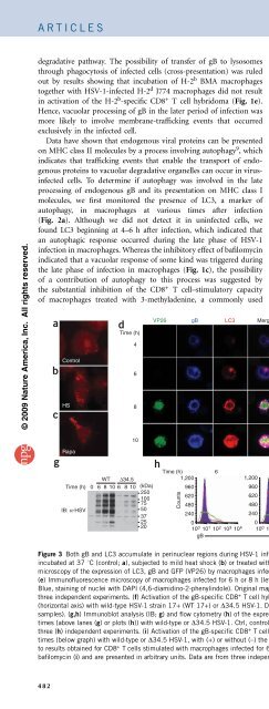
Figure 3 Both gB and LC3 accumulate in perinuclear regions during HSV-1 infection. (a–c) Immunofluorescence microscopy of uninfected macrophages<br />
incubated at 37 1C (control; a), subjected to mild heat shock (b) or treated with rapamycin (c), then stained with anti-LC3. (d) Immunofluorescence<br />
microscopy of the expression of LC3, gB and GFP (VP26) by macrophages infected for various times (left margin) with HSV-1. White indicates colocalization.<br />
(e) Immunofluorescence microscopy of macrophages infected for 6 h or 8 h (left margin) with wild-type HSV-1 (WT) or HSV-1 lacking ICP34.5 (D34.5).<br />
Blue, staining of nuclei with DAPI (4,6-diamidino-2-phenylindole). Original magnification, 100 (a–c) or 63 (d,e). Results in a–e are representative of<br />
three independent experiments. (f) Activation of the gB-specific CD8 + T cell hybridoma (as described in Fig. 1a) by macrophages infected for various times<br />
(horizontal axis) with wild-type HSV-1 strain 17+ (WT 17+) or D34.5 HSV-1. Data are from three independent experiments (mean and s.e.m. of triplicate<br />
samples). (g,h) Immunoblot analysis (IB; g) and flow cytometry (h) of the expression of HSV-1 proteins (g) and gB (h) in macrophages infected for various<br />
times (above lanes (g) or plots (h)) with wild-type or D34.5 HSV-1. Ctrl, control (uninfected BMA macrophages; h). Data are representative of two (g) or<br />
three (h) independent experiments. (i) Activation of the gB-specific CD8 + T cell hybridoma (as described in Fig. 1a) by macrophages infected for various<br />
times (below graph) with wild-type or D34.5 HSV-1, with (+) or without (–) the addition of bafilomycin A at 2 h after infection. Results in f,i are normalized<br />
to results obtained for CD8 + T cells stimulated with macrophages infected for 6 h with wild-type virus (f) or with infected macrophages incubated without<br />
bafilomycin (i) and are presented in arbitrary units. Data are from three independent experiments (mean and s.e.m. of triplicate samples).<br />
482 VOLUME 10 NUMBER 5 MAY 2009 NATURE IMMUNOLOGY<br />
β-galactosidase<br />
activity (relative)