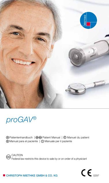
proGAV® - Christoph Miethke GmbH & Co. KG
proGAV® - Christoph Miethke GmbH & Co. KG
proGAV® - Christoph Miethke GmbH & Co. KG

You also want an ePaper? Increase the reach of your titles
YUMPU automatically turns print PDFs into web optimized ePapers that Google loves.
proGAV ®<br />
USA<br />
Patientenhandbuch | GB Patient Manual | Manuel du patient<br />
Manual para el paciente | Manuale per il paziente<br />
D F<br />
E I<br />
CAUTION<br />
Federal law restricts this device to sale by or on order of a physician!<br />
CHRISTOPH MIETHKE GMBH & CO. <strong>KG</strong> 0297
Abb. 1, Fig. 1<br />
Abb. 9, Fig. 9<br />
1<br />
2<br />
3<br />
4<br />
5<br />
6<br />
1<br />
2<br />
3<br />
6<br />
6<br />
4<br />
5<br />
3
Das Unternehmen<br />
Die <strong>Christoph</strong> <strong>Miethke</strong> <strong>GmbH</strong> & <strong>Co</strong>. <strong>KG</strong> ist ein<br />
Brandenburger Unternehmen, das sich mit der<br />
Entwicklung, der Produktion und dem Vertrieb von<br />
innovativen neurochirurgischen Implantaten zur<br />
Behandlung des Hydrocephalus beschäftigt. Wir<br />
arbeiten hierbei erfolgreich mit Kliniken weltweit<br />
zusammen.<br />
Diese Broschüre soll Ihnen und Ihrer Familie einen<br />
Einblick in die Behandlung des Hydrocephalus geben.<br />
Erst seit den 50er Jahren ist es möglich, diese<br />
Krankheit erfolgreich zu behandeln. Der Techniker<br />
John D. Holter hatte in einem dramatischen<br />
Wettlauf um das Leben seines an Hydrocephalus<br />
leidenden Sohnes Casey in Philadelphia in wenigen<br />
Wochen ein Silikon-Ventil entwickelt. Obwohl<br />
sich dieses Ventil nach seiner Implantation im März<br />
1956 klinisch bewährt hatte und einen großen<br />
Schritt in der Behandlung dieser Krankheit darstellt,<br />
gibt es bis heute eine erhebliche Anzahl von<br />
Patienten, die mit Ventilsystemen große Probleme<br />
haben.<br />
Die <strong>Christoph</strong> <strong>Miethke</strong> <strong>GmbH</strong> & <strong>Co</strong>. <strong>KG</strong> hat die Erkenntnisse<br />
von 50 Jahren Ventilbehandlung aufgegriffen<br />
und durch die Verwendung des Werkstoffs<br />
Titan eine neue Generation von hochpräzisen<br />
Ventilen entwickelt. Erstmals stehen Ventilsysteme<br />
zur Verfügung, die konsequent die physikalischen<br />
Randbedingungen der Hirnwasserableitung berücksichtigen<br />
und so einen physiologischen Hirndruck<br />
unabhängig von der Körperlage einstellen.<br />
Abb. 1: Anatomische Darstellung des Schädels<br />
(siehe Umschlaginnenseite)<br />
1) Schädeldecke<br />
2) Gehirn<br />
3) Hirnwasser (Liquor)<br />
4) Seitlicher Ventrikel<br />
5) Dritter Ventrikel<br />
6) Vierter Ventrikel<br />
anatomische GrUnDlaGen<br />
Das menschliche Gehirn (Abb. 1) ist von einer<br />
speziellen Flüssigkeit, dem Hirnwasser (Liquor),<br />
umgeben. Im Inneren des Kopfes befinden sich<br />
mehrere Hirnkammern, so genannte Ventrikel,<br />
in denen das Hirnwasser produziert wird. Die<br />
Ventrikel sind durch Kanäle untereinander verbunden<br />
und stellen ein komplexes Ableitungs-<br />
system dar. Das Wasser zirkuliert durch diese Hirnkammern<br />
und wird schließlich in das venöse Blut<br />
abgegeben. Die Aufgabe des Hirnwassers besteht<br />
Patientenhandbuch | D<br />
darin, das Gehirn vor mechanischer Schädigung<br />
zu schützen. Zusätzlich regelt es den Hirninnendruck,<br />
hält das Hirngewebe feucht und transportiert<br />
die Stoffwechselprodukte.<br />
KranKheitsbilD<br />
Beim gesunden Menschen existiert ein Gleichgewicht<br />
zwischen Produktion und Resorption des<br />
Hirnwassers. Die täglich produzierte Flüssigkeitsmenge<br />
liegt beim Säugling bei ca. 100 ml, beim<br />
Kleinkind bei ca. 250 ml und beim Erwachsenen<br />
bei ca. 500 ml. Wird mehr Liquor gebildet als abgebaut<br />
werden kann, kommt es zur Vergrößerung der<br />
Hirnkammern, dem so genannten Hydrocephalus<br />
(Abb. 2). Der Begriff Hydrocephalus beschreibt einen<br />
Zustand, bei dem „Wasser“ (Hydro) im „Kopf“<br />
(Cephalus) ständig an Volumen zunimmt. Dieser<br />
Zustand besteht oft schon bei der Geburt (angeborener<br />
Hydrocephalus). Er kann sich aber auch<br />
im späteren Leben ausbilden, z.B. durch eine Entzündung<br />
oder Blutung, durch eine schwere Verletzung<br />
am Kopf oder infolge einer Hirnoperation. In<br />
diesen Fällen spricht man von einem erworbenen<br />
Hydrocephalus.<br />
Man unterscheidet außerdem zwischen dem Hydrocephalus<br />
occlusus (nicht kommunizierender<br />
Hydrocephalus) und dem Hydrocephalus communicans<br />
(kommunizierender Hydrocephalus). Beim<br />
Hydrocephalus occlusus ist die Verbindung zwischen<br />
den Hirnkammern unterbrochen, so dass<br />
sie nicht miteinander „kommunizieren“ können.<br />
Wenn die Ventrikel miteinander frei verbunden sind,<br />
aber eine Störung der Hirnwasserresorption besteht,<br />
liegt ein Hydrocephalus communicans vor.<br />
a) b)<br />
Abb. 2: Ventrikelgröße<br />
a) normal b) Hydrocephalus<br />
5
D | Patientenhandbuch Patientenhandbuch | D<br />
KranKheitssymptome<br />
Im Säuglingsalter sind die Schädelknochen noch<br />
nicht fest verwachsen. Das zunehmende Hirnwasser<br />
führt hier zu einer Zunahme des Kopfumfangs<br />
unter gleichzeitigem Abbau von Hirngewebe. Ab<br />
einem Alter von ca. 2 Jahren wird durch den harten<br />
Schädel eine Vergrößerung des Kopfumfangs<br />
verhindert. Hier führt die Flüssigkeitszunahme zu<br />
einem enormen Druckanstieg, wodurch sich die<br />
Hirnkammern erweitern und das Gehirn komprimiert<br />
wird. Sowohl beim Säugling als auch beim<br />
Erwachsenen können irreversible Gehirnschäden<br />
auftreten. Je nach Grad der Störung kommt es zu<br />
Übelkeit, Kopfschmerzen, Erbrechen, Koordinationsstörung,<br />
Schläfrigkeit und schließlich Bewusstlosigkeit.<br />
DiaGnose Der erKranKUnG<br />
Dem Arzt stehen heute verschiedene Möglichkeiten<br />
zur Diagnose eines Hydrocephalus zur<br />
Verfügung. Mittels bildgebender Verfahren (z. B.<br />
<strong>Co</strong>mputertomographie, Ultraschall oder Magnetresonanztomographie)<br />
wird die Größe der Ventrikel<br />
bestimmt.<br />
<strong>Co</strong>mputertomographie (CT)<br />
Bei dieser schnellen und schmerzlosen Untersuchung<br />
werden durch Röntgenstrahlung Abbildungen<br />
der verschiedenen Schichten des Kopfes<br />
erzeugt.<br />
Magnetresonanztomographie (MRT)<br />
Dieses schmerzlose bildgebende Verfahren liefert<br />
durch elektromagnetische Wellen sehr feine<br />
Schichtbilder des Kopfes. Es wird auch als Kernspinresonanztomographie<br />
bezeichnet.<br />
Ultraschall<br />
Nur bei kleinen Kindern kann bei diesem Verfahren<br />
durch die offene Fontanelle das Kopfinnere untersucht<br />
werden.<br />
Durch Druckmessungen kann eine Erhöhung des<br />
Hirndrucks festgestellt werden. Kontrastmitteluntersuchungen<br />
dienen der Untersuchung der Hirnwasserzirkulation.<br />
behanDlUnGsmethoDen<br />
Obwohl es immer Bemühungen gab, alternative<br />
Therapiemöglichkeiten zur Ventilimplantation zu<br />
finden, beispielsweise durch die Behandlung mit<br />
Medikamenten oder in jüngster Zeit auch durch<br />
minimalinvasive chirurgische Eingriffe, gibt es bis<br />
heute in den meisten Fällen keine Alternative zur<br />
Implantation eines Ableitungssystems, des so<br />
genannten „Shunts“. Die Operation ist im Allgemeinen<br />
weder gefährlich noch schwierig. Die Ableitungssyteme<br />
(siehe Abb. 3) bestehen aus Kathetern,<br />
durch die das Hirnwasser abgeführt wird, und<br />
einem Ventil zur Regulierung des Hirninnendrucks.<br />
Es wird zwischen ventrikulo-peritonealer (vom Kopf<br />
in die Bauchhöhle) und ventrikulo-atrialer (vom<br />
Kopf in den rechten Vorhof des Herzens) Ableitung<br />
unterschieden.<br />
1 Rechter Herzvorhof<br />
2 Herzkatheter (atrialer Katheter)<br />
3 Ventil<br />
4 Reservoir<br />
5 Hirnkammerkatheter (Ventrikelkatheter)<br />
6 Hirnkammern<br />
7Bauchhöhlenkatheter (Peritonealkatheter)<br />
8 Bauchhöhle<br />
4<br />
5<br />
6<br />
3<br />
2<br />
1<br />
Abb. 3: Ableitungssysteme<br />
a) ventrikulo-atrial b) ventrikulo-perioneal<br />
therapie-KompliKationen<br />
Die Behandlung des Hydrocephalus mit einem<br />
Shuntsystem ist nicht immer komplikationslos. Es<br />
kann wie bei jedem chirurgischen Eingriff zu einer<br />
Infektion kommen. Leider treten auch teilweise<br />
Probleme auf, die direkt oder indirekt mit dem implantierten<br />
Ventilsystem in Verbindung stehen können.<br />
Solche Komplikationen sind Verstopfungen<br />
des Ableitungssystems oder die ungewollt erhöhte<br />
Ableitung des Hirnwassers. Um den physika-<br />
4<br />
3<br />
7<br />
8<br />
lischen Hintergrund zu verstehen, warum sich Ihr<br />
Arzt in Ihrem Fall für das proGAV entschieden hat,<br />
wird im Kapitel “physikalische Grundlagen” erklärt.<br />
Verhalten nach Der operation<br />
Die Patienten, die mit Ventilsystemen versorgt werden,<br />
sind im Normalfall in ihrem täglichen Leben<br />
nicht eingeschränkt. Es sollte dennoch von erhöhten<br />
Anstrengungen (körperlich schwere Arbeit,<br />
Sport) abgesehen werden. Treten beim Patienten<br />
starke Kopfschmerzen, Schwindelanfälle, unnatürlicher<br />
Gang oder Ähnliches auf, sollte unverzüglich<br />
ein Arzt aufgesucht werden.<br />
physiKalische GrUnDlaGen<br />
Beim gesunden Menschen ist der Hirninnendruck<br />
(hier dargestellt durch Wasserspiegel im Hirnkammerbehälter)<br />
in der liegenden Körperposition leicht<br />
positiv und in der stehenden 0 oder sogar leicht<br />
negativ, siehe Abb. 4.<br />
Besteht ein Hydrocephalus, ist der Hirn-<br />
innendruck unabhängig von der Körperlage erhöht,<br />
die Hirnkammern sind erweitert, siehe Abb. 5.<br />
Es ist jetzt dringend erforderlich, den Hirninnendruck<br />
unabhängig von der Körperhaltung zu senken<br />
und ihn in normalen Grenzen zu halten.<br />
Hierzu wird ein „Shunt“ implantiert, der eine Verbindungzwischen<br />
dem Kopf und der Bauchhöhle herstellt,<br />
um das überschüssige Hirnwasser abzuleiten.<br />
Aufgrund von Änderungen der Körperposition<br />
kommt es ständig zu erheblichen physikalischen<br />
Veränderungen im Ableitungssystem.<br />
1 Hirnkammerbehälter<br />
2 Hirnkammer<br />
6 7<br />
0<br />
1 2<br />
Abb. 4a: Hirnkammerdruck beim gesunden Menschen in<br />
liegender Position<br />
0<br />
1<br />
Abb. 4b: Hirnkammerdruck beim gesunden Menschen in<br />
stehender Position<br />
1 Hirnkammerbehälter<br />
2 Erweiterte Hirnkammer<br />
0<br />
1 2<br />
Abb. 5a: Hirnkammerdruck beim kranken<br />
Menschen in liegender Position<br />
1<br />
0<br />
Abb. 5b: Hirnkammerdruck beim kranken<br />
Menschen in stehender Position<br />
Sowohl die Bauchhöhle als auch die Hirnkammern<br />
können vereinfacht als offene Gefäße angesehen<br />
werden, die durch einen Schlauch verbunden sind.<br />
Solange der Patient liegt (Kopf und Bauch befinden<br />
sich in der gleichen Höhe) und kein Ventil in das<br />
Ableitungssystem integriert ist, haben beide Wasseroberflächen<br />
die gleiche Höhe, es handelt sich<br />
um kommunizierende Gefäße (Abb. 6).
D | Patientenhandbuch Patientenhandbuch | D<br />
0<br />
1 Hirnkammerbehälter<br />
2 Hirnkammer<br />
3 Ableitungsschlauch<br />
4 Bauchhöhle<br />
5 Bauchhöhlenbehälter<br />
6 Bluterguss<br />
7 verkleinerte Hirnkammern<br />
a) 1 2 3 4 5<br />
1<br />
5<br />
0<br />
0<br />
b)<br />
Abb. 6: Hirnwasserableitung nur mit Schlauch, ohne Ventil<br />
a) liegend b) stehend<br />
Die Bauchhöhle kann vereinfacht als Überlaufgefäß<br />
aufgefasst werden. Wenn in den Behälter, der die<br />
Hirnkammern darstellt, zusätzlich Flüssigkeit gefüllt<br />
wird, bleibt der Wasserspiegel im Hirnkammerbehälter<br />
gleich, denn die Flüssigkeit wird schnell in die<br />
Bauchhöhle abgeleitet. Steht der Patient auf, befinden<br />
sich die Hirnkammern wesentlich höher als die<br />
Bauchhöhle. Es kommt jetzt so lange zu einer Ableitung<br />
des Hirnwassers durch den Schlauch, bis<br />
beide Wasseroberflächen wieder die gleiche Höhe<br />
haben. In diesem Fall ist aber der Hirnkammerbehälter<br />
völlig leer gelaufen. Da die Hirnkammern<br />
keine starren Behälter sind, führt das Leerlaufen<br />
zum Zusammenziehen der Hirnkammern. Eine<br />
Folge hiervon kann der angesprochene Verschluss<br />
des Ableitungssystems sein. Das Hirnwasser<br />
wird übermäßig herausgesaugt, das Gehirn wird<br />
deformiert. Durch diese Überdrainage kann es im<br />
Gehirn zu Wasser- oder Blutansammlungen zwischen<br />
Gehirn und Schädelknochen kommen.<br />
Wird ein konventionelles Ventil in das Ableitungssystem<br />
eingesetzt, bewirkt dies eine Erhöhung<br />
des Wasserspiegels im Hirnkammerbehälter<br />
um den Öffnungsdruck des Ventils. Jetzt wir-<br />
6<br />
7<br />
3<br />
4<br />
ken die Hirnkammerbehälter und die Bauchhöhlenbehälter<br />
erst dann zusammen wenn das<br />
Ventil geöffnet ist. Steht der Patient auf, wird<br />
so lange Hirnwasser abgeleitet, bis die Höhendifferenz<br />
zwischen den beiden Behältern der<br />
liegenden Körperlage erreicht ist. Der Öffnungsdruck<br />
des Ventils, der für die liegende Position<br />
ausgelegt ist, liegt aber wesentlich unter der<br />
schon beschriebenen Höhendifferenz zwischen<br />
den Hirnkammern und der Bauchhöhle. Auch in<br />
diesem Fall werden die Hirnkammern leergesaugt<br />
und es kommt zu den angesprochenen Problemen<br />
(Abb. 7).<br />
1 Hirnkammerbehälter<br />
2 Hirnkammer<br />
3 Ableitungsschlauch<br />
4 Bauchhöhle<br />
5 Bauchhöhlenbehälter<br />
6 Bluterguss<br />
7 verkleinerte Hirnkammern<br />
8 Differenzdruckventil<br />
x<br />
a) 1 2 3 4 5<br />
b)<br />
1<br />
5<br />
8<br />
0<br />
x<br />
6<br />
7<br />
Abb. 7: Hirnwasserableitung mit konventionellem Ventil<br />
a) liegend b) stehend<br />
Das einfache Schema macht deutlich, wie wichtig<br />
es ist, ein Ventil zu implantieren, das für die stehende<br />
Position einen wesentlich höheren Öffnungsdruck<br />
hat (entsprechend dem Abstand zwischen<br />
Gehirn und Bauch) als für die liegende Position.<br />
Das proGAV ist ein solches Ventil. In jeder Körperposition<br />
stellt es den für den Patienten erforderlichen<br />
Hirninnendruck ein. Die beschriebenen<br />
Probleme und Komplikationen werden vermieden,<br />
indem die ungewollte Ableitung einer erhöhten<br />
Menge von Hirnwasser verhindert wird (Abb. 8).<br />
3<br />
4<br />
x Ventilöffnungsdruck in der liegenden<br />
Position<br />
1 Hirnkammerbehälter<br />
2 Hirnkammer<br />
3 Ableitungsschlauch<br />
4 Bauchhöhle<br />
5 Bauchhöhlenbehälter<br />
6 proGAV<br />
Abb. 8: Hirnwasserableitung mit dem proGAV<br />
a) liegend b) stehend<br />
8 9<br />
x<br />
b)<br />
proGAV<br />
0...20<br />
CHRISTOPH MIETHKE<br />
a) 1 2 3 4 5<br />
1<br />
6<br />
5<br />
0<br />
0<br />
VentilmechanismUs<br />
Das proGAV ist ein lageabhängig arbeitendes<br />
Hydrocephalusventil. Es besteht aus einem Federventil<br />
mit verstellbarem Ventilöffnungsdruck<br />
(Verstelleinheit) und einem Gravitationsventil (Gravitationseinheit).<br />
Auf diese Weise kann in jeder<br />
Körperposition eine für den individuellen Patienten<br />
optimale Liquor-Ableitung sichergestellt werden.<br />
Abb. 9: Funktionszeichnung des proGAV<br />
(siehe Umschlaginnenseite)<br />
1 Saphirkugel<br />
2 Stabfeder<br />
3 Rotor<br />
4 Tantalkugel<br />
5 Saphirkugel<br />
6 Konnektor unter dem Katheter<br />
Die Verstelleinheit steuert den Hirndruck, wenn<br />
der Patient liegt. Dieser Öffnungsdruck kann auch<br />
nach der Implantation durch die Haut verändert<br />
werden, ohne dass eine erneute Operation notwendig<br />
ist.<br />
proGAV<br />
0...20<br />
CHRISTOPH MIETHKE<br />
2<br />
3<br />
4<br />
Sobald sich der Patient aufrichtet, wird eine Tantalkugel<br />
aktiviert, die zusätzlich durch ihre Schwerkraft<br />
eine Erhöhung des Ventilöffnungsdrucks realisiert.<br />
Je aufrechter der Oberkörper des Patienten<br />
ist, desto größer ist der Öffnungsdruck des gesamten<br />
proGAV.<br />
Das proGAV besteht aus ausschließlich hochwertigen<br />
Materialien, die für die Anwendung als<br />
Implantatwerkstoffe erprobt und normiert sind,<br />
Hauptbestandteil ist Titan. Durch das stabile Gehäuse<br />
werden Einflüsse (z. B. Druck von außen)<br />
auf die Ventilfunktion auf ein vernachlässigbares<br />
Minimum reduziert. Somit sind eine hohe Funktionssicherheit<br />
und damit eine lange Lebensdauer<br />
garantiert.<br />
18 mm<br />
Abb. 10: proGAV im Maßstab 1:1<br />
15 mm<br />
Warnhinweis: Das Ventilsystem kann ein pumpbares<br />
Reservoir enthalten. Da häufiges Pumpen<br />
zu einer übermäßigen Wasserableitung<br />
und damit zu sehr ungünstigen Druckverhältnissen<br />
führen kann, sollte dieser Vorgang dem<br />
Arzt vorbehalten bleiben.<br />
patientenpass<br />
Jedem proGAV liegt ein Patientenpass bei. Dieser<br />
wird vom behandelnden Arzt ausgefüllt und enthält<br />
so wichtige Informationen für die Nachuntersuchungen.
D | Patientenhandbuch Patientenhandbuch | D<br />
KlEInES PATIEnTEnlExIKOn<br />
Anatomie<br />
Lehre vom Bau der Körperteile<br />
Arachnoidea Spinnwebenhaut;<br />
bindegewebige Membran, die sich über Furchen<br />
und Windungen des Gehirns und das Rückenmark<br />
zieht<br />
<strong>Co</strong>mputer-Tomographie (CT)<br />
Bildgebendes Verfahren, bei dem durch Röntgenstrahlung<br />
Schichtbilder erzeugt werden<br />
Drainage<br />
Ableitung einer Flüssigkeitsansammlung<br />
Dura mater<br />
Harte Hirnhaut<br />
Fontanelle<br />
Bindegewebige Knochenlücke am kindlichen<br />
Schädel, die später verknöchert<br />
Hirnventrikel<br />
Mit Hirnwasser gefüllte Gehirnkammer<br />
Implantat<br />
Produkt, das zur Erfüllung bestimmter Ersatzfunktionen<br />
für einen begrenzten Zeitraum oder auf Lebenszeit<br />
in den menschlichen Körper eingebracht<br />
wird<br />
Katheter<br />
Schlauch<br />
Kommunizierende Gefäße<br />
Gefäße, die über einen Kanal miteinander verbunden<br />
sind<br />
Leptomeninx<br />
Weiche Hirnhaut, die sich unterteilt in Arachnoidea<br />
und Pia mater<br />
Liquor (liquor cerebrospinalis)<br />
Gehirn-Rückenmark-Flüssigkeit oder Hirnwasser<br />
Liquorbestandteile<br />
Hirnwasserbestandteile<br />
Lumbalpunktion<br />
Punktion des Rückenmarkskanals am unteren Teil<br />
der Wirbelsäule<br />
Lumbo-peritoneale Ableitung<br />
Ableitung des Hirnwassers aus der Hirnkammer<br />
über den Lendenwirbelbereich in die Bauchhöhle<br />
Meningen<br />
Hirn- bzw. Rückenmarkshäute<br />
Meningitis<br />
Entzündung der Hirnhaut<br />
Minimalinvasiv<br />
Minimal eindringend<br />
Peritoneum<br />
Haut, die die Bauch- und Beckenhöhle auskleidet<br />
Pia mater<br />
Gefäßführender Teil der weichen Hirnhaut<br />
Punktion<br />
Einstich einer Hohlnadel oder eines Trokars in Gefäße<br />
zur Entnahme von Flüssigkeiten<br />
Resorption<br />
Aufsaugung bzw. Aufnahme von Stoffen über<br />
Haut, Schleimhaut oder Gewebe<br />
Rückenmark<br />
Im Wirbelkanal eingeschlossener Teil des Zentralen<br />
Nervensystems<br />
Shunt<br />
Kurzschlussverbindung, hier Katheterableitungs-<br />
14system mit integriertem Ventil<br />
Subdurales Hämatom<br />
Blutgerinnsel zwischen Gehirn und Schädeldecke<br />
Subkutandruck<br />
Druck unter der Haut<br />
Überdrainage<br />
Ungewollter, erhöhter Abfluss von Hirnwasser<br />
Ventrikulo-peritoneale Ableitung<br />
Ableitung des Hirnwassers aus der Hirnkammer<br />
direkt in die Bauchhöhle (Bauchhöhlenkatheter)<br />
nACHUnTERSUCHUnGEn<br />
Eine Nachuntersuchung ist in jedem Fall erforderlich.<br />
Datum Behandlung<br />
Notizen und Anmerkungen<br />
10 11
GB<br />
| Patient manual Patient manual |<br />
the company<br />
<strong>Christoph</strong> <strong>Miethke</strong> <strong>GmbH</strong> & <strong>Co</strong>. <strong>KG</strong> is a Brandenburg-based<br />
company that develops, manufactures<br />
and markets innovative neurosurgical implants for<br />
the treatment of hydrocephalus. In this, we work<br />
in successful partnerships with numerous hospitals<br />
worldwide.<br />
The purpose of this booklet is to provide you and<br />
your family with some understanding of the treatment<br />
of hydrocephalus. The successful treatment<br />
of this condition has only been possible since the<br />
1950s. In a dramatic race against time to save the<br />
life of his son, Casey, who suffered from hydrocephalus,<br />
a technician named John D. Holter developed,<br />
in only a few weeks, a novel silicone valve.<br />
Despite the fact that, since its first implantation in<br />
March 1956, this valve has proven to be clinically<br />
effective and a giant step in the treatment of this<br />
condition, there are many patients today who experience<br />
considerable problems with hydrocephalus<br />
valve systems.<br />
<strong>Christoph</strong> <strong>Miethke</strong> picked up the knowledge gained<br />
in 50 years of valve treatment and developed a<br />
new generation of highprecision valves made of<br />
the metal titanium. For the first time, there are valve<br />
systems available that consistently take into account<br />
the physical conditions of brain fluid drainage<br />
and can thus maintain a physiological brain<br />
pressure, independent of the body position of the<br />
patient.<br />
Fig. 1: Anatomic sketch of the cranium (inner cover page)<br />
1) skull pan<br />
2) brain<br />
3) cerebrospinal fluid<br />
4) lateral ventricle<br />
5) third ventricle<br />
6) fourth ventricle<br />
basic anatomy<br />
The human brain (Fig. 1) is surrounded by a special<br />
fluid known as cerebrospinal fluid (CSF). Cerebrospinal<br />
fluid is produced in several chambers,<br />
so-called ventricles, that are found within the brain.<br />
The channels, by which the ventricles are interconnected,<br />
constitute a complex drainage system.<br />
The fluid in the brain circulates through these<br />
ventricles and eventually flows into the venous<br />
blood. The function of this fluid is to protect the<br />
brain from mechanical damage. The CSF also<br />
regulates the internal brain pressure (intracranial<br />
pressure, ICP), keeps the brain tissue moist and<br />
transports the products of metabolism.<br />
clinical pictUre of the conDition<br />
In healthy humans, a balance exists between the<br />
production and resorption of cerebrospinal fluid.<br />
In infants, approx. 100 ml of this fluid is produced<br />
every day; in small children, the daily production<br />
is approx. 250 ml, in adults approx. 500 ml. If the<br />
amount of fluid produced exceeds the amount resorbed,<br />
the ventricles expand, leading to the condition<br />
known as hydrocephalus (Fig. 2). The term<br />
hydrocephalus refers to the continuous increase<br />
of the volume of “water” (hydro) in the “head” (cephalus).<br />
This condition is often observed at birth<br />
(congenital hydrocephalus), but it can also develop<br />
later in life, e.g., as the result of inflammation,<br />
hemorrhage or severe head injury, or after brain<br />
surgery. Such cases are referred to as acquired<br />
hydrocephalus.<br />
A further distinction is made between obstructive<br />
hydrocephalus and communicating hydrocephalus.<br />
In obstructive hydrocephalus, the links between<br />
the ventricles of the brain are interrupted<br />
so that the ventricles cannot “communicate” with<br />
each other. Cases in which the ventricles are interlinked<br />
through open channels, but resorption of<br />
cerebrospinal fluid is impaired, are diagnosed as<br />
communicating hydrocephalus.<br />
a) b)<br />
Fig. 2: Ventricle size<br />
a) normal, b) hydrocephalus<br />
clinical symptoms of the conDition<br />
In infants, the cranial bones have not adhered solidly<br />
yet. The increasing volume of cerebrospinal<br />
fluid causes the head to increase in circumference<br />
while, at the same time, brain tissue disintegrates.<br />
From the age of about 2, the hardened skull prevents<br />
any growth of the head's circumference. In<br />
that situation, the increase in fluid volume leads to<br />
a massive pressure rise, resulting in the expansion<br />
of the brain ventricles and the compression of the<br />
brain itself. The consequence for infants and adults<br />
can be irreversible brain damage. Symptoms (de-<br />
pending on the severity of the disorder) include<br />
nausea, headache, vomiting, impaired coordination,<br />
drowsiness and, in the end, unconsciousness.<br />
DiaGnosis of the conDition<br />
Doctors have a variety of ways at their disposal<br />
to diagnose hydrocephalus. The ventricle size<br />
is measured through imaging procedures (e.g.<br />
computerized tomography, ultrasound or NMRtomography).<br />
<strong>Co</strong>mputerized tomography (CT)<br />
This quick and painless diagnostic procedure produces<br />
X-ray images of different layers of the head.<br />
nuclear Magnetic Resonance (nMR) tomography<br />
This painless electromagnetic imaging process<br />
produces images of very fine layers of the head.<br />
It is also known as NMR, MRT, or MRI scanning.<br />
Ultrasound<br />
This procedure, in which the interior of the head is<br />
examines through the open fontanel, can only be<br />
applied to small children.<br />
Another way of diagnosing hydrocephalusis<br />
through pressure measurements showing an increased<br />
brain pressure. The circulation of cerebrospinal<br />
fluid is investigated through examinations with<br />
contrast agents.<br />
methoDs of treatment<br />
For all the efforts to find therapeutic alternatives to<br />
valve implantation (e. g. through pharmaceutical<br />
treatment or, most recently, by minimally invasive<br />
surgery), there is currently no alternative, in most<br />
cases, to the implantation of a drainage system,<br />
referred to as a shunt. The operation is usually neither<br />
risky nor difficult. The drainage system (Fig. 3)<br />
is comprised of catheters that drain off the cerebrospinal<br />
fluid and a valve that regulates intracranial<br />
pressure. In many cases, a reservoir is implanted<br />
between the ventricle catheter and the valve. The<br />
doctor uses this reservoir for checking the patency<br />
of the valve, to siphon off CSF-samples or to inject<br />
medicines.<br />
A distinction is made between two types of drainage:<br />
ventriculo-peritoneal (from the head to the<br />
abdominal cavity) and ventriculo-atrial (from the<br />
head to the right atrium of the heart). In special<br />
cases, there is also the option of implanting a lumbo-peritoneal<br />
shunt (from the spine canal into the<br />
abdominal cavity).<br />
1 right atrium<br />
2 heart catheter (atrial catheter)<br />
3 valve<br />
4 reservoir<br />
5 ventricular catheter<br />
6 ventricles<br />
7 abdominal catheter (peritoneal catheter)<br />
8 abdominal cavity<br />
Fig. 3: Drainage systems for hydrocephalus patients<br />
a) ventriculo-atrial, b) ventriculo-peritoneal<br />
12 13<br />
4<br />
a)<br />
5<br />
6<br />
3<br />
2<br />
1<br />
b)<br />
therapy complications<br />
The treatment of hydrocephalus with a shunt system<br />
can sometimes arise complications. As is the<br />
case for any surgical intervention, there is a risk of<br />
infection. There can also be complications that are<br />
directly or indirectly related to the implanted valve<br />
system. Such complications include blockages<br />
of the drainage system or inadvertently increased<br />
fluid drainage. To give you an understanding why<br />
your physician decided for the proGAV, the physics<br />
of drainage is explained in the chapter “Physics<br />
background“.<br />
after the operation<br />
As a rule, the everyday activities of patients with<br />
shunt implants are not restricted. However, patients<br />
should abstain from major physical exertion<br />
(e. g. hard physical work, strenuous sports). Hydrocephalus<br />
patients who experience headache,<br />
dizziness, unnatural gait or similar symptoms<br />
should consult a physician without delay. Apart<br />
4<br />
3<br />
7<br />
8<br />
GB
GB<br />
| Patient manual Patient manual |<br />
from that, we recommend medical check-ups at<br />
regular intervals. The patient should avoid knocks<br />
or pressure on the valve and catheters. The valve<br />
has been designed to be resistant against magnetic<br />
fields.<br />
physics bac<strong>KG</strong>roUnD<br />
In the following chapter we describe the pressure<br />
conditions relevant for hydrocephalus drainage.<br />
The ventricle pressure and the pressure in the abdominal<br />
cavity are represented by water levels.<br />
In a healthy human, the ventricular pressure (water<br />
level in the ventricle container) is positive (slightly<br />
above 0) in the horizontal position and negative<br />
(slightly below 0) in the upright (vertical) position<br />
(Fig. 4).<br />
1 ventricle container<br />
2 ventricles<br />
0<br />
1 2<br />
Fig. 4a: Ventricle pressure in a healthy<br />
human in horizontal position<br />
0<br />
1<br />
Fig. 4b: Ventricle pressure in a healthy<br />
human in vertical position<br />
In hydrocephalus patients the ventricular pressure<br />
is always increased (water level in the ventricle container<br />
far above 0) irrespective of the body position.<br />
The ventricles are expanded (see Fig. 5).<br />
1 ventricle container<br />
2 expanded ventricles<br />
0<br />
1 2<br />
Fig. 5a: Ventricle pressure in a hydrocephalus patient in<br />
horizontal position<br />
1<br />
0<br />
Fig. 5b: Ventricle pressure in a hydrocephalus patient in<br />
vertical position<br />
Now there is an urgent need to lower the intracranial<br />
pressure und keep it within normal limits, independent<br />
of the body position. Finally, excessive<br />
cerebrospinal fluid is drained into the abdominal<br />
cavity. Due to changes in the patient’s body position,<br />
the drainage system is subject to incessant,<br />
considerable physical changes. Fig. 6 shows the<br />
effects on the intracranial pressure when a tube is<br />
implanted, although there has been no valve integrated<br />
in the drainage system yet.<br />
For simplicity, the abdominal pressure as well as<br />
the ventricles can be regarded as open vessels,<br />
which are now connected by a tube. As long as<br />
the patient is lying down (head and abdomen at<br />
the same height) and no valve is integrated in the<br />
drainage system, both water levels are at the same<br />
height, too: It is a system of communicating vessels.<br />
In a simplified picture, the abdomen can be<br />
regarded as an overflow vessel. Even if more fluid is<br />
filled into the ventricle container, the water level in it<br />
will remain at the same height, because the fluid is<br />
instantly drained into the abdomen. When the pa-<br />
tient stands up, the ventricles are at a significantly<br />
higher level than the abdomen. In this case, the<br />
fluid is drained through the tube until both water levels<br />
are at the same height. This means, however,<br />
that the ventricle container is emptied completely.<br />
Since the ventricles do not have rigid walls, this<br />
emptying leads to a contraction of the ventricles.<br />
This, in turn, can result in the above mentioned<br />
blockage of the drainage system. The cerebrospinal<br />
fluid is sucked out of the ventricles and the<br />
brain suffers deformation. And when the brain contracts,<br />
the resulting open space between brain and<br />
skull bone can fill up with water or blood (Fig. 6).<br />
1 ventricle container<br />
2 ventricle<br />
3 drainage tube<br />
4 abdominal cavity<br />
5 abdominal container<br />
6 accumulated water or blood<br />
7 contracted ventricles<br />
Fig. 6: Ventricle drainage without a valve<br />
a) horizontal, b) vertical<br />
14 15<br />
0<br />
a) 1 2 3 4 5<br />
b)<br />
1<br />
5<br />
0<br />
0<br />
A conventional valve integrated into the drainage<br />
system causes a rise of the water level in the ventricle<br />
container, by exactly the opening pressure of<br />
the valve. Now, the two containers “communicate”<br />
only when the valve is open. When the patient<br />
stands up, cerebrospinal fluid is drained off until<br />
the height difference between the two containers<br />
for the horizontal position is reached. However, the<br />
opening pressure of the valve, which was adjusted<br />
for the horizontal position, is considerably lower<br />
6<br />
7<br />
3<br />
4<br />
than the pressure corresponding to the height difference<br />
between the ventricles and the abdomen.<br />
Hence, the ventricles will still drained empty, resulting<br />
in the above mentioned problems (Fig. 7).<br />
1 ventricle container<br />
2 ventricle<br />
3 drainage tube<br />
4 abdominal cavity<br />
5 abdominal container<br />
6 accumulated water or blood<br />
7 contracted ventricles<br />
8 valve<br />
x<br />
a) 1 2 3 4 5<br />
0<br />
1<br />
b)<br />
x<br />
5<br />
8<br />
Fig. 7: Ventricle drainage with conventional valve<br />
a) horizontal, b) vertical<br />
The following sketch demonstrates the importance<br />
of implanting a valve that offers a significantly higher<br />
opening pressure for the vertical position (corresponding<br />
to the distance between the brain and<br />
the abdomen) than for the horizontal position.<br />
The proGAV is such a valve. It sets the required intracranial<br />
pressure for the patient in every body position.<br />
The problems and complications described<br />
above are avoided by preventing any unintentional<br />
overdrainage of cerebrospinal fluid (Fig. 8).<br />
6<br />
7<br />
3<br />
4<br />
GB
GB<br />
| Patient manual Patient manual |<br />
x opening pressure in horizontal position<br />
1 ventricle container<br />
2 ventricles<br />
3 drainage tube<br />
4 abdominal cavity<br />
5 abdominal container<br />
6 proGAV<br />
x<br />
proGAV<br />
0...20<br />
Fig. 8: Ventricle drainage with proGAV<br />
valve a) horizontal, b) vertical<br />
CHRISTOPH MIETHKE<br />
a) 1 2 3 4 5<br />
1<br />
6<br />
5<br />
b)<br />
0<br />
0<br />
technical specifications of the ValVe<br />
The proGAV is a combination of an adjustable valve<br />
and a gravitational valve.<br />
Fig. 9: Schematic cross section of the proGAV (see inner<br />
cover page)<br />
1 sapphire ball<br />
2 torsion bar<br />
3 rotor<br />
4 tantalum ball<br />
5 sapphire ball<br />
6 connector underneath the silicone catheter<br />
In particular cases, the adjustable valve can be implanted<br />
on its own. The dimensions are shown in<br />
Fig. 10.<br />
proGAV<br />
0...20<br />
CHRISTOPH MIETHKE<br />
2<br />
3<br />
4<br />
18 mm<br />
Fig. 10 proGAV, Scale 1:1<br />
15 mm<br />
The proGAV is a hydrocephalus valve whose mode<br />
of operation depends on its position. It consists of<br />
a spring valve with adjustable opening pressure,<br />
and a gravitational valve. In this way it can be ensured<br />
that optimal CSF drainage is maintained for<br />
every individual patient, independent of the body<br />
position of the patient.<br />
The adjustable valve controls the intracranial pressure<br />
if the patient is in a horizontal position. This<br />
opening pressure can be changed post-operative<br />
through the skin, without necessitating any further<br />
surgical intervention. As soon as the patient moves<br />
to an upright position, a tantalum ball is activated,<br />
which, by its weight, sets the opening pressure of<br />
the valve to a higher value. The closer the upper<br />
body of the patient is to the vertical position, the<br />
higher the total opening pressure of the proGAV.<br />
The proGAV is made only of high-grade materials<br />
that have been tested and standardized for use in<br />
implants. The main material is titanium. The robust<br />
housing of the valve ensures that any influences<br />
(e. g. external pressure) on the functioning of the<br />
valve are reduced to an insignificant minimum. This<br />
guarantees a high level of functional safety and a<br />
long service life.<br />
Warning note: The shuntsystem may comprise<br />
a reservoir that can be pumped. Since frequent<br />
pumping may lead to overdrainage and thus to<br />
very unfavourable pressure conditions, such<br />
pumping should only be carried out by the physician.<br />
patient iD<br />
Each proGAV is supplied with an individual patient<br />
ID. The ID is filled out by the attending physician<br />
and contains important information for the followup<br />
examinations.<br />
A BRIEf PATIEnT GlOSSARy<br />
Anatomy<br />
A guide to the structure of body components<br />
Arachnoid<br />
<strong>Co</strong>nnective tissue in the brain that lies between the<br />
dura mater and the pia mater<br />
16 17<br />
Catheter<br />
Tube<br />
Cerebrospinal fluid<br />
Watery spinal fluid in the brain<br />
<strong>Co</strong>mmunicating vessels<br />
Vessels that are connected by a channel<br />
<strong>Co</strong>mputed tomography (CT)<br />
Imaging technique whereby „slices“ of the body are<br />
recorded with an X-ray scanner<br />
Drainage<br />
Drainage of accumulated fluid<br />
Dura mater<br />
The hardest component of the meninx<br />
Fluid component<br />
Cerebrospinal fluid component<br />
Fontanel<br />
<strong>Co</strong>nnective tissue opening in a young infant’s skull<br />
that later ossifies<br />
Implant<br />
Substance that is placed in the human body to replace<br />
a particular function for a limited period of<br />
time or for the rest of the patient’s life<br />
Lumbar puncture<br />
Puncture of the spinal channel at the lower spine<br />
Lumboperitoneal drainage<br />
Drainage of cerebrospinal fluid from the ventricle<br />
of the brain, by way of the region of the lumbar<br />
vertebrae in the abdominal cavity<br />
Meninges<br />
Membrane found in the brain and spine<br />
Meningitis<br />
nflammation of the meninx<br />
Minimally invasive<br />
Minimally infiltrating<br />
Overdrainage<br />
Undesirable outward flow of cerebrospinal fluid<br />
Peritoneum<br />
Membrane that covers the pelvic and abdominal<br />
cavities<br />
Pia mater<br />
<strong>Co</strong>mponent of the soft meninx containing blood<br />
vessels<br />
Piaarachnoid<br />
Soft component of the meninx that is divided into<br />
the arachnoidea and pia mater<br />
Puncture<br />
Insertion of a hollow needle or a trocar into a vessel<br />
for the purpose of removing fluid<br />
Resorption<br />
Suctioning or removal of material through skin, mucosa,<br />
or tissue<br />
Shunt<br />
A passage between two channels – here a catheter<br />
drainage system with an integrated valve<br />
Spinal column<br />
Element of the central nerous system located<br />
within the vertebral channel<br />
Subcutaneous pressure<br />
Pressure beneath the skin<br />
Subdural hematoma<br />
An accumulation of blood between the brain and<br />
cranium<br />
Ventricle of the brain<br />
Intracranial space containing cerebrospinal fluid<br />
Ventricular peritoneal drainage<br />
Drainage of cerebrospinal fluid from the ventricle<br />
of the brain directly into the abdominal<br />
cavity (abdominal catheter)<br />
GB
GB<br />
18<br />
| Patient manual<br />
fOllOW-UP ExAMInATIOnS<br />
A follow-up examination must be carried out in all cases.<br />
Date Treatment<br />
Notes and comments<br />
la société<br />
La société <strong>Christoph</strong> <strong>Miethke</strong> <strong>GmbH</strong> & <strong>Co</strong>. <strong>KG</strong> est<br />
une entreprise de Berlin-Brandebourg qui travaille<br />
dans le développement, la production et la commercialisation<br />
d’implants neurochirurgicaux novateurs<br />
pour le traitement de l’hydrocéphalie. Dans<br />
cette optique, nous coopérons avec succès avec<br />
différentes cliniques à travers le monde.<br />
Cette brochure a but de vous fournir, ainsi qu’à<br />
votre famille, des informations sur le traitement de<br />
l’hydrocéphalie. Ce n‘est que depuis les années<br />
50 Il n’est possible de traiter cette maladie avec<br />
des résultats positifs. À l’époque, à Philadelphie,<br />
le technicien John D. Holter avait mis au point une<br />
valve en silicone en quellques semaines, dans une<br />
dramatique course contre la montre pour sauver la<br />
vie de son fils Casey atteint d’hydrocéphalie. Bien<br />
que cette valve, après sa première implantation en<br />
mars 1956, ait fait ses preuves cliniques et constitué<br />
une étape considérable dans le traitement de<br />
cette maladie, il existe aujourd‘hui encore un très<br />
grand nombre de patients souffrant d‘importants<br />
problèmes liés aux systèmes de valves utilisés.<br />
La société <strong>Christoph</strong> <strong>Miethke</strong> <strong>GmbH</strong> & <strong>Co</strong>. <strong>KG</strong><br />
s‘est inspirée des connaissances tirées de 50 années<br />
de traitement par valve et a mis au point une<br />
nouvelle génération de valves de très haute précision,<br />
en utilisant comme matériau le titane.<br />
Nous disposons ainsi pour la première fois de<br />
systèmes de valves qui tiennent compte des contraintes<br />
physique de dérivation du liquide céphalorachidien,<br />
et qui règlent une pression intracrânienne<br />
indépendamment de la position du corps.<br />
Fig. 1: Représentation anatomique du crâne<br />
(couverture intérieure)<br />
1) Calotte crânienne<br />
2) Cerveau<br />
3) Liquide céphalo-rachidien (LCR)<br />
4) Ventricule latéral<br />
5) Troisième ventricule<br />
6) Quatrième ventricule<br />
Données anatomiqUes De base<br />
Le cerveau humain (fig. 1) baigne dans un liquide<br />
biologique particulier, appelé le liquide céphalorachidien<br />
(LCR). L’intérieur du crâne comprend<br />
plusieurs cavités cérébrales, appelées ventricules,<br />
dans lesquelles est produit le LCR. Les ventricules<br />
sont reliés entre eux par des canaux et constituent<br />
un système de dérivation complexe. Le LCR circu-<br />
manuel du patient | F<br />
le à travers ces cavités cérébrales pour être enfin<br />
évacué dans le système veineux. Le rôle du LCR<br />
est permettre la protection mécanique du cerveau.<br />
Le LCR régule en outre la pression intracrânienne,<br />
maintient la teneur en humidité des tissus cérébraux<br />
et assure la distribution métaboliques.<br />
siGnes cliniqUes<br />
Chez le sujet en bonne santé, la production et la<br />
résorption du liquide céphalorachidien est équilibrée.<br />
La quantité de LCR produite quotidiennement<br />
est d’env. 100 ml chez le nourrisson, d’env.<br />
250 ml chez le petit enfant et d’env. 500 ml chez<br />
l’adulte. Lorsque la quantité de LCR produite<br />
excède les capacités de résorption. Il se produit<br />
une dilatation des ventricules cérébraux, c‘est ce<br />
que l‘on appelle l‘hydrocéphalie (fig. 2). Le terme<br />
d’hydrocéphalie décrit un état dans lequel le liquide<br />
(hydro-: eau) occupe un volume croissant dans la<br />
tête (:-céphalie). Cet état se rencontre souvent dès<br />
la naissance (hydrocéphalie congénitale). Il peut<br />
toutefois également survenir plus tard, p. ex. à la<br />
suite d’un état inflammatoire ou d’un saignement,<br />
d’une blessure grave à la tête ou d’une opération<br />
du cerveau.<br />
a) b)<br />
Fig. 2: Taille du ventricule<br />
a) normale, b) avec hydrocéphalie<br />
On parle dans ces cas d’hydrocéphalie acquise.<br />
On distingue deux types d‘hydrocephalie:<br />
l’hydrocéphalie obstructive (non communicante)<br />
et l’hydrocéphalie normotensive (communicante).<br />
Dans le cadre de l’hydrocéphalie obstructive, la liaison<br />
entre les cavités cérébrales est interrompue,<br />
de sorte qu’elles ne communiquent plus entre elles.<br />
Lorsque les ventricules communiquent librement<br />
entre eux, mais qu’il y a un dysfonctionnement de<br />
la résorption du LCR, il s‘agit d’une hydrocéphalie<br />
communicante.<br />
19
F<br />
| manuel du patient manuel du patient | F<br />
symptômes De l‘hyDrocéphalie<br />
Chez le nourrisson, les os du crâne ne sont pas<br />
encore fermement soudés. L’augmentation de<br />
LCR entraîne ici une augmentation du volume de<br />
la tête, avec diminution simultanée du tissu cérébral.<br />
À partir de l’âge de 2 ans env., la calotte<br />
crânienne durcie empêche un accroissement du<br />
volume de la tête. Dans ce cas, l’augmentation<br />
de LCR entraîne une hausse considérable de la<br />
pression intracrânienne, qui dilate les ventricules<br />
et comprime le cerveau. Il peut en résulter chez le<br />
nourrisson comme chez l’adulte des lésions cérébrales<br />
irréversibles. Selon son degré de gravité, la<br />
maladie peut entraîner des nausées, des maux de<br />
tête, des vomissements, des troubles de l‘équilibre<br />
et de la marche, des états de somnolence et enfin<br />
des évanouissements.<br />
DiaGnostic De la malaDie<br />
A l‘heure actuelle, les médecins disposent de plusieurs<br />
moyens pour diagnostiquer une hydrocéphalie.<br />
Les procédés d’imagerie (par. ex. scanner,<br />
écographie ou IRM) permettent de déterminer la<br />
taille des ventricules.<br />
Tomographie informatisée (scanner)<br />
Cet examen rapide et indolore génère par rayonnement<br />
radiologique des images des différentes<br />
couches de la tête.<br />
Imagerie par résonance magnétique (IRM)<br />
Ce procédé d’imagerie indolore fournit par<br />
des ondes électromagnétiques des images<br />
stratifiées très fines de la tête. On l’appelle<br />
également tomographie à spin nucléaire ou remnographie.<br />
Écographie<br />
Ce procédé ne peut être utilisé que sur les tout<br />
petits enfants pour examiner l’intérieur de la tête<br />
par la fontanelle ouverte.<br />
Enfin, des mesures de pression permettent de<br />
constater une hausse de la pression intracrânienne.<br />
Les produits de contraste servent à examiner<br />
la circulation du liquide céphalo-rachidien.<br />
méthoDe De traitement<br />
Bien que la recherche ait toujours tenté de trouver<br />
des alternatives thérapeutiques à l’implantation de<br />
valve (par exemple, avec un traitement médical ou<br />
plus récemment avec une intervention chirurgicale<br />
invasive minimale, il n’existe aujourd’hui dans la plupart<br />
des cas pas d’autre solution que l’implantation<br />
d’un système de dérivation, appelé “shunt”.<br />
L’opération n‘est en général ni dangereuse ni difficile.<br />
Les systèmes de dérivation (voir fig. 3) sont<br />
composés de cathéters, par lesquels le LCR est<br />
évacué, et d’une valve servant à la régulation de la<br />
pression intracrânienne.<br />
On implante souvent un réservoir entre le cathéter<br />
ventriculaire et la valve. Ce réservoir sert au médecin<br />
pour contrôler la perméabilité de la dérivation, pour<br />
prélever du LCR ou injecter des médicaments.<br />
On distingue la dérivation ventriculo-péritonéale<br />
(de la tête à la cavité abdominale) et la dérivation<br />
ventriculo-atriale (de la tête à l’oreillette droite du<br />
coeur). Dans des cas particuliers, un shunt lombopéritonéal<br />
(allant du canal de la moelle épinière à la<br />
cavité abdominale) peut être implanté.<br />
1 Oreillette droite<br />
2 Cathéter cardiaque (cathéter atrial)<br />
3 proGAV<br />
4 Réservoir<br />
5 Cathéter ventriculaire<br />
6 Ventricules<br />
7 Cathéter abdominal (cathéter péritonéal)<br />
8 Cavité abdominale<br />
4<br />
5<br />
6<br />
3<br />
2<br />
1<br />
a) b)<br />
Fig. 3: Dérivations chez les patients atteints ’hydrocéphalie<br />
a) ventriculo-atriale, b) ventriculo-péritonéale<br />
4<br />
3<br />
7<br />
8<br />
complications<br />
Le traitement de l’hydrocéphalie avec un système<br />
de shunt n’est pas toujours exempt de complications.<br />
<strong>Co</strong>mme dans toutes les interventions chirurgicales,<br />
il y a un risque d’infection. Malheureusement<br />
peuvent également survenir des problèmes<br />
liés directement ou indirectement au système de<br />
valve implanté. Ces complications consistent notamment<br />
dans des obturations du système de<br />
dérivation, dans la nécessité d’adapter le système<br />
à la croissance de l‘enfant ou un problème de<br />
surdrainage inattendu du LCR. Pour comprendre<br />
pourquoi votre médecin a choisi la valve proGAV,<br />
les principes physiques d’une dérivation sont présentés<br />
à la Section „Données physiques de base“.<br />
comportement postopératoire<br />
Les patients traités avec un système de valve ne sont<br />
généralement pas gênés dans leur vie quotidienne.<br />
Les efforts intenses (travail physique dur, sport) doivent<br />
cependant être évités. En cas d’apparition chez<br />
le patient de forts maux de tête, d’accès de<br />
vertige, d’une démarche inhabituelle, etc., consulter<br />
immédiatement consulter un médecin. On recommande<br />
par ailleurs de se soumettre régulièrement à<br />
des examens médicaux de contrôle. Le patient doit<br />
éviter les chocs et les pressions sur la valve et ses<br />
cathéters. La valve est conçue de manière à être<br />
insensible aux champs magnétiques.<br />
Données physiqUes De base<br />
Ce chapitre présente les rapports de pression dans<br />
le cas du drainage hydrocéphalique. La pression<br />
intraventriculaire comme la pression à l’intérieur<br />
de la cavité abdominale sont symbolisées par le<br />
niveau d’eau (niveau hydrostatique).<br />
Chez un patient en bonne santé, la pression intraventriculaire<br />
(niveau hydrostatique du récipient<br />
ventriculaire) est légèrement positive à l’horizontale<br />
(un peu au-dessus de 0) et légèrement négative à<br />
la verticale (un peu en dessous de 0), voir fig. 4.<br />
En cas d’hydrocéphalie, la pression intraventriculaire<br />
est accrue indépendamment de la position du<br />
corps (le niveau hydrostatique du récipient ventriculaire<br />
est bien supérieur à 0). Les ventricules sont<br />
représentés sous forme dilatée, voir fig. 5.<br />
1 Récipient ventriculaire<br />
2 Ventricules crâniens<br />
20 21<br />
0<br />
1 2<br />
Fig. 4a: Pression intraventriculaire chez le sujet sain<br />
pour la position couchée<br />
0<br />
1<br />
Fig. 4b: Pression intraventriculaire<br />
chez le sujet sain pour la position debout<br />
1 Récipient ventriculaire<br />
2 Ventricules dilatés<br />
0<br />
1 2<br />
Fig. 5a: Pression intraventriculaire chez le sujet<br />
atteint d’hydrocéphalie pour la position couchée<br />
1<br />
0<br />
Fig. 5b: Pression intraventriculaire chez le sujet<br />
atteint d’hydrocéphalie pour la position debout
F<br />
| manuel du patient manuel du patient | F<br />
Il faut à présent faire baisser de toute urgence la<br />
pression intraventriculaire indépendamment de la<br />
position du corps et la maintenir à la limite normale.<br />
Pour pouvoir baisser la pression intracrânienne, le<br />
liquide céphalo-rachidien en excédent est drainé<br />
vers la cavité abdominale.<br />
Les changements de position entraînent en permanence<br />
d’importantes fluctuations d’ordre physique<br />
dans le système de dérivation.<br />
x Pression d’ouverture pour la position couchée<br />
1 Récipient ventriculaire<br />
2 Ventricules crâniens<br />
3 Tube de dérivation<br />
4 Cavité abdominale<br />
5 Récipient de la cavité abdominale<br />
6 Accumulation de liquide ou de sang<br />
7 Ventricules contractés (rétrécis)<br />
0<br />
a) 1 2 3 4 5<br />
b)<br />
1<br />
5<br />
0<br />
Fig. 6: Dérivation ventriculaire sans valve<br />
a) couché, b) debout<br />
La fig. 6 montre les effets sur la pression intracrânienne<br />
d‘implantation d’un tube sans valve, il n’y a<br />
pas encore de valve intégrée au système de dérivation.<br />
La cavité abdominale ainsi que les ventricules<br />
cérébraux peuvent être considérés de façon<br />
simplifiée comme des récipients ouverts, qui sont<br />
maintenant reliés entre eux par un tube. Lorsque<br />
que le patient est couché (tête et abdomen sont à<br />
la même hauteur) et que le système de dérivation<br />
ne possède pas de valve, c’est le principe des vases<br />
communicants.<br />
La cavité abdominale peut être considérée de façon<br />
simplifiée comme un réservoir de trop-plein.<br />
Lorsque du liquide est ajouté dans le récipient<br />
3<br />
4<br />
qui représente les ventricules, le niveau hydrostatique<br />
dans le récipient ventriculaire reste le<br />
même, parce que le liquide est rapidement évacué<br />
vers la cavité abdominale. Lorsque le patient<br />
se lève, les ventricules se trouvent à un niveau<br />
beaucoup plus élevé que la cavité abdominale. Il<br />
s’ensuit une dérivation du LCR par le tube jusqu’à<br />
ce que les deux niveaux hydrostatiques soient<br />
équilibrés. Mais dans ce cas, le récipient ventriculaire<br />
est entièrement vidé. Les ventricules n‘étant<br />
pas rigides, cet écoulement entraîne la contraction<br />
des dernières. Ceci peut entraîner l’obturation<br />
susmentionnée du système de dérivation. Le LCR<br />
est aspiré vers l‘extérieur, le cerveau subit une déformation.<br />
Mais lorsque le cerveau se contracte, il<br />
peut se former par compensation des accumulations<br />
de liquide ou de sang entre le cerveau et les<br />
os du crâne.<br />
x Pression d’ouverture pour la position couchée<br />
1 Récipient ventriculaire<br />
2 Ventricules crâniens<br />
3 Tube de dérivation<br />
4 Cavité abdominale<br />
5 Récipient de la cavité abdominale<br />
6 Accumulation de liquide ou de sang<br />
7 Ventricules contractés (rétrécis)<br />
8 valve<br />
x<br />
a) 1 2 3 4 5<br />
0<br />
1<br />
b)<br />
x<br />
5<br />
8<br />
Fig. 7: Dérivation ventriculaire avec une valve normale<br />
a) couché, b) debout<br />
Quand une valve conventionnelle avec une<br />
seule pression d’ouverture est mise en place<br />
dans le système de dérivation, il en résulte une<br />
hausse du niveau hydrostatique dans le récipient<br />
ventriculaire exactement égale à la pres-<br />
6<br />
7<br />
3<br />
4<br />
sion d’ouverture de la valve. Les récipients ne<br />
sont alors plus en interaction que lorsque la<br />
valve est ouverte. Lorsque le patient se lève, la<br />
dérivation du LCR a lieu jusqu’à ce que soit atteinte<br />
entre les deux récipients la différence de niveau<br />
correspondant à la position couchée.<br />
La pression d’ouverture de la valve réglée pour la<br />
position couchée est cependant nettement inférieure<br />
à la différence de niveau précédemment évoquée<br />
entre ventricule et cavité abdominale. Dans<br />
ce cas également, les ventricules sont vidés par<br />
aspiration et les problèmes susmentionnés apparaissent<br />
(fig. 7).<br />
x Pression d’ouverture pour la position couchée<br />
1 Récipient ventriculaire<br />
2 Ventricules crâniens<br />
3 Tube de dérivation<br />
4 Cavité abdominale<br />
5 Récipient de la cavité abdominale<br />
6 proGAV<br />
Fig.8: Dérivation ventriculaire avec une vproGAV<br />
a) couché, b) debout<br />
22 23<br />
x<br />
proGAV<br />
0...20<br />
CHRISTOPH MIETHKE<br />
a) 1 2 3 4 5<br />
b)<br />
1<br />
6<br />
5<br />
0<br />
0<br />
Ce schéma simple met en évidence l‘importance<br />
de l‘implantation d‘une valve dont la pression<br />
d’ouverture est nettement plus élevée pour la position<br />
debout conformément à la distance entre cerveau<br />
et abdomen, que pour la position couchée.<br />
La proGAV est ce type de valve. Elle régle la pression<br />
intracrânienne requise pour le patient,dans<br />
toutes les positions du corps de celui-ci. On peut<br />
ainsi éviter les problèmes et les complications<br />
décrit ci-dessus en empêchant les problèmes de<br />
surdrainage involontaire du LCR (fig. 8).<br />
proGAV<br />
0...20<br />
CHRISTOPH MIETHKE<br />
2<br />
3<br />
4<br />
caractéristiqUes techniqUes De la<br />
ValVe<br />
La proGAV combine une valve programmable et<br />
une valve gravitationnelle. Dans les cas particuliers,<br />
il est aussi possible de n’implanter que la valve ajustable.<br />
Fig. 9: La proGAV en coupe (couverture intérieure)<br />
1 Valve conique á bille<br />
2 Ressort á tige<br />
3 Rotor<br />
4 Bille en tantale<br />
5 Bille en saphir<br />
6 <strong>Co</strong>nnecteur sous le cathéter<br />
Les dimensions de la proGAV sont indiquées à la<br />
Fig. 10.<br />
18 mm<br />
Fig. 10: proGAV, Échelle 1:1<br />
15 mm<br />
La proGAV est une valve pour l` hydrocéphalie qui<br />
travaille dépendamment de la position du corps du<br />
patient. Elle est composée d’une valve à ressort<br />
avec pression d’ouverture réglable et d’une valve<br />
gravitationnelle. De cette manière, on assure à<br />
chaque patient une dérivation optimale du liquide<br />
céphalorachidien dans toutes les positions du<br />
corps. La valve ajustable commande la pression<br />
intracrânienne quand le patient est couché. Cette<br />
pression d’ouverture peut également être modifiée<br />
à travers la peau après l’implantation, sans qu’une<br />
nouvelle opération ne soit nécessaire. Dès que le<br />
patient se redresse, une bille en tantale est activée<br />
permettant d’obtenir par sa gravité une hausse additionnelle<br />
de la pression d’ouverture de la valve.<br />
Plus le torse du patient est redressé, plus grande<br />
est la pression d’ouverture de la valve proGAV<br />
dans son ensemble.<br />
La valve proGAV est uniquement fabriquée dans<br />
des matériaux de haute qualité, qui ont été testés<br />
et mis aux normes pour être utilisés comme matériaux<br />
d’implants; son constituant principal est le<br />
titane. Le boîtier robuste réduit à un minimum négligeable<br />
les influences (p. ex. pression extérieure)<br />
exercées sur le fonctionnement des valves. On garantit<br />
ainsi une sécurité de fonctionnement élevée<br />
et par conséquent une grande longévité.
F<br />
| manuel du patient manuel du patient | F<br />
Avertissement: le système de valve peut<br />
contenir un réservoir de pompage. <strong>Co</strong>mme un<br />
pompage fréquent peut entraîner une dérivation<br />
excessive de liquide et donc des rapports<br />
de pression très préjudiciables. C‘est la raison<br />
pour laquelle, cette procédure devrait être réservée<br />
à un médecin.<br />
iDentification DU patient<br />
Chaque valve proGAV est accompagnée d‘une<br />
carte d´identification du patient. Celle-ci est rempli<br />
par le médecin traitant et contient des informations<br />
importantes pour les examens de contrôle.<br />
PETIT GlOSSAIRE à l’USAGE DES PATIEnTS<br />
À invasion minimale<br />
Avec une pénétration minimale<br />
Anatomie<br />
Étude de la structure desparties du corps<br />
Arachnoïde<br />
Membrane conjonctive qui recouvre les sillons et<br />
les circonvolutions du cerveau ainsi que la moelle<br />
épinière<br />
Cathéter<br />
Cathéter Tuyau<br />
Dérivation lombopéritonéale<br />
Dérivation du liquide céphalo-rachidien hors<br />
du ventricule directement vers la cavité<br />
abdominale (cathéter péritonéal)<br />
Dérivation ventriculopéritonéale<br />
Dérivation du liquide céphalo-rachidien hors du<br />
ventricule directement vers la cavité abdominale<br />
(cathéter péritonéal)<br />
Drainage<br />
Dérivation d’une accumulation de liquide<br />
Dure-mère<br />
La plus résistante des méninges<br />
Fontanelle<br />
Espace membraneux compris entre les os du<br />
crâne du jeune enfant, qui s’ossifie ultérieurement<br />
Hématome sous-dural<br />
Caillot de sang entre le cerveau et la calotte crânienne<br />
Implant<br />
Produit mis en place dans le corps humain pour<br />
remplir certaines fonctions de substitution pour<br />
une période limitée ou à vie<br />
Leptoméninges<br />
Méninges molles qui se divisent en arachnoïde et<br />
pie-mère<br />
Liquide céphalorachidien (LCR)<br />
Liquide contenu dans le cerveau et la moelle épinière<br />
Méningite<br />
Inflammation des méninges<br />
Moelle épinière<br />
Partie du système nerveux central enfermée dans<br />
le canal rachidien<br />
Particules de LCR<br />
Particules transportées par le LCR<br />
Péritoine<br />
Membrane qui tapisse la cavité abdominale et pelvienne<br />
Pie-mère<br />
Partie des méninges molles directement au<br />
contact du cerveau et de la moelle épinière<br />
Ponction<br />
Piqûre réalisée avec une aiguille creuse ou un trocart<br />
dans un réceptacle pour le prélèvement de<br />
liquide<br />
Ponction lombaire<br />
Ponction du canal de la moelle épinière dans la<br />
partie inférieure de la colonne vertébrale<br />
Pression sous-cutanée<br />
Pression en présence sous la peau<br />
Résorption<br />
Disparition ou absorption de substances par la<br />
peau, les muqueuses ou les tissus<br />
Shunt<br />
Liaison de court-circuit, ici système de dérivation<br />
par cathéter avec valve intégrée<br />
Surdrainage<br />
Écoulement excessif et involontaire de liquide céphalo-rachidien<br />
Tomographie informatisée (scanographie)<br />
Procédé d’imagerie permettant d’obtenir la radiographie<br />
de couches de tissus ou d’organes<br />
Vases communicants<br />
Réceptacles reliés entre eux par des canaux<br />
Ventricule cérébral<br />
Cavité cérébrale remplie de liquide céphalo-rachidien<br />
24 25
F<br />
26<br />
| manuel du patient<br />
ExAMEnS DE COnTRôlE<br />
Un examen de contrôle est nécessaire dans tous les cas.<br />
Date Traitement<br />
Notes et remarques<br />
Una empresa<br />
<strong>Christoph</strong> <strong>Miethke</strong> <strong>GmbH</strong> & <strong>Co</strong>. <strong>KG</strong> es una empresa<br />
de Berlín dedicada al diseño, fabricación y distribución<br />
de innovadores implantes neuroquirúrgicos<br />
para el tratamiento de la hidrocefalia. Para ello<br />
contamos con la colaboración de distintas clínicas<br />
en el mundo entero.<br />
<strong>Co</strong>n este prospecto, usted y su familia podrán formarse<br />
una idea general acerca del tratamiento de<br />
la hidrocefalia. Hasta los años 50 no empezaron a<br />
verse los primeros resultados del tratamiento de<br />
esta enfermedad.<br />
En Filadelfia el científico John D. Holter fabricó<br />
en pocas semanas una válvula de silicona, en un<br />
intento desesperado de salvar la vida de su hijo<br />
Casey que padecía de hidrocefalia. La eficacia de<br />
esta válvula fue probada clínicamente tras su implantación<br />
en marzo de 1956 y, aunque supuso un<br />
gran avance en el tratamiento de la enfermedad,<br />
hoy en día sigue habiendo una gran cantidad de<br />
pacientes que tienen serios problemas al utilizar los<br />
sistemas de válvulas habituales.<br />
Basándose en los conocimientos adquiridos a lo<br />
largo de 50 años en la utilización de válvulas para<br />
el tratamiento de la hidrocefalia, <strong>Christoph</strong> <strong>Miethke</strong><br />
<strong>GmbH</strong> & <strong>Co</strong>. <strong>KG</strong> ha creado una nueva generación<br />
de válvulas de alta precisión fabricadas con titanio.<br />
Fig. 1: Representación anatómica del cerebro<br />
(la contrapartada)<br />
1) Cráneo<br />
2) Cerebro<br />
3) Líquido cefalorraquídeo<br />
4) Ventrículo lateral<br />
5) Tercer ventrículo<br />
6) Cuarto ventrículo<br />
fUnDamentos anatómicos<br />
El cerebro humano (fig. 1) está rodeado de un líquido<br />
especial, el líquido cefalorraquídeo. En el<br />
interior del cerebro humano existen varias cámaras,<br />
los denominados ventrículos cerebrales, en<br />
los que se produce el líquido cefalorraquídeo. Los<br />
ventrículos están unidos entre sí por canales formando<br />
un complejo sistema de drenaje. El líquido<br />
circula a través de estos ventrículos y desemboca<br />
finalmente en el sistema venoso. La tarea del líquido<br />
cefalorraquídeo consiste en proteger al cerebro<br />
de daños mecánicos. Además, regula la presión<br />
intracraneal, mantiene el tejido cerebral húmedo y<br />
transporta los productos del metabolismo.<br />
cUaDro clínico<br />
Manual para el paciente | E<br />
En las personas sanas existe un equilibrio entre<br />
la producción y la resorción de líquido cefalorraquídeo.<br />
La cantidad de líquido producida diariamente<br />
en un recién nacido es de aproximadamente<br />
100 ml, en un niño pequeño de 250 ml y<br />
en un adulto de 500 ml. Si se produce más líquido<br />
del que se puede eliminar, se produce un aumento<br />
del tamaño de las cavidades cerebrales, la denominada<br />
hidrocefalia (fig. 2). El término hidrocefalia<br />
describe un estado, en el cual el volumen de<br />
„agua“ (hidro) en la „cabeza“ (cefalia) incrementa<br />
constantemente. A menudo este estado ya se<br />
presenta al nacer (hidrocefalia congénita). Pero<br />
también puede desarrollarse posteriormente, p. ej.<br />
debido a una inflamación, una herida grave en la<br />
cabeza, un tumor canceroso o como secuela de<br />
una meningitis. En estos casos se suele hablar de<br />
una hidrocefalia adquirida.<br />
a) b)<br />
Fig. 2: Tamaño de los ventrículos<br />
a) normal, b) hidrocefalia<br />
Además, se diferencia entre la hidrocefalia obstructiva<br />
(hidrocefalia no comunicante) y la hidrocefalia<br />
comunicante. En la hidrocefalia obstructiva, la<br />
conexión entre los ventrículos está obstruida, de<br />
modo que no pueden „comunicarse“ entre ellos.<br />
Cuando el paso entre los ventrículos está libre<br />
pero existe un trasstorno de la resorción del líquido<br />
cefalorraquídeo, se habla de una hidrocefalia comunicante.<br />
síntomas<br />
En el caso de los recién nacidos, el crecimiento<br />
del cráneo todavía no se ha completado. Por esta<br />
razón el aumento del volumen de líquido cefalorraquídeo<br />
provoca un aumento del tamaño de la<br />
cabeza y, al mismo tiempo, la atrofia del tejido cerebral.<br />
En los adultos, la dureza del cráneo impide<br />
el aumento del tamaño de la cabeza. En este caso,<br />
27
E<br />
| Manual para el paciente Manual para el paciente | E<br />
la acumulación de líquido provoca un aumento de<br />
presión y, en consecuencia, una dilatación de los<br />
ventrículos cerebrales y una compresión del cerebro.<br />
Este aumento del volumen de líquido cefalorraquídeo<br />
puede aparecer de forma brusca (p. ej.<br />
debido a un accidente) o gradual (hidrocefalia a<br />
presión normal).<br />
Tanto en los niños de pecho como en los adultos,<br />
la hidrocefalia puede provocar un daño irreversible<br />
en el cerebro. En función del trastorno pueden<br />
manifestarse náuseas, dolor de cabeza, vómitos,<br />
trastornos de coordinación y somnolencia.<br />
DiaGnóstico De la enfermeDaD<br />
El médico dispone en la actualidad de diferentes<br />
posibilidades para diagnosticar la hidrocefalia.<br />
Mediante procedimientos de diagnóstico por la<br />
imagen (p.ej., tomografía computerizada, ecografía<br />
o resonancia magnética) se puede determinar<br />
el tamaño de los ventrículos.<br />
Tomografía computerizada (TAC)<br />
<strong>Co</strong>n este método rápido e indoloro se producen<br />
mediante rayos X imágenes de las diferentes capas<br />
del cerebro.<br />
Resonancia magnética (RM)<br />
<strong>Co</strong>n este método indoloro se obtienen mediante<br />
ondas electromagnéticas unas imágenes muy precisas<br />
de secciones del cerebro. También se conoce<br />
como resonancia magnética nuclear.<br />
Ecografía<br />
Este método sólo se puede utilizar en niños pequeños,<br />
en los que se puede analizar el cerebro a<br />
través de la fontanela abierta.<br />
Además, midiendo la presión se puede determinar<br />
un aumento de la presión cerebral. Los análisis con<br />
medios de contraste sirven para analizar la circulación<br />
del líquido cefalorraquídeo.<br />
tratamiento<br />
A pesar del esfuerzo realizado para encontrar métodos<br />
terapéuticos alternativos a la implantación<br />
de una válvula, p. ej. mediante un tratamiento a<br />
base de medicamentos o, recientemente, mediante<br />
intervenciones quirúrgicas de invasión mínima,<br />
en la mayoría de casos hoy en día no existe ningu-<br />
na alternativa a la implantación de un sistema de<br />
derivación, los denominados „Shunt“.<br />
Generalmente la operación no es peligrosa ni complicada.<br />
Los sistemas de derivación (fig. 3) están<br />
formados por catéteres, por los que se drena el<br />
líquido cefalorraquídeo, y por una válvula que regula<br />
la presión intracraneal. Se distingue entre la<br />
derivación ventriculoperitoneal (desde la cabeza<br />
a la cavidad abdominal) y la derivación ventriculoatrial<br />
(desde la cabeza a la aurícula derecha del<br />
corazón).<br />
1 Aurícula derecha<br />
2 Catéter cardiaco (catéter auricular)<br />
3 proGAV<br />
4 Depósito<br />
5 Catéter en el ventrículo cerebral (catéter ventricular)<br />
6 Ventrículos cerebrales<br />
7 Catéter en la cavidad abdominal (catéter peritoneal)<br />
8 Cavidad abdominal<br />
4<br />
5<br />
6<br />
3<br />
2<br />
1<br />
a) b)<br />
Fig. 3: Drenajes para pacientes con hidrocefalia<br />
a) ventrículo-auricular, b) ventrículo-peritoneal<br />
complicaciones Del tratamiento<br />
El tratamiento de la hidrocefalia mediante un sistema<br />
de derivación no siempre está exento de<br />
complicaciones. <strong>Co</strong>mo en el caso de cualquier<br />
intervención quirúrgica, puede producirse una infección.<br />
Lamentablemente parte de los problemas<br />
que surgen están directa o indirectamente relacionados<br />
con el sistema de válvula implantado. Las<br />
complicaciones que pueden surgir son la obturación<br />
del sistema de derivación, la adaptación del<br />
sistema al crecimiento del niño y un sobredrenaje<br />
de líquido cefalorraquídeo. Para entender mejor la<br />
razón por la que su médico se ha decidido por el<br />
sistema proGAV puede consultar la explicación física<br />
de la derivación en el capítulo Principio físico.<br />
4<br />
3<br />
7<br />
8<br />
comportamiento DespUés De la<br />
operación<br />
Generalmente, los pacientes que llevan una válvula<br />
pueden seguir haciendo una vida normal. Sin<br />
embargo, deben evitarselos esfuerzos excesivos<br />
(trabajo físico duro, deporte). Si el paciente sufre<br />
fuertes dolores de cabeza, mareos, dificultad para<br />
caminar o similares, deberá consultar a un médico<br />
inmediatamente.<br />
fUnDamentos físicos<br />
En el siguiente capítulo se describen las relaciones<br />
de presión que existen en el drenaje de la<br />
hidrocefalia. Tanto la presión intraventricular como<br />
la presión en la cavidad abdominal se simbolizan<br />
mediante el nivel de agua.<br />
En una persona sana, la presión ventricular (nivel<br />
de agua en el recipiente ventricular) en posición<br />
tumbada es positiva (ligeramente superior a 0) y de<br />
pie es ligeramente negativa (ligeramente inferior a<br />
0), véase la fig. 4.<br />
En caso de hidrocefalia, la presión ventricular está<br />
aumentada, independientemente de la posición<br />
del cuerpo (el nivel de agua en el recipiente ventricular<br />
es bastante mayor de 0). Los ventrículos<br />
están aumentados, véase la fig. 5.<br />
Ahora, es imprescindible reducir la presión intracraneal,<br />
independientemente de la posición corporal<br />
y restaurarla a los valores normales.<br />
Para poder reducir la presión intracraneal, el líquido<br />
cefalorraquídeo sobrante se drena hasta la<br />
cavidad abdominal. Debido a los cambios de posición<br />
se producen constantemente importantes<br />
cambios físicos en el sistema de drenaje.<br />
La fig. 6 muestra el efecto de la colocación de<br />
un tubo sobre la presión intracraneal, antes de<br />
insertar la válvula en el sistema de drenaje. Tanto<br />
la cavidad abdominal como los ventrículos cerebrales<br />
pueden considerarse de forma simplificada<br />
como recipientes abiertos que están conectados<br />
entre sí por un tubo.<br />
1 Recipiente ventricular<br />
2 Ventrículos<br />
28 29<br />
0<br />
1 2<br />
Fig. 4a: Presión ventricular en una persona sana en<br />
posición tumbada<br />
0<br />
1<br />
Fig. 4b: Presión ventricular en una persona sana en<br />
posición vertical<br />
1 Recipiente ventricular<br />
2 Ventrículos dilatados<br />
0<br />
1 2<br />
Fig. 5a: Presión ventricular en un paciente con<br />
hidrocefalia en posición tumbada<br />
1<br />
0<br />
Fig. 5b: Presión ventricular en un paciente con hidrocefalia<br />
en posición vertical
E<br />
| Manual para el paciente Manual para el paciente | E<br />
1 Recipiente ventricular<br />
2 Ventrículos cerebrales<br />
3 Tubo de drenaje<br />
4 Cavidad abdominal<br />
5 Recipiente de la cavidad de abdominal<br />
6 Acumulatión de agua o sangre<br />
7 Ventrículo reducido<br />
0<br />
a) 1 2 3 4 5<br />
b)<br />
1<br />
5<br />
0<br />
Fig. 6: Drenaje del ventrículo sin válvula<br />
a) en posición tumbada b) de pie<br />
Mientras el paciente permanece tumbado (la cabeza<br />
y el estómago se encuentran a la misma altura)<br />
y no se ha insertado ninguna válvula en el sistema<br />
de drenaje, los dos niveles de agua están a la<br />
misma altura y constituyen un sistema de vasos<br />
comunicantes. La cavidad abdominal se puede<br />
contemplar de forma simplificada como un recipiente<br />
de rebosamiento.<br />
Si el recipiente que representa el ventrículo se llena<br />
con más líquido, el nivel de agua del recipiente del<br />
ventrículo sigue igual y el líquido se drena rápidamente<br />
hacia la cavidad abdominal. Si el paciente<br />
se levanta, los ventrículos se encuentran bastante<br />
por encima de la cavidad abdominal.<br />
En este momento se produce un drenaje de líquido<br />
cefalorraquídeo a través del tubo hasta<br />
que los dos niveles de agua tienen la misma altura.<br />
En este caso el recipiente del ventrículo se<br />
ha vaciado totalmente. <strong>Co</strong>mo los ventrículos no<br />
son recipientes rígidos, el vaciado produce la<br />
retracción de los ventrículos. Una consecuencia de<br />
esto puede ser el mencionado cierre del sistema<br />
de drenaje.<br />
3<br />
4<br />
El líquido cefalorraquídeo es succionado y el cerebro<br />
se deforma. Pero si el cerebro se contrae,<br />
puede producirse como compensación una acumulación<br />
de agua o de sangre entre el cerebro y<br />
los huesos craneales.<br />
Si en el sistema de drenaje se implanta una válvula<br />
convencional con un mecanismo de apertura unidireccional,<br />
aumentará el nivel de agua en el depósito<br />
ventricular exactamente hasta la presión de apertura<br />
de la válvula. Ahora los recipientes interactúan entre<br />
sí cuando la válvula se abre. Si el paciente se<br />
levanta, se drena líquido cefalorraquídeo hasta que<br />
se alcanza la diferencia de altura entre ambos recipientes<br />
de la posición corporal tumbada. La presión<br />
de apertura de la válvula escogida para la posición<br />
tumbada está sin embargo bastante por debajo de<br />
la diferencia de altura descrita entre los ventrículos y<br />
la cavidad abdominal. En este caso los recipientes<br />
ventriculares también se vacían y se producen los<br />
problemas citados (fig. 7).<br />
1 Recipiente ventricular<br />
2 Ventrículos cerebrales<br />
3 Tubo de drenaje<br />
4 Cavidad abdominal<br />
5 Recipiente de la cavidad de abdominal<br />
6 Acumulatión de agua o sangre<br />
7 Ventrículo reducido<br />
8 válvula convencional<br />
x<br />
a) 1 2 3 4 5<br />
0<br />
1<br />
b)<br />
x<br />
5<br />
8<br />
Fig. 7: Drenaje del ventrículo con una válvula convencional<br />
a) en posición tumbada b) de pie<br />
El esquema simplificado ilustra la importancia de<br />
implantar una válvula con una presión de apertura<br />
6<br />
7<br />
3<br />
4<br />
considerablemente mayor para la posición de pie<br />
(que corresponde a la distancia entre el cerebro<br />
y el abdomen) que para la posición tumbada. La<br />
proGAV es una válvula de este tipo. Para cada<br />
posición corporal determina la presión intracraneal<br />
necesaria para el paciente. Se evitan así los<br />
problemas y complicaciones descritas, impidiendo<br />
el drenaje accidental excesivo de líquido cefalorraquídeo<br />
(fig. 8).<br />
x Presión de apertura para la posición vertical<br />
1 Recipiente ventricular<br />
2 Ventrículos cerebrales<br />
3 Tubo de drenaje<br />
4 Cavidad abdominal<br />
5 Recipiente de la cavidad abdominal<br />
6 proGAV<br />
Fig. 8: Drenaje del ventrículo con proGAV<br />
a) en posición tumbada, b) de pie<br />
30 31<br />
x<br />
proGAV<br />
0...20<br />
CHRISTOPH MIETHKE<br />
a) 1 2 3 4 5<br />
b)<br />
1<br />
6<br />
5<br />
0<br />
mecanismo De la VálVUla<br />
La proGAV es una combinación de una válvula<br />
ajustable y una válvula de gravedad. En algunos<br />
casos sólo se implanta la válvula ajustable (sin la<br />
válvula de gravedad).<br />
Fig. 9: Sección transversal de la proGAV<br />
(la contrapartada)<br />
1 Bola zafiro<br />
2 Resorte en arco<br />
3 Rotor<br />
4 Bola tántalo<br />
5 Bola zafiro<br />
6 Un conector situado bajo el catéter<br />
proGAV<br />
0...20<br />
CHRISTOPH MIETHKE<br />
2<br />
3<br />
4<br />
Las medidas se muestran en la Fig. 10.<br />
18 mm<br />
Fig. 10: proGAV, Escala 1:1<br />
15 mm<br />
La proGAV es una válvula para hidrocefalia con<br />
funcionamiento dependiente de la posición.<br />
<strong>Co</strong>nsta de una válvula de muelle con una presión<br />
de apertura de la válvula ajustable y de una válvula<br />
de gravedad.<br />
De este modo, se puede garantizar para cada posición<br />
corporal un drenaje de fluido óptimo para<br />
cada paciente. La válvula ajustable controla la<br />
presión craneal cuando el paciente está tumbado.<br />
Después de la implantación esta presión de<br />
apertura puede modificarse también a través de la<br />
piel sin que sea necesaria una nueva intervención<br />
quirúrgica. Cuando el paciente se levanta, se activa<br />
una bola de tantalio que provoca por gravedad<br />
un aumento adicional de la presión de apertura de<br />
la válvula. Cuanto más vertical se encuentre la parte<br />
superior del cuerpo del paciente, mayor será la<br />
presión de apertura de toda la proGAV.<br />
La proGAV está fabricada exclusivamente con materiales<br />
de alta calidad, que cumplen todas las normas<br />
para su uso como materiales para implantes.<br />
El componente principal es el titanio. Gracias a su<br />
estructura estable se ha reducido al mínimo la influencia<br />
de factores externos (p. ej., presión exterior)<br />
sobre la función de la válvula. De este modo<br />
queda garantizada una alta seguridad funcional y<br />
con ello una larga duración.<br />
Advertencia: El sistema valvular puede contener<br />
un depósito bombeable. <strong>Co</strong>mo el bombeo<br />
frecuente puede producir un drenaje excesivo<br />
de líquido y como consecuencia unas<br />
relaciones de presión demasiado desfavorables,<br />
este procedimiento debería realizarlo exclusivamente<br />
un médico.<br />
iDentificación Del paciente<br />
Toda proGAV incluye una tarjeta de identificación<br />
del paciente. El médico responsable debe rellenar<br />
los datos de la tarjeta, con lo que contendrá información<br />
importante para exámenes posteriores.
E<br />
| Manual para el paciente Manual para el paciente | E<br />
BREVE DICCIOnARIO DEl PACIEnTE<br />
Anatomía<br />
Estudio de la estructura de las partes del cuerpo<br />
humano<br />
Aracnoides<br />
Membrana de tejido conectivo que cubre los surcos<br />
y repliegues del encéfalo y la médula espinal<br />
Catéter<br />
Tubo<br />
<strong>Co</strong>mponentes del líquido cefalorraquídeo<br />
Elementos que constituyen el líquido cefalorraquídeo<br />
Derivación ventriculoperitoneal<br />
Derivación del líquido cefalorraquídeo desde el<br />
ventrículo cerebral directamente a la cavidad abdominal<br />
(catéter para cavidad abdominal)<br />
Drenaje<br />
Derivación de una acumulación de líquido<br />
Duramadre<br />
Meninge dura<br />
Fontanela<br />
Espacio de tejido conectivo sin osificar en el cráneo<br />
del niño, que posteriormente se cierra<br />
Hematoma subdural<br />
<strong>Co</strong>águlo de sangre entre el cerebro y la bóveda<br />
craneal<br />
Implante<br />
Elemento que se coloca en el cuerpo humano con<br />
el fin de eemplazar la falta de ciertos órganos y<br />
desempeñar su función por un tiempo limitado o<br />
durante toda la vida<br />
Invasión mínima<br />
Penetración mínima del organismo<br />
Leptomeninge<br />
Meninge blanda formada por la aracnoides y la<br />
piamadre<br />
Líquido cefalorraquídeo<br />
Líquido contenido en los ventrículos cerebrales y el<br />
conducto medular<br />
Médula espinal<br />
Porción intrarraquídea del sistema nervioso central<br />
Meninges<br />
Membranas que envuelven el encéfalo y la médula<br />
espinal<br />
Meningitis<br />
Inflamación de las meninges<br />
Peritoneo<br />
Membrana que reviste las cavidades abdominal y<br />
pélvica<br />
Piamadre<br />
Membrana vascular, meninge blanda<br />
Presión subcutánea<br />
Presión producida bajo la piel<br />
Punción<br />
Incisión en un vaso mediante una cánula o un trocar<br />
para la extracción de líquido<br />
Punción lumbar<br />
Punción del canal medular en la parte inferior de la<br />
columna vertebral<br />
Resorción<br />
Absorción y asimilación de sustancias a través de<br />
la piel, la mucosa o los tejidos<br />
Shunt<br />
<strong>Co</strong>rtocircuito, aquí sistema de derivación por catéter<br />
con válvula integrada<br />
Sobredrenaje<br />
Salida excesiva y no deseada de líquido cefalorraquídeo<br />
Tomografía computadorizada (CT)<br />
Pro cedimiento de creación de imagen, basado en<br />
radiografías seriadas por planos paralelos<br />
Vasos comunicantes<br />
Vasos conectados entre sí por un conducto<br />
Ventrículo cerebral<br />
Cavidad cerebral llena de líquido cefalorraquídeo<br />
ExáMEnES POSTERIORES<br />
En todos los casos se requiere un examen posterior.<br />
Fecha Tratamiento<br />
Notas y observaciones<br />
32 33
I<br />
| manuale per il paziente manuale per il paziente | I<br />
l’AzIEnDA<br />
<strong>Christoph</strong> <strong>Miethke</strong> <strong>GmbH</strong> & <strong>Co</strong>. <strong>KG</strong> è un’azienda<br />
berlinese che si occupa dello sviluppo, la produzione<br />
e la distribuzione di innovativi impianti neurochirurgici<br />
per il trattamento dell’idrocefalo. In tale<br />
contesto collaboriamo con successo con numerosissime<br />
cliniche in tutto il mondo.<br />
La presente brochure si propone di fornire al paziente<br />
ed alla Sua famiglia un quadro complessivo<br />
del trattamento dell’idrocefalo. Questa patologia<br />
può essere trattata con successo soltanto dagli<br />
anni Cinquanta. In quel periodo, infatti, il tecnico<br />
di Philadelphia John D. Holter sviluppò, nel giro di<br />
poche settimane ed in una drammatica lotta contro<br />
il tempo per salvare la vita del figlio Casey malato<br />
di idrocefalo, una valvola in silicone. Benché dopo<br />
l’impianto avvenuto nel marzo 1956 la valvola si<br />
sia dimostrata clinicamente valida, permettendo<br />
quindi di compiere un grossissimo passo avanti nel<br />
trattamento di questa malattia, ancor oggi un cospicuo<br />
numero di pazienti ha grossi problemi con i<br />
sistemi di valvole utilizzati.<br />
<strong>Christoph</strong> <strong>Miethke</strong> <strong>GmbH</strong> & <strong>Co</strong>. <strong>KG</strong> ha sfruttato<br />
l’esperienza maturata in 50 anni di trattamento a<br />
mezzo valvole per creare una nuova generazione di<br />
valvole di alta precisione realizzate in titanio.<br />
Fig. 1: Rappresentazione anatomica del cranio<br />
1) Volta cranica<br />
2) Encefalo<br />
3) Fluido cerebro-spinale (liquor)<br />
4) Ventricolo laterale<br />
5) Terzo ventricolo<br />
6) Quarto ventricolo<br />
fOnDAMEnTI AnATOMICI<br />
Il cervello umano (fig. 1) è circondato uno speciale<br />
fluido detto fluido cerebro-spinale (liquor).<br />
All’interno della testa sono presenti diverse camere<br />
cerebrali, i cosiddetti ventricoli, in cui è prodotto<br />
il liquor. I ventricoli sono collegati l’uno all’altro da<br />
canali che creano un sistema di deflusso estremamente<br />
complesso. Il liquor circola attraverso<br />
le camere cerebrali ed è infine escreto nel circolo<br />
venoso. La funzione del liquor è quella di proteggere<br />
il cervello da ogni danno meccanico. Inoltre,<br />
esso regola la pressione intracranica, mantiene<br />
umidi i tessuti cerebrali e trasporta i prodotti del<br />
metabolismo.<br />
QUADRO PATOlOGICO<br />
Nelle persone sane esiste un equilibrio tra produzione<br />
e riassorbimento del fluido cerebrospinale.<br />
La quantità di liquor prodotta quotidianamente<br />
è di circa 100 ml nel neonato, circa 250 ml nel<br />
bambino piccolo e circa 500 ml nell’adulto. Se la<br />
quantità di liquor prodotta supera quella che può<br />
essere smaltita, si instaura un ingrossamento delle<br />
camere cerebrali, il cosiddetto idrocefalo (fig. 2). Il<br />
termine idrocefalo indica quindi una condizione in<br />
cui l’“acqua“ (idro) presente nella „testa“ (cefalo)<br />
aumenta costantemente di volume. Spesso tale<br />
condizione è presente già alla nascita (idrocefalo<br />
congenito), ma può instaurarsi anche nelle successive<br />
età della vita, ad es. a causa di infiammazioni,<br />
traumi cranici gravi, patologie tumorali o quale esito<br />
di una meningite. In questi casi si parla di idrocefalo<br />
acquisito.<br />
a) b)<br />
Fig. 2: Dimensioni dei ventricoli<br />
a) normali b) idrocefalo<br />
Inoltre, si distingue tra Hydrocephalus occlusus<br />
(idrocefalo non comunicante) ed Hydrocephalus<br />
communicans (idrocefalo comunicante).<br />
Nell’Hydrocephalus occlusus il collegamento tra le<br />
camere cerebrali è interrotto, con la conseguenza<br />
che esse non riescono a „comunicare“. Se i canali<br />
di collegamento tra i ventricoli sono invece liberi,<br />
ma sussiste un disturbo del riassorbimento del liquor,<br />
si parla di Hydrocephalus communicans.<br />
SInTOMI DEllA MAlATTIA<br />
Nell’età neonatale le ossa del cranio non sono ancora<br />
saldate. L’incremento della quantità di liquor<br />
determina quindi un aumento della circonferenza<br />
della testa, con conseguenti pregiudizi a carico dei<br />
tessuti cerebrali. Nell’adulto invece, la rigidità del<br />
cranio impedisce l’ingrossamento della circonferenza<br />
cranica. In questo caso, l’accumulo di fluido<br />
induce invece un enorme innalzamento della pres-<br />
sione, con conseguente dilatazione delle camere<br />
cerebrali e compressione del cervello.Tale innalzamento<br />
della pressione può avere un’insorgenza<br />
acuta (ad es. a seguito di un incidente) o insidiosa<br />
(idrocefalo a pressione normale). Sia nel neonato<br />
che nell’adulto possono instaurarsi danni cerebrali<br />
irreversibili. A seconda del grado della malattia si<br />
manifestano nausea, cefalea, vomito, disturbi della<br />
coordinazione e sonnolenza.<br />
DIAGnoSI DEllA mAlAttIA<br />
Al giorno d’oggi i medici dispongono di diverse<br />
metodiche per diagnosticare l‘idrocefalo. Le dimensioni<br />
dei ventricoli possono infatti essere determinate<br />
mediante procedimenti di imaging, quali<br />
ad es. tomografia computerizzata, ecografia e tomografia<br />
a risonanza magnetica.<br />
Tomografia computerizzata (CT)<br />
Quest’indagine, rapida ed indolore, permette di ottenere<br />
medianti raggi X immagini stratificate della testa.<br />
Tomografia a risonanza magnetica (TRM)<br />
Questo procedimento di imaging assolutamente<br />
indolore consente di ottenere mediante apposite<br />
onde elettromagnetiche immagini stratificate della<br />
testa estremamente precise. E’ chiamato anche tomografia<br />
in risonanza magnetica nucleare.<br />
Ecografia<br />
Questo procedimento diagnostico può essere<br />
utilizzato soltanto per i bambini piccoli, nei quali è<br />
possibile esaminare l‘interno della testa attraverso<br />
la fontanella aperta.<br />
Inoltre l’aumento della pressione intracranica può<br />
essere accertato mediante apposite misurazioni.<br />
Per esa-minare la circolazione del liquor si possono<br />
utilizzare indagini con mezzi di contrasto.<br />
METODI DI TRATTAMEnTO<br />
Nonostante tutti gli sforzi fatti per trovare terapie<br />
alternative all‘impianto di una valvola, come ad es.<br />
trattamenti farmacologici o più recentemente anche<br />
interventi chirurgici mini-invasivi, sino ad oggi in gran<br />
parte dei casi non vi sono alternative all‘impianto di<br />
un sistema di deviazione, un cosiddetto „shunt“. In<br />
generale l’intervento non è né difficile, né pericoloso.<br />
I sistemi di shunt (vedere la fig. 3) sono composti<br />
da cateteri attraverso cui è fatto defluire il fluido<br />
cerebrospinale e da una valvola che presiede alla<br />
regolazione della pressione intracranica. Spesso tra<br />
il catetere per le camere cerebrali e la valvola è impiantato<br />
un reservoir che può essere utilizzato dal<br />
medico per verificare la pervietà della deviazione,<br />
eseguire prelievi di fluido cerebrospinale o iniettare<br />
farmaci.Therapie-Komplikationen Si distingue tra<br />
deviazioni ventricolo-peritoneali (dalla testa alla cavità<br />
addominale) e ventricolo-atriali (dalla testa all‘atrio<br />
cardiaco destro). In casi particolari è possibile impiantare<br />
anche un shunt lombo-peritoneale (dal canale<br />
spinale nella cavità addominale).<br />
1 Atrio cardiaco destro<br />
2 Catetere cardiaco (catetere atriale)<br />
3 proGAV<br />
4 Reservoir<br />
5 Catetere per camere cerebrali (catetere ventricolare)<br />
6 Camere cerebrali<br />
7 Catetere addominale (catetere peritoneale)<br />
8 Cavità addominale<br />
Fig. 3: Deviazione per i pazienti idrocefali<br />
a) ventriculo-atrial, b) ventriculo-peritoneal<br />
34 35<br />
4<br />
5<br />
6<br />
3<br />
2<br />
1<br />
a) b)<br />
COMPlICAnzE DEllA TERAPIA<br />
Il trattamento dell’idrocefalo mediante sistemi<br />
di shunt non è sempre scevro da complicazioni.<br />
<strong>Co</strong>me per ogni intervento chirurgico, possono<br />
infatti insorgere infezioni. Purtroppo, talora si manifestano<br />
anche problemi che, direttamente o indirettamente,<br />
possono essere messi in relazione con<br />
il sistema di valvole impiantato. Tali complicanze<br />
sono costituite da intasamenti del sistema di deviazione,<br />
dal necessario adattamento del sistema<br />
alla crescita del bambino, nonché da inauspicati<br />
sovradrenaggi del liquor. Per comprendere perché<br />
il Vostro medico ha scelto proGAV, nel capitolo<br />
Fondamenti fisici sono illustrati i fondamenti fisici<br />
del sistema di deviazione.<br />
4<br />
3<br />
7<br />
8
I<br />
| manuale per il paziente manuale per il paziente | I<br />
COMPORTAMEnTO DOPO l’InTERVEnTO<br />
Di norma i pazienti trattati con i sistemi di valvole<br />
non sono soggetti ad alcuna restrizione della vita<br />
normale, eccetto per il fatto che devono evitare<br />
sforzi eccessivi (lavori fisici o sport pesanti).<br />
Se il paziente accusa forti emicranie, attacchi di<br />
vertigini, andatura incerta o quant’altro deve consultare<br />
immediatamente un medico.<br />
fOnDAMEnTI fISICI<br />
Nel capitolo seguente sono illustrati i rapporti di<br />
pressione nel drenaggio dell‘idrocefalo.<br />
Sia la pressione nelle camere cerebrali che quella<br />
nella cavità addominale sono simboleggiate dal<br />
livello del liquor. Nei soggetti sani in posizione stesa<br />
la pressione nelle camere cerebrali (livello del<br />
fluido cerebro-spinale nel vaso della camera cerebrale)<br />
è positiva (leggermente superiore allo 0),<br />
mentre in posizione eretta è leggermente negativa<br />
(leggermente inferiore allo 0), vedere la fig. 4.<br />
1 Vaso delle camere cerebrali<br />
2 Camere cerebrali<br />
0<br />
1<br />
2<br />
Fig. 4a: Pressione nelle camere cerebrali<br />
dei soggetti sani in posizione eretta<br />
0<br />
1<br />
Fig. 4b: Pressione nelle camere cerebrali<br />
dei soggetti sani in posizione stesa<br />
Nei casi di idrocefalo la pressione intracerebrale è<br />
aumentata a prescindere dalla posizione corporea (il<br />
livello del liquor nel vaso della camera cerebrale è ampiamente<br />
superiore allo 0). Le camere cerebrali sono<br />
rappresentate dilatate, vedere la fig. 5.<br />
Pertanto è assolutamente necessario abbassare<br />
e mantenere la pressione intracranica entro i limiti<br />
normali, a prescindere dalla posizione corporea.<br />
Per abbassare la pressione intracerebrale, il fluido<br />
cerebrospinale in eccesso è deviato nella cavità<br />
addominale. A seguito dei cambi di posizione, si<br />
determinano costantemente notevoli variazioni fisiche<br />
all‘interno del sistema di deviazione.<br />
1 Vaso delle camere cerebrali<br />
2 Camere cerebrali dilatate<br />
0<br />
1 2<br />
Fig. 5a: Pressione nelle camere cerebrali<br />
dei pazienti affetti da idrocefalo in posizione stesa<br />
1<br />
0<br />
Fig. 5b: Pressione nelle camere cerebrali<br />
dei pazienti affetti da idrocefalo in posizione eretta<br />
La fig. 6 mostra gli effetti sulla pressione intracranica<br />
dell‘impianto di un tubicino, nel caso in cui il<br />
sistema di deviazione non integri ancora alcuna<br />
valvola. Sia la cavità addominale che le camere<br />
cerebrali possono essere viste, semplificando,<br />
come dei vasi aperti, ora collegati da un tubicino.<br />
Finché il paziente è in posizione stesa (testa e addome<br />
si trovano alla stessa altezza) ed il sistema di<br />
deviazione non integra alcuna valvola, i due livelli<br />
del liquor hanno la stessa altezza e pertanto si<br />
tratta di vasi comunicanti. La cavità addominale<br />
può essere vista, semplificando, come un vaso di<br />
tracimazione. Quando nel vaso che rappresenta<br />
le camere cerebrali si aggiunge dell‘altro liquido,<br />
i livello del liquor nel vaso delle camere cerebrali<br />
rimane uguale, in quanto il fluido è rapidamente<br />
deviato nella cavità addominale. Quando il paziente<br />
si alza in piedi, le camere cerebrali vengono<br />
a trovarsi ad un‘altezza notevolmente superiore<br />
rispetto alla cavità addominale. In tale condizione,<br />
il fluido cerebrospinale è fatto defluire attraverso il<br />
tubicino finché i due livelli del liquor sono alla stessa<br />
altezza. In questo caso, tuttavia, il vaso delle<br />
camere cerebrali si svuota completamente. Dato<br />
che le camere cerebrali non sono dei contenitori<br />
rigidi, il loro svuotamento ne determina la contrazione.<br />
Una conseguenza di ciò può essere la<br />
predetta otturazione del sistema di deviazione. Il<br />
fluido cerebrospinale è aspirato ed il cervello si deforma.<br />
Tuttavia, se quest’ultimo si restringe, quale<br />
compensazione, possono formarsi degli accumuli<br />
d’acqua o sangue tra l’encefalo e le ossa craniche.<br />
1 Vaso delle camere cerebrali<br />
2 Camere cerebrali<br />
3 Tubicino di deviazione<br />
4 Cavità addominale<br />
5 Vaso della cavità addominale<br />
6 Accumulo di acqua o sangue<br />
7 Camere cerebrali rimpicciolite<br />
Fig. 6: Deviazione dalle camere cerebrali senza valvola<br />
a) in posizione stesa, b) in posizione eretta<br />
36 37<br />
0<br />
a) 1 2 3 4 5<br />
b)<br />
1<br />
0<br />
5<br />
3<br />
4<br />
Se nel sistema di deviazione è inserita una valvola<br />
convenzionale con un’unica pressione di apertura,<br />
questa determina un incremento del livello del fluido<br />
nel vaso delle camere cerebrali esattamente<br />
pari alla pressione di apertura della valvola. In tale<br />
situazione i vasi interagiscono soltanto se la valvola<br />
è aperta. Quando il paziente si alza in piedi, il fluido<br />
cerebrospinale è deviato fino a raggiungere il dislivello<br />
tra i due vasi nella posizione corporea stesa.<br />
La pressione di apertura della valvola, che è concepita<br />
per la posizione stesa, è tuttavia notevolmente<br />
inferiore al predetto dislivello tra camere cerebrali e<br />
cavità addominale.<br />
Anche in questo caso, le camere cerebrali sono aspirate<br />
fino allo svuomento e quindi si determinano i<br />
problemi già illustrati (fig. 7).<br />
1 Vaso delle camere cerebrali<br />
2 Camere cerebrali<br />
3 Tubicino di deviazione<br />
4 Cavità addominale<br />
5 Vaso della cavità addominale<br />
6 Accumulo di acqua o sangue<br />
7 Camere cerebrali rimpicciolite<br />
8 valvola<br />
x<br />
a) 1 2 3 4 5<br />
0<br />
1<br />
b)<br />
x<br />
5<br />
8<br />
Fig. 7: Deviazione dalle camere cerebrali con una valvola<br />
normale a) in posizione stesa b) in posizone eretta<br />
Questo semplice schema evidenzia come sia importante<br />
impiantare una valvola che presenti una<br />
pressione d‘apertura notevolmente superiore per<br />
6<br />
7<br />
3<br />
4
I<br />
| manuale per il paziente manuale per il paziente | I<br />
la postura eretta (corrispondente alla distanza tra<br />
il cervello e l‘addome) che non per quella stesa. La<br />
proGAV è una valvola siffatta. Per ogni posizione<br />
corporea essa instaura infatti la pressione intracerebrale<br />
necessaria per il paziente, prevenendo così<br />
i problemi e le complicanze precedentemente illustrate,<br />
in quanto impedisce inopportune deviazioni<br />
di una maggior quantità di fluido cerebrospinale<br />
(fig. 8).<br />
x Pressione di apertura (pos. orizzontale)<br />
1 Vaso delle camere cerebrali<br />
2 Camere cerebrali<br />
3 Tubicino di deviazione<br />
4 Cavità addominali<br />
5 Vaso della cavità addominale<br />
6 proGAV<br />
x<br />
proGAV<br />
0...20<br />
CHRISTOPH MIETHKE<br />
a) 1 2 3 4 5<br />
0<br />
1<br />
6<br />
5<br />
0<br />
b)<br />
Fig. 8: Deviazione dalle camere cerebrali<br />
con proGAV a) in posizione stesa b) in posizone eretta<br />
meccanismo Della ValVola<br />
La proGAV è composta da una valvola a regolazione<br />
combinata con una a gravitazione. In singoli<br />
casi è impiantata anche soltanto la valvola a regolazione<br />
(senza quella a gravitazione).<br />
Fig. 9: La proGAV in sezione (prima pagina)<br />
1 sfera in zaffiro<br />
2 molla<br />
3 rotore<br />
4 sfera in tantalio<br />
5 sfera in zaffiro<br />
6 connettore silicone<br />
proGAV<br />
0...20<br />
CHRISTOPH MIETHKE<br />
2<br />
3<br />
4<br />
Le misure sono indicate nella Fig. 10.<br />
18 mm<br />
Fig. 10: proGAV, Scala 1:1<br />
15 mm<br />
La proGAV è una valvola per idrocefalo operante<br />
in maniera indipendente dalla posizione. Essa è<br />
composta da una valvola a molla con pressione di<br />
apertura regolabile e da una valvola a gravitazione<br />
che, in combinazione, assicurano una deviazione<br />
del fluido cerebrospinale ottimale per il singolo<br />
paziente in ogni posizione corporea. La valvola a<br />
regolazione controlla la pressione intracerebrale<br />
quando il paziente è in posizione stesa. Tale pressione<br />
di apertura può essere modificata, anche<br />
dopo l'impianto attraverso la cute, senza che sia<br />
richiesto un nuovo intervento. Non appena il paziente<br />
si porta in posizione eretta è attivata una sfera<br />
in tantalio che, con la propria forza di gravità aggiuntiva,<br />
induce un aumento della pressione di apertura<br />
della valvola. Quanto più il busto del paziente è<br />
verticale, tanto maggiore è la pressione di apertura<br />
dell’intera proGAV.<br />
La proGAV è fabbricata esclusivamente con materiali<br />
di altissima qualità, sperimentati e normalizzati<br />
per l'impiego quali materiali da impianto. Il costituente<br />
principale è il titanio. La robusta custodia riduce<br />
ad un minimo trascurabile gli influssi (quale ad<br />
es. la pressione esterna) che possono disturbare<br />
il funzionamento della valvola. In questo modo è<br />
possibile garantire un'elevata sicurezza funzionale<br />
e quindi una lunga vita di servizio.<br />
avvertenza: il sistema di valvole può comprendere<br />
un reservoir pompabile. pompaggi<br />
frequenti possono tuttavia causare deviazioni<br />
eccessive del fluido cerebrospinale che,<br />
a loro volta, possono determinare rapporti di<br />
pressione inadeguati. pertanto l'esecuzione<br />
di questa procedura è riservata al medico.<br />
libretto del paziente<br />
Ogni proGAV è fornita corredata da un libretto del<br />
paziente. Questo è compilato dal medico curante<br />
con i dati salienti rilevanti per le successive visite<br />
di controllo.<br />
GlOSSARIETTO PER Il PAzIEnTE<br />
Anatomia<br />
Scienza che studia la struttura delle parti del corpo<br />
Aracnoide<br />
Membrana di tessuto connettivo simile ad una<br />
ragnatela che avvolge i solchi e le circonvoluzioni<br />
dell’encefalo e del midollo spinale<br />
38 39<br />
Catetere<br />
Tubicino<br />
<strong>Co</strong>mponenti del liquor<br />
<strong>Co</strong>mponenti del fluido cerebro-spinale<br />
Deviazione lomboperitoneale<br />
Devia zione del fluido cerebro-spinale dalla camera<br />
cerebrale lungo il tratto lombare del rachide sino<br />
alla cavità addominale<br />
Deviazione ventricoloperitoneale<br />
Deviazione del fluido cerebro-spinale dalla camera<br />
cerebrale direttamente nella cavità addominale (catetere<br />
addominale)<br />
Drenaggio<br />
Deviazione di una raccolta di liquido<br />
Dura madre<br />
Meninge esterna fibrosa<br />
Ematoma subdurale<br />
Travaso di sangue tra encefalo e volta cranica<br />
Fontanella<br />
Zona non ossificata di tessuto connettivo del cranio<br />
dei bambini destinata ad ossificarsi successivamente<br />
Impianto<br />
Prodotto inserito nel corpo umano al fine di assolvere<br />
determinate funzioni sostitutive per un periodo<br />
di tempolimitato oppure a vita<br />
Leptomeninge<br />
Sottile meninge che si suddivide in aracnoide e pia<br />
madre<br />
Liquor (fluido cerebro-spinale)<br />
Fluido presente nell’encefalo e nel midollo spinale<br />
Meningi<br />
Membrane che rivestono l’en cefalo ed il midollo spinale<br />
Meningite<br />
Infiammazione delle meningi<br />
Midollo spinale<br />
Parte del sistema nervoso Centrale inclusa nel canale<br />
verte brale<br />
Mini-invasivo<br />
A scarsa invasività, che causa un trauma minimo.<br />
Peritoneo<br />
Membrana che riveste la cavità addominale e del<br />
bacino<br />
Pia madre<br />
Porzione percorsa da vasi sanguigni delle meningi<br />
sottili<br />
Pressione sottocutanea<br />
Pressione sotto la pelle<br />
Puntura<br />
Inserzione nei vasi di un ago cavo o di un trocar<br />
finalizzata al prelievo di fluidi<br />
Puntura lombare<br />
Punzione del canale del midollo spinale eseguita<br />
nel tratto inferiore della colonna vertebrale<br />
Riassorbimento<br />
Assorbimento di sos tanze tramite la cute, le cose<br />
o i tessuti<br />
Shunt<br />
<strong>Co</strong>llegamento di ”corto-circuito”, in questo caso<br />
sistema di deviazione a mezzo catetere con valvola<br />
integrata<br />
Sovradrenaggio<br />
<strong>Co</strong>ndizione inauspicabile in cui vi è un aumento<br />
deflusso di fluido cerebro-spinale<br />
Tomografia computerizzata (TC)<br />
Procedimento di diagnostica per immagini in cui<br />
immagini stratificate sono generate mediante<br />
raggi X<br />
Vasi comunicanti<br />
Vasi collegati l’uno all’altro tramite un canale<br />
Ventricolo cerebrale<br />
Camera del cervello piena di fluido cerebro-spinale
I<br />
40<br />
| manuale per il paziente<br />
SUCCESSIVI COnTROllI<br />
Successivi controlli sono assolutamente necessari in tutti i casi.<br />
Data Trattamento<br />
Appunti ed annotazioni
0297<br />
CE-Kennzeichnung gemäß Richtlinie 93/42/EWG<br />
CE marking according to directive 93/42/EEC<br />
Label CE conforme à la directive 93/42/CEE<br />
Identificatión CE en conformidad con la directriz 93/42/CEE<br />
Marchio CE conforme alla direttiva 93/42/CEE<br />
Technische Änderungen vorbehalten<br />
Technical alterations reserved<br />
Sous réserve de modifications techniques<br />
Sujeto a modificationes técnicas<br />
<strong>Co</strong>n riserva di modifiche tecniche<br />
Manufacturer acc. MDD 93/42/EEC:<br />
CHRISTOPH MIETHKE GMBH & CO. <strong>KG</strong><br />
<strong>Christoph</strong> <strong>Miethke</strong> <strong>GmbH</strong> & <strong>Co</strong>. <strong>KG</strong> | Ulanenweg 2 | 14469 Potsdam | Germany<br />
Phone +49 (0) 331 62 083-0 | Fax +49 (0) 331 62 083-40 | www.miethke.com<br />
Distributed by:<br />
Aesculap AG | Am Aesculap-Platz | 78532 Tuttlingen | Germany<br />
Phone +49 (0) 7461 95-0 | Fax +49 (0) 74 61 95-26 00 | www.aesculap.com<br />
Aesculap - a B. Braun company<br />
TA 011545 PH08081012