proGAV® - Christoph Miethke GmbH & Co. KG
proGAV® - Christoph Miethke GmbH & Co. KG
proGAV® - Christoph Miethke GmbH & Co. KG

You also want an ePaper? Increase the reach of your titles
YUMPU automatically turns print PDFs into web optimized ePapers that Google loves.
GB<br />
| Patient manual Patient manual |<br />
the company<br />
<strong>Christoph</strong> <strong>Miethke</strong> <strong>GmbH</strong> & <strong>Co</strong>. <strong>KG</strong> is a Brandenburg-based<br />
company that develops, manufactures<br />
and markets innovative neurosurgical implants for<br />
the treatment of hydrocephalus. In this, we work<br />
in successful partnerships with numerous hospitals<br />
worldwide.<br />
The purpose of this booklet is to provide you and<br />
your family with some understanding of the treatment<br />
of hydrocephalus. The successful treatment<br />
of this condition has only been possible since the<br />
1950s. In a dramatic race against time to save the<br />
life of his son, Casey, who suffered from hydrocephalus,<br />
a technician named John D. Holter developed,<br />
in only a few weeks, a novel silicone valve.<br />
Despite the fact that, since its first implantation in<br />
March 1956, this valve has proven to be clinically<br />
effective and a giant step in the treatment of this<br />
condition, there are many patients today who experience<br />
considerable problems with hydrocephalus<br />
valve systems.<br />
<strong>Christoph</strong> <strong>Miethke</strong> picked up the knowledge gained<br />
in 50 years of valve treatment and developed a<br />
new generation of highprecision valves made of<br />
the metal titanium. For the first time, there are valve<br />
systems available that consistently take into account<br />
the physical conditions of brain fluid drainage<br />
and can thus maintain a physiological brain<br />
pressure, independent of the body position of the<br />
patient.<br />
Fig. 1: Anatomic sketch of the cranium (inner cover page)<br />
1) skull pan<br />
2) brain<br />
3) cerebrospinal fluid<br />
4) lateral ventricle<br />
5) third ventricle<br />
6) fourth ventricle<br />
basic anatomy<br />
The human brain (Fig. 1) is surrounded by a special<br />
fluid known as cerebrospinal fluid (CSF). Cerebrospinal<br />
fluid is produced in several chambers,<br />
so-called ventricles, that are found within the brain.<br />
The channels, by which the ventricles are interconnected,<br />
constitute a complex drainage system.<br />
The fluid in the brain circulates through these<br />
ventricles and eventually flows into the venous<br />
blood. The function of this fluid is to protect the<br />
brain from mechanical damage. The CSF also<br />
regulates the internal brain pressure (intracranial<br />
pressure, ICP), keeps the brain tissue moist and<br />
transports the products of metabolism.<br />
clinical pictUre of the conDition<br />
In healthy humans, a balance exists between the<br />
production and resorption of cerebrospinal fluid.<br />
In infants, approx. 100 ml of this fluid is produced<br />
every day; in small children, the daily production<br />
is approx. 250 ml, in adults approx. 500 ml. If the<br />
amount of fluid produced exceeds the amount resorbed,<br />
the ventricles expand, leading to the condition<br />
known as hydrocephalus (Fig. 2). The term<br />
hydrocephalus refers to the continuous increase<br />
of the volume of “water” (hydro) in the “head” (cephalus).<br />
This condition is often observed at birth<br />
(congenital hydrocephalus), but it can also develop<br />
later in life, e.g., as the result of inflammation,<br />
hemorrhage or severe head injury, or after brain<br />
surgery. Such cases are referred to as acquired<br />
hydrocephalus.<br />
A further distinction is made between obstructive<br />
hydrocephalus and communicating hydrocephalus.<br />
In obstructive hydrocephalus, the links between<br />
the ventricles of the brain are interrupted<br />
so that the ventricles cannot “communicate” with<br />
each other. Cases in which the ventricles are interlinked<br />
through open channels, but resorption of<br />
cerebrospinal fluid is impaired, are diagnosed as<br />
communicating hydrocephalus.<br />
a) b)<br />
Fig. 2: Ventricle size<br />
a) normal, b) hydrocephalus<br />
clinical symptoms of the conDition<br />
In infants, the cranial bones have not adhered solidly<br />
yet. The increasing volume of cerebrospinal<br />
fluid causes the head to increase in circumference<br />
while, at the same time, brain tissue disintegrates.<br />
From the age of about 2, the hardened skull prevents<br />
any growth of the head's circumference. In<br />
that situation, the increase in fluid volume leads to<br />
a massive pressure rise, resulting in the expansion<br />
of the brain ventricles and the compression of the<br />
brain itself. The consequence for infants and adults<br />
can be irreversible brain damage. Symptoms (de-<br />
pending on the severity of the disorder) include<br />
nausea, headache, vomiting, impaired coordination,<br />
drowsiness and, in the end, unconsciousness.<br />
DiaGnosis of the conDition<br />
Doctors have a variety of ways at their disposal<br />
to diagnose hydrocephalus. The ventricle size<br />
is measured through imaging procedures (e.g.<br />
computerized tomography, ultrasound or NMRtomography).<br />
<strong>Co</strong>mputerized tomography (CT)<br />
This quick and painless diagnostic procedure produces<br />
X-ray images of different layers of the head.<br />
nuclear Magnetic Resonance (nMR) tomography<br />
This painless electromagnetic imaging process<br />
produces images of very fine layers of the head.<br />
It is also known as NMR, MRT, or MRI scanning.<br />
Ultrasound<br />
This procedure, in which the interior of the head is<br />
examines through the open fontanel, can only be<br />
applied to small children.<br />
Another way of diagnosing hydrocephalusis<br />
through pressure measurements showing an increased<br />
brain pressure. The circulation of cerebrospinal<br />
fluid is investigated through examinations with<br />
contrast agents.<br />
methoDs of treatment<br />
For all the efforts to find therapeutic alternatives to<br />
valve implantation (e. g. through pharmaceutical<br />
treatment or, most recently, by minimally invasive<br />
surgery), there is currently no alternative, in most<br />
cases, to the implantation of a drainage system,<br />
referred to as a shunt. The operation is usually neither<br />
risky nor difficult. The drainage system (Fig. 3)<br />
is comprised of catheters that drain off the cerebrospinal<br />
fluid and a valve that regulates intracranial<br />
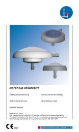
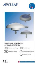
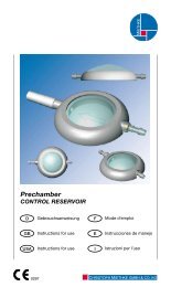
pressure. In many cases, a reservoir is implanted<br />
between the ventricle catheter and the valve. The<br />
doctor uses this reservoir for checking the patency<br />
of the valve, to siphon off CSF-samples or to inject<br />
medicines.<br />
A distinction is made between two types of drainage:<br />
ventriculo-peritoneal (from the head to the<br />
abdominal cavity) and ventriculo-atrial (from the<br />
head to the right atrium of the heart). In special<br />
cases, there is also the option of implanting a lumbo-peritoneal<br />
shunt (from the spine canal into the<br />
abdominal cavity).<br />
1 right atrium<br />
2 heart catheter (atrial catheter)<br />
3 valve<br />
4 reservoir<br />
5 ventricular catheter<br />
6 ventricles<br />
7 abdominal catheter (peritoneal catheter)<br />
8 abdominal cavity<br />
Fig. 3: Drainage systems for hydrocephalus patients<br />
a) ventriculo-atrial, b) ventriculo-peritoneal<br />
12 13<br />
4<br />
a)<br />
5<br />
6<br />
3<br />
2<br />
1<br />
b)<br />
therapy complications<br />
The treatment of hydrocephalus with a shunt system<br />
can sometimes arise complications. As is the<br />
case for any surgical intervention, there is a risk of<br />
infection. There can also be complications that are<br />
directly or indirectly related to the implanted valve<br />
system. Such complications include blockages<br />
of the drainage system or inadvertently increased<br />
fluid drainage. To give you an understanding why<br />
your physician decided for the proGAV, the physics<br />
of drainage is explained in the chapter “Physics<br />
background“.<br />
after the operation<br />
As a rule, the everyday activities of patients with<br />
shunt implants are not restricted. However, patients<br />
should abstain from major physical exertion<br />
(e. g. hard physical work, strenuous sports). Hydrocephalus<br />
patients who experience headache,<br />
dizziness, unnatural gait or similar symptoms<br />
should consult a physician without delay. Apart<br />
4<br />
3<br />
7<br />
8<br />
GB