La Célula Animal - American 3B Scientific
La Célula Animal - American 3B Scientific
La Célula Animal - American 3B Scientific

Create successful ePaper yourself
Turn your PDF publications into a flip-book with our unique Google optimized e-Paper software.
English<br />
Introduction<br />
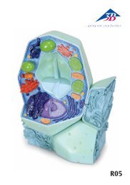
The <strong>Animal</strong> Cell<br />
Cells in animal multicellular organisms principally only occur in groups of similar cells or together with<br />
other differentiated cells, or embedded in the intercellular substrate (intercellular substance, extracellular<br />
matrix). The surrounding environment of the unicellular and primitive multicellular organisms (the “primordial<br />
soup“, so to speak) also surrounds the cells of more complex highly organized animal (human)<br />
organisms, and ensures its nutrition via the blood vessels that penetrate throughout the tissues (down to<br />
the capillaries).<br />
The following basic characteristics distinguish the cells of living organisms: they possess higher complexity<br />
of organization than their surroundings, they can react to stimuli from within and from their environment,<br />
and they have the ability to reproduce (reduplication).<br />
Overview of the construction and function of cells<br />
The cell membrane (plasma membrane) encloses the cell and also provides a barrier to the external environment<br />
allowing the maintenance of its own internal environment. Within the cell, certain structures and<br />
small organs (organelles, see list below) are also enclosed by a plasma membrane. The plasma membrane<br />
itself consists of polar lipids that form a semi-permeable membrane. Thus the individual compartments<br />
and organelles are separated from one another and from the specific molecules and ions they contain.<br />
The plasma membrane is also connected to a fine framework of structural proteins, the filaments of the<br />
cell skeleton (cytoskeleton). This cytoskeleton consists of fine actin filaments (7 nm diameter), hollow<br />
microtubules (25 nm diameter) and, lying in between in diameter, the intermediary filaments. The micro-<br />
®<br />
tubules develop from an organization centre, usually the centriole. They are also responsible for transport<br />
processes along their length, to and from the organization centre (directional active transport, which also<br />
occurs in the axons of nerve cells). The centriole itself is an organelle consisting of two groups of tubes perpendicular<br />
to one another, from which the microtubules extend – this also occurs in newly formed cells.<br />
During cell division the separation of the chromosomes is carried out by the “marionette threads”, the<br />
microtubules emanating from the centriole.<br />
As the name implies, the cytoskeleton ensures overall stability for the cell along with a corresponding<br />
degree of flexibility. Furthermore the cytoskeleton enables extreme versatility in the active movements of<br />
the cell: from stretching out foot-like appendages (e.g. filopodia) to make major changes in shape of the<br />
entire cell (also the basis of active muscle contraction for example) to active movement of the cell (cell<br />
migration). Moreover, the elements of the cytoskeleton propagate the tension lines within a cell via the<br />
so-called cell-cell connections (e.g. desmosomes, see below) to the neighbouring cells and so mechanically<br />
connect different areas of cells e.g. in the epidermis of the skin – particularly clear in the prickle cells.<br />
Within the cell-cell connections (intercellular contact) structures with predominantly mechanical function<br />
(contact adhesion: zonula; punctum; fascia adhaerens; macula adhaerens = desmosome) can be distinguished<br />
from those with an active metabolic and electro-coupling function (nexus, macula communicans<br />
= gap junction; synapse). Finally, there are cell connections that seal off the intercellular area (contact<br />
barrier: zonula occludens). Connections to the extracellular membrane form focal contacts and to the basal<br />
membrane the hemidesmosome.<br />
All proteins, which make up the components of the cytoskeleton, are made by the “sewing machine“ of<br />
the proteins, the ribosomes. These can be suspended in the cytoplasm or may be bound onto the vacuole<br />
system of the rough endoplasmic reticulum (rough ER). Information is carried to the ribosomes from the<br />
cell nucleus, where genetic information is stored on the chromosomes by means of the mRNA. The ribosome<br />
couples amino acid to amino acid to order and “sews” them onto a peptide or protein. Peptides and<br />
proteins are further modified by auxiliary proteins within the ER, e.g. sugar groups may be added to the<br />
protein (glycosylation). The smooth ER can synthesize lipids (cholesterol, triglycerides, steroid hormones),<br />
lipoproteins and phospholipids. Furthermore the smooth ER makes fat-soluble compounds water-soluble