Modelo de la célula vegetal - 3B Scientific
Modelo de la célula vegetal - 3B Scientific
Modelo de la célula vegetal - 3B Scientific

You also want an ePaper? Increase the reach of your titles
YUMPU automatically turns print PDFs into web optimized ePapers that Google loves.
R05
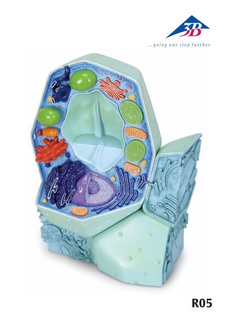
Mo<strong>de</strong>l of a P<strong>la</strong>nt Cell<br />
(Magnification approx. 500,000-1,000,000)<br />
English<br />
The history of cytology<br />
Cytology is an in<strong>de</strong>pen<strong>de</strong>nt science in botany and <strong>de</strong>als with the structure and function of p<strong>la</strong>nt cells. The<br />
term cell (Latin: cellu<strong>la</strong> = chamber, compartment, cell) was coined in 1665 by Robert Hooke, after he had<br />
discovered and recor<strong>de</strong>d the cells within the tissue of a bottle cork with the help of one of the earliest light<br />
microscopes. At the beginning of the 19th Century, Franz Meyen (1804-1840) recognised cells as the elementary<br />
units of p<strong>la</strong>nt organs. In 1838/1839, Matthias Jacob Schlei<strong>de</strong>n and Theodor Schwann establish cell<br />
theory: “Cells are the basis for all p<strong>la</strong>nts and animals.” In 1845, Karl Theodor Ernst von Siebold, based on<br />
his observations on protozoa (unicellu<strong>la</strong>r organisms), wrote that cells can exist in<strong>de</strong>pen<strong>de</strong>ntly and represent<br />
the smallest unit of life. At the same time, Louis Pasteur and other scientists refuted the prevailing theory<br />
of the time which stated that cells can originate spontaneously out of <strong>de</strong>ad organic matter (generatio<br />
spontanea). In 1855, Rudolf Virchow confirmed Meyen’s theory which stated that every cell originates from<br />
another cell (“omnis cellu<strong>la</strong> ex cellu<strong>la</strong>”). In 1879, Eduard Strasburger discovered the division of the nucleus<br />
in p<strong>la</strong>nts. An important breakthrough in un<strong>de</strong>rstanding the structure and function of cells was achieved by<br />
E. Ruska and H. Mahl in 1940, thanks to the <strong>de</strong>velopment of the transmission electron microscope.<br />
As in the animal world, p<strong>la</strong>nt cells, too, are characterised by the following:<br />
• They have a more complex structure than their environment<br />
• They react to inner and outer stimuli<br />
• They have the ability to reproduce<br />
Differences between animal and p<strong>la</strong>nt cells<br />
In spite of the consistency regarding the cellu<strong>la</strong>r structure of animal and p<strong>la</strong>nt cells – which had been<br />
<strong>de</strong>tected by Schlei<strong>de</strong>n and Schwann in 1838 – there still are important differences in their basic structural<br />
p<strong>la</strong>n. The following three features characterise the differences between most p<strong>la</strong>nt ® cells and animal cells:<br />
1. P<strong>la</strong>nt cells are enclosed by a cell wall which is responsible for resisting the inner osmotic pressure of the<br />
cell (turgor pressure), thereby giving it rigidity and increased stability.<br />
2. As organelles, only p<strong>la</strong>nt cells possess p<strong>la</strong>stids. These inclu<strong>de</strong>, for example, the green chlorop<strong>la</strong>sts, the<br />
scene of photosynthesis.<br />
3. They possess the sap vacuoles characteristic of p<strong>la</strong>nts, in which dissolved substances are stored and<br />
macromolecules broken down.<br />
P<strong>la</strong>nt cells have an average size of 10-100 µm and can be observed by using a simple light microscope. A<br />
tree is ma<strong>de</strong> up of 10 13 (= 10 trillion) cells! In multicellu<strong>la</strong>r organisms, they form groups of homogenous,<br />
in part strongly differentiated and specialised cells (= tissues).<br />
Structure and function of a p<strong>la</strong>nt cell<br />
(For numbering see diagram)<br />
Note: Unlike the mo<strong>de</strong>l presented, all components of a living cell are in a state of constant motion!!!<br />
The individual cell components have differences in their composition, e.g. proteins/enzymes, ionic milieu,<br />
etc., and can best be c<strong>la</strong>ssified according to their functions. An important term in p<strong>la</strong>nt cytology is protop<strong>la</strong>st,<br />
which refers to a cell surroun<strong>de</strong>d by a p<strong>la</strong>sma membrane in which the cell wall has been removed.<br />
Cytop<strong>la</strong>sm with cytoskeleton (1)<br />
In the course of evolution, a kind of division of work originated in a cell. This division of work is called<br />
compartmentation. It is achieved when special reaction complexes, the organelles (Greek: organon =<br />
tool), are surroun<strong>de</strong>d and <strong>de</strong>fined by membranes. These organelles can be <strong>de</strong>tected, with the help of a<br />
light microscope, in the fluid and colourless cytop<strong>la</strong>sm of protop<strong>la</strong>sts (60 to 90% water, proteins, lipids,<br />
nucleic acids). The cell membrane (2) forms the boundary of the cell, marking it off from its exterior surroundings.<br />
The cell membrane consists of monomolecu<strong>la</strong>r <strong>la</strong>yers of phospholipids and proteins which can<br />
move in the lipid matrix (“fluid mosaic” – mo<strong>de</strong>l). Inci<strong>de</strong>ntally, all p<strong>la</strong>nt and animal membranes are based<br />
on the same elementary principle (unit membrane).
English<br />
Mo<strong>de</strong>l of a P<strong>la</strong>nt Cell<br />
(Magnification approx. 500,000-1,000,000)<br />
This membrane is responsible for controlling the selective transport into and out of the cell. It has the<br />
same function with regard to the organelles, too. The cytoskeleton in the cytop<strong>la</strong>sm, which is ma<strong>de</strong> up<br />
of proteins, guarantees not just the stability of the cell but also the most diverse intracellu<strong>la</strong>r movements<br />
(e.g. visible p<strong>la</strong>sma streams).<br />
Nucleus (3a) with nucleolus (3b)<br />
The nucleus (approx. 5-25 µm) is the information centre of the cell. It is enclosed within a double membrane<br />
lining with <strong>de</strong>fined channels (= nuclear pores for controlling the metabolic flow between the nucleus<br />
and the cytop<strong>la</strong>sm) and contains the main part of the cell’s genetic information, present in the form of<br />
chromatin. Only during cell division is the chromatin (normally not visible un<strong>de</strong>r a light microscope) transformed<br />
into its more compact form, viz. chromosomes. In the process, the DNA, which is bound by proteins,<br />
is strongly reduced by con<strong>de</strong>nsation and spiralisation. The nucleoli occur exclusively in the interior of<br />
the nucleus and are the site for the synthesis of preliminary stages of cytop<strong>la</strong>smic ribosomes (5).<br />
Endop<strong>la</strong>smic Reticulum (smooth ER (4a) and rough ER (4b)) Ribosomes (5)<br />
All proteins of the cell are built at the “sewing machine” of proteins, the ribosomes. These extremely small<br />
organelles (approx. 20-30 nm) can float freely in the cytop<strong>la</strong>sm or get attached to the sacciform or tubu<strong>la</strong>r<br />
membrane system of the endop<strong>la</strong>smic reticulum (rough ER). Insi<strong>de</strong> the ER´s compartment, proteins are in<br />
part transformed into helper proteins, commonly known as molecu<strong>la</strong>r chaperones, and are transported<br />
to their biophase. The smooth ER, without ribosomes attached, is mainly responsible for the synthesis of<br />
lipids. The structure of the ER is extremely dynamic and always subject to constant reorganisation. The ER<br />
is also connected to the membrane coating of the nucleus. That is to say, the membrane and the lumens of<br />
both of the organelles blend directly with one another. ®<br />
P<strong>la</strong>smo<strong>de</strong>smata (6)<br />
P<strong>la</strong>smo<strong>de</strong>smata constitute contact structures between neighbouring p<strong>la</strong>nt cells. In the process they form<br />
connections, in the form of fine channels, between the living protop<strong>la</strong>sts through the cell wall and the<br />
middle <strong>la</strong>mel<strong>la</strong>. The coupling is built through tubu<strong>la</strong>r ER cisternae of both of the cells and its function is to<br />
transfer low-molecu<strong>la</strong>r substances between cells.<br />
P<strong>la</strong>stids<br />
P<strong>la</strong>stids are compartments typical to p<strong>la</strong>nt cells. They are always surroun<strong>de</strong>d by a double membrane. The<br />
inner membrane is formed for the purpose of en<strong>la</strong>rging the reactive surface into the interior of the p<strong>la</strong>stids.<br />
P<strong>la</strong>stids emerge on their own from the division of young prop<strong>la</strong>stids and spread themselves during<br />
mitosis into daughter cells. Chlorop<strong>la</strong>sts possess their own genetic information (ring-shaped, extrachromosomal<br />
genome; p<strong>la</strong>stids DNA).<br />
The green chlorop<strong>la</strong>sts (7) are the site of photosynthesis and the synthesis of numerous p<strong>la</strong>nt constituents<br />
(e.g. fatty acids). The colourless and fluid matrix is <strong>de</strong>noted as stroma; the en<strong>la</strong>rged <strong>la</strong>mel<strong>la</strong>r/sacciform<br />
inner membranes are called thy<strong>la</strong>koids. The stacked membrane areas are called grana thy<strong>la</strong>koids. The<br />
protein-bound pigments responsible for photosynthesis are found in these membranes (chiefly chlorophyll<br />
and carotenoids). These photosynthesis pigments are also responsible for the Hill reaction. The Calvin-<br />
Benson cycle or the photosynthetic carbon reduction cycle (PCR) CO 2 fixation as well as the formation of<br />
carbohydrates and starch take p<strong>la</strong>ce in the stroma region.<br />
Other p<strong>la</strong>stids:<br />
Chromop<strong>la</strong>sts: are inactive photosynthesis p<strong>la</strong>stids responsible for the colouration of p<strong>la</strong>nt organs<br />
Leucop<strong>la</strong>sts: are responsible for storing starch (amylop<strong>la</strong>sts), proteins (proteinop<strong>la</strong>sts), oils (e<strong>la</strong>iop<strong>la</strong>sts)<br />
Etiop<strong>la</strong>sts: are the preliminary stage of chlorop<strong>la</strong>sts and originate in the dark<br />
Gerontop<strong>la</strong>sts: are all p<strong>la</strong>stids at a very mature age<br />
Mitochondria (8)<br />
Mitochondria are the organelles responsible for cell respiration and energy conversion. They are therefore<br />
the “power p<strong>la</strong>nt” of every cell. Only mitochondria can give rise to mitochondria. Just like p<strong>la</strong>stids,
Mo<strong>de</strong>l of a P<strong>la</strong>nt Cell<br />
(Magnification approx. 500,000-1,000,000)<br />
English<br />
mitochondria, too, are enclosed in a double membrane coating and possess their own genetic information.<br />
The components/proteins responsible for the respiratory chain (ATP synthesis) are located on the inner si<strong>de</strong><br />
of the membrane. The citrate cycle and the fatty acid oxidation cycle take p<strong>la</strong>ce insi<strong>de</strong> the mitochondrial<br />
matrix.<br />
Endosymbiont theory<br />
The endosymbiont theory attempts to exp<strong>la</strong>in the origin of mitochondria and p<strong>la</strong>stids. According to this<br />
theory, mitochondria and p<strong>la</strong>stids go back to intercellu<strong>la</strong>r protozoan (bacterial) symbiosis. In other words:<br />
p<strong>la</strong>stids have <strong>de</strong>veloped from cyanobacteria, and mitochondria have <strong>de</strong>veloped from purple bacteria. At<br />
some point in the course of evolution, a “prototype“ cell with a nucleus (urocyte) incorporated prokaryotes<br />
and integrated them into its cellu<strong>la</strong>r functions. A strong indication of this is the fact that mitochondria and<br />
p<strong>la</strong>stids have the following in common:<br />
• A double membrane coating (inner and outer membranes are quite different in their chemical composition;<br />
the inner membrane resembles bacterial membranes)<br />
• Inherent ring-shaped genome<br />
• Inherent ribosomes (correspond to bacterial ribosomes, differ from cytop<strong>la</strong>smic ribosomes)<br />
Dictyosomes/Golgi apparatus (9)<br />
Dictyosomes are disc-shaped, membranous hollow cavities (cisternae). The sum of all dictyosomes in a cell<br />
are termed the Golgi apparatus. They are closely connected to the ER and are responsible for the conversion,<br />
storage and transfer of the products of the ER. Consequently, a distinction can be drawn between a<br />
generation si<strong>de</strong> (facing the ER, regeneration from the ER) and a secretion si<strong>de</strong> (facing away from the ER)<br />
which forms a significant cellu<strong>la</strong>r transport system responsible for exocytosis ® (elimination of substances<br />
from the cell), the construction of biomembranes and is also involved in cell-wall formation.<br />
Vacuole (10)<br />
The vacuole is an organelle only to be found in p<strong>la</strong>nt cells. It is a space filled with fluid and is surroun<strong>de</strong>d<br />
by a simple membrane (= tonop<strong>la</strong>st). In mature p<strong>la</strong>nt cells, the volume of the central vacuole can constitute<br />
up to 80% of the total volume of the cell. In the cell, vacuoles serve as reaction, storage (e.g. of ions,<br />
organic acids, sacchari<strong>de</strong>s, proteins, pigments), transport and <strong>de</strong>posit compartments (for substances that<br />
can be harmful to the cell, e.g. toxins, tanning agents). The breakdown of macromolecules (lytic compartment)<br />
is also carried out in the vacuoles.<br />
Microsomes/Microbodies (11)<br />
Microsomes are organelles with a homogenous structure (simple membrane, spherical, size: 1 µm, granu<strong>la</strong>r<br />
matrix) on the one hand, and strong biochemical and functional differences on the other.<br />
Different functions:<br />
− Lysosomes: are responsible for the break-up of proteins, polysacchari<strong>de</strong>s and nucleic acids<br />
− Glyoxysomes: p<strong>la</strong>y an important part in converting <strong>de</strong>pot fats to carbohydrates<br />
− Oleosomes (oil globules): are responsible for the break-up of fats and oils<br />
− Peroxisomes: p<strong>la</strong>y an important part in photorespiration. Peroxisomes also break up the glycol<strong>la</strong>te<br />
which is inevitably created during CO 2 fixation. Carbon is fed back into the photosynthesis cycle, and two<br />
amino acids are produced for protein synthesis.<br />
Cell wall (12)<br />
Possessing a rigid cell wall is an additional feature which distinguishes p<strong>la</strong>nt cells from animal cells. The<br />
cell wall gives the p<strong>la</strong>nt cell rigidity and form (exoskeleton) by resisting the interior osmotic pressure<br />
(= turgor pressure) of the cell. It is a product secreted by the protop<strong>la</strong>sts (apop<strong>la</strong>st). From a chemical point<br />
of view, the cell wall is ma<strong>de</strong> up of polysacchari<strong>de</strong>s and proteins.<br />
The cell wall is ma<strong>de</strong> up of up to three <strong>la</strong>yers.<br />
Middle <strong>la</strong>mel<strong>la</strong>: a ge<strong>la</strong>tinous <strong>la</strong>yer, only a few nm in thickness, ma<strong>de</strong> up of pectin compounds with a low<br />
quantity of proteins. It has no fibril structure and is therefore e<strong>la</strong>stic.
English<br />
Primary cell wall: a ge<strong>la</strong>tinous base substance (matrix) ma<strong>de</strong> up of pectin compounds, hemicellulose components<br />
and proteins. In the matrix, fibril structures can be <strong>de</strong>tected (10-25%) which are arranged in an<br />
irregu<strong>la</strong>r, scattered texture (e<strong>la</strong>sticity still present).<br />
Secondary wall: is chiefly composed of 90% cellulose fibrils. The arrangement of the fibrils is primarily in<br />
a parallel texture. There are often <strong>de</strong>posits of lignin, tanning agents, CaCO 3 , SiO 2 or colouring agents. Cells<br />
having a marked secondary wall are no longer capable of growth.<br />
Author: Dr. Gerd Vogg, University of Würzburg, Germany<br />
Mo<strong>de</strong>l of a P<strong>la</strong>nt Cell<br />
(Magnification approx. 500,000-1,000,000)<br />
Numbering:<br />
1 Cytop<strong>la</strong>sm with cytoskeleton<br />
2 Cell membrane<br />
3 a Nucleus<br />
3 b Nucleolus<br />
4 a Smooth Endop<strong>la</strong>smic Reticulum (smooth ER)<br />
4 b Rough Endop<strong>la</strong>smic Reticulum (rough ER)<br />
5 Ribosomes<br />
6 P<strong>la</strong>smo<strong>de</strong>smata<br />
7 Chlorop<strong>la</strong>sts<br />
8 Mitochondria<br />
9 Dictyosomes/Golgi apparatus<br />
10 Vacuole<br />
11 Microsomes/Microbodies<br />
12 Cell wall (<strong>la</strong>yered structure)<br />
®
Mo<strong>de</strong>ll <strong>de</strong>r Pf<strong>la</strong>nzenzelle<br />
(Vergrößerung etwa 500.000 - 1.000.000-fach)<br />
Deutsch<br />
Historisches zur pf<strong>la</strong>nzlichen Zelllehre (Zytologie)<br />
Die Zytologie ist eine eigenständige Wissenschaft innerhalb <strong>de</strong>r Botanik, die sich mit <strong>de</strong>r Struktur und<br />
<strong>de</strong>n Funktionen <strong>de</strong>r pf<strong>la</strong>nzlichen Zelle beschäftigt. Den Begriff Zelle (<strong>la</strong>t. cellu<strong>la</strong> = Kämmerchen) prägte<br />
im Jahre 1665 Robert Hooke, nach<strong>de</strong>m er diese im Gewebe <strong>de</strong>s F<strong>la</strong>schenkorks mit Hilfe eines <strong>de</strong>r ersten<br />
Lichtmikroskope ent<strong>de</strong>ckte und <strong>de</strong>tailliert aufzeichnete. Zu Beginn <strong>de</strong>s 19. Jahrhun<strong>de</strong>rts wur<strong>de</strong> die Zelle<br />
von Franz Meyen (1804 – 1840) als Elementareinheit <strong>de</strong>r Pf<strong>la</strong>nzenorgane erkannt. 1838/1839 begrün<strong>de</strong>n<br />
Matthias Jacob Schlei<strong>de</strong>n und Theodor Schwann die Zellentheorie: „Pf<strong>la</strong>nzen und Tiere sind gleichermaßen<br />
stets von Zellen aufgebaut“. 1845 veröffentlichte Karl Theodor Ernst von Siebold aufgrund <strong>de</strong>r<br />
Beobachtung an Protozoen (Einzeller), dass Zellen unabhängig voneinan<strong>de</strong>r leben können und die kleinste<br />
lebensfähige Einheit darstellen. Zur gleichen Zeit wi<strong>de</strong>rlegten Louis Pasteur und an<strong>de</strong>re die damals<br />
gelten<strong>de</strong> Theorie, dass Zellen spontan aus toter organischer Materie (generatio spontanea) entstehen<br />
können. 1855 bestätigte Rudolf Virchow die Theorie Meyens, dass je<strong>de</strong> Zelle aus einer an<strong>de</strong>ren entsteht<br />
(„omnis cellu<strong>la</strong> ex cellu<strong>la</strong>“). 1879 ent<strong>de</strong>ckte Eduard Strasburger die Kernteilung bei Pf<strong>la</strong>nzen. Ein wichtiger<br />
Schritt im Verständnis von Bau und Funktion <strong>de</strong>r Zelle wur<strong>de</strong> mit <strong>de</strong>r Entwicklung <strong>de</strong>s Transmissions-<br />
Elektronenmikroskopes im Jahr 1940 durch E. Ruska und H. Mahl erzielt.<br />
Wie im tierischen System zeichnen sich auch die pf<strong>la</strong>nzlichen Zellen dadurch aus,<br />
• dass sie komplexer organisiert sind als ihre Umgebung<br />
• dass sie auf Reize aus ihrem Inneren und aus ihrer Umgebung reagieren<br />
• dass sie die Fähigkeit haben, sich zu vermehren.<br />
Unterschie<strong>de</strong> im Grundbaup<strong>la</strong>n von pf<strong>la</strong>nzlichen und tierischen Zellen<br />
Trotz <strong>de</strong>r 1838 von Schlei<strong>de</strong>n und Schwann gefun<strong>de</strong>nen Übereinstimmung im zellulären Aufbau von<br />
Pf<strong>la</strong>nzen und Tieren gibt es wichtige Unterschie<strong>de</strong> in <strong>de</strong>ren Grundbaup<strong>la</strong>n. ® So unterschei<strong>de</strong>t sich die<br />
Mehrzahl <strong>de</strong>r Pf<strong>la</strong>nzenzellen von <strong>de</strong>n tierischen Zellen in folgen<strong>de</strong>n drei Merkmalen:<br />
1. Die pf<strong>la</strong>nzlichen Zellen sind von einer Zellwand umhüllt, die <strong>de</strong>m osmotischen Innendruck <strong>de</strong>r Zelle<br />
(= Turgor) entgegensteht und ihr dadurch eine hohe Festigkeit verleiht.<br />
2. Nur die pf<strong>la</strong>nzlichen Zellen besitzen als Organellen die P<strong>la</strong>sti<strong>de</strong>n, dazu gehören z.B. die grünen<br />
Chlorop<strong>la</strong>sten, die Orte <strong>de</strong>r Photosynthese.<br />
3. Sie besitzen die pf<strong>la</strong>nzentypischen Zellsaftvakuolen, in <strong>de</strong>nen gelöste Stoffe gespeichert und<br />
Makromoleküle abgebaut wer<strong>de</strong>n.<br />
Die pf<strong>la</strong>nzliche Zelle besitzt eine durchschnittliche Größe von 10 - 100 µm und kann mit einfachen<br />
Lichtmikroskopen beobachtet wer<strong>de</strong>n. Ein Baum besteht aus bis zu 10 13 (= 10 Billionen) Zellen! In mehrzelligen<br />
Organismen bil<strong>de</strong>n sie Verbän<strong>de</strong> aus gleichartigen, zum Teil stark differenzierten und dadurch<br />
spezialisierten Zellen (= Gewebe).<br />
Bau und Funktion <strong>de</strong>r pf<strong>la</strong>nzlichen Zelle<br />
(Nummerierung siehe Abbildung)<br />
Wichtig: Im Gegensatz zum vorliegen<strong>de</strong>n Mo<strong>de</strong>ll sind in einer leben<strong>de</strong>n Zelle alle Bestandteile ständig<br />
in Bewegung und im Fluss !!!<br />
Die einzelnen Zellbestandteile, besitzen unterschiedliche Ausstattungen z.B. an Proteinen/Enzymen,<br />
Ionenmilieu etc. und <strong>la</strong>ssen sich am sinnvollsten entsprechend ihrer Funktionen einteilen. Ein wichtiger<br />
Begriff in <strong>de</strong>r pf<strong>la</strong>nzlichen Zytologie ist <strong>de</strong>r Protop<strong>la</strong>st, darunter versteht man eine von einer<br />
Zytop<strong>la</strong>smamembran umgebenen Zelle bei <strong>de</strong>r die Zellwand entfernt wur<strong>de</strong>.<br />
Zytop<strong>la</strong>sma mit Zytoskelett (1)<br />
Im Laufe <strong>de</strong>r Evolution wur<strong>de</strong> eine Art Arbeitsteilung innerhalb einer Zelle eingeführt, die man<br />
Kompartimentierung nennt. Dies wird erreicht in<strong>de</strong>m spezielle Reaktionsbereiche, die Organellen (griechisch:<br />
Organon = Werkzeug), durch Membranen umhüllt und abgegrenzt wer<strong>de</strong>n. Diese Organellen<br />
kann man im flüssigen und farblosen Zytop<strong>la</strong>sma <strong>de</strong>s Protop<strong>la</strong>sten (60 bis 90 % Wasser, Proteine,
Deutsch<br />
Mo<strong>de</strong>ll <strong>de</strong>r Pf<strong>la</strong>nzenzelle<br />
(Vergrößerung etwa 500.000 - 1.000.000-fach)<br />
Lipi<strong>de</strong>, Nukleinsäuren) bereits mit <strong>de</strong>m Lichtmikroskop erkennen. Nach außen wird die Zelle durch die<br />
Zellmembran (2) abgegrenzt. Diese besteht aus zwei monomoleku<strong>la</strong>ren Schichten von Phospholipi<strong>de</strong>n und<br />
Proteinen, welche sich in <strong>de</strong>r Lipidmatrix bewegen können (’fluid mosaic’ - Mo<strong>de</strong>ll). Im Übrigen basieren<br />
alle pf<strong>la</strong>nzlichen und tierischen Membranen auf diesem gleichen Grundbauprinzip (= Einheitsmembran).<br />
Durch die Membranen wird <strong>de</strong>r selektive Transport in und aus <strong>de</strong>r Zelle sowie in und aus <strong>de</strong>n Organellen<br />
kontrolliert. Das aus Proteinen aufgebaute Zytoskelett im Zytop<strong>la</strong>sma gewährleistet die Stabilität <strong>de</strong>r Zelle<br />
aber auch vielfältigste intrazelluläre Bewegungen (z. B. sichtbare P<strong>la</strong>smaströmungen).<br />
Zellkern (Nucleus) (3a) mit Kernkörperchen (Nucleolus) (3b)<br />
Der Zellkern (ca. 5-25 µm) ist das Informationszentrum für die Zelle. Er wird von einer doppelten<br />
Membranhülle mit <strong>de</strong>finierten Kanälen (= Kernporen zur Steuerung <strong>de</strong>s Stoffflusses zwischen Kern und<br />
Zytop<strong>la</strong>sma) umgeben und enthält <strong>de</strong>n Hauptanteil <strong>de</strong>r genetischen Information <strong>de</strong>r Zelle in Form <strong>de</strong>s<br />
Chromatins. Nur für eine Kernteilung wird das ansonsten lichtmikroskopisch nicht sichtbare Chromatin in<br />
die kompakte Transportform, die Chromosomen, umgewan<strong>de</strong>lt. Dabei wird die an Proteine gebun<strong>de</strong>ne<br />
DNA durch Kon<strong>de</strong>nsation und Spiralisierung stark verkürzt. Die Kernkörperchen (Nucleoli) treten ausschließlich<br />
im Innern <strong>de</strong>s Kerns auf und sind <strong>de</strong>r Ort <strong>de</strong>r Synthese von Vorstufen <strong>de</strong>r cytop<strong>la</strong>smatischen<br />
Ribosomen (5).<br />
Endop<strong>la</strong>smatisches Retikulum (g<strong>la</strong>ttes ER (4a) und raues ER (4b)) und Ribosomen (5)<br />
Alle Proteine (Eiweiße) <strong>de</strong>r Zelle wer<strong>de</strong>n an <strong>de</strong>n „Nähmaschinen“ <strong>de</strong>r Proteine, <strong>de</strong>n Ribosomen, gebil<strong>de</strong>t.<br />
Diese sehr kleinen Organellen (ca. 20 x 30 nm) können frei im Zytop<strong>la</strong>sma liegen o<strong>de</strong>r an das<br />
sack- o<strong>de</strong>r röhrenförmige Membransystem <strong>de</strong>s Endop<strong>la</strong>smatischen Retikulums ® gebun<strong>de</strong>n sein (raues ER).<br />
Im Kompartiment <strong>de</strong>s ER wer<strong>de</strong>n die Proteine zum Teil durch Helferproteine verän<strong>de</strong>rt und an ihren<br />
Wirkort transportiert. Das g<strong>la</strong>tte ER, ohne aufge<strong>la</strong>gerte Ribosomen, ist vor allem für die Synthese von<br />
Lipi<strong>de</strong>n zuständig. Die Struktur <strong>de</strong>s ER ist sehr dynamisch und einer ständigen Reorganisation unterworfen.<br />
Weiterhin steht das ER mit <strong>de</strong>r Membranhülle <strong>de</strong>s Zellkerns in Verbindung. Das heißt, sowohl die<br />
Membranen als auch das Lumen bei<strong>de</strong>r Organellen gehen direkt ineinan<strong>de</strong>r über.<br />
P<strong>la</strong>smo<strong>de</strong>smata (6)<br />
Die P<strong>la</strong>smo<strong>de</strong>smata stellen Kontaktstrukturen zwischen benachbarten pf<strong>la</strong>nzlichen Zellen dar. Sie verbin<strong>de</strong>n<br />
dabei als feine Kanäle die leben<strong>de</strong>n Protop<strong>la</strong>sten durch die Zellwand und Mittel<strong>la</strong>melle hindurch.<br />
Die Verbindung wird durch sch<strong>la</strong>uchförmige ER–Zisternen bei<strong>de</strong>r Zellen gebil<strong>de</strong>t. Die Funktion ist <strong>de</strong>r<br />
Stofftransport nie<strong>de</strong>rmoleku<strong>la</strong>rer Substanzen zwischen <strong>de</strong>n Zellen.<br />
P<strong>la</strong>sti<strong>de</strong>n<br />
Die P<strong>la</strong>sti<strong>de</strong>n sind für die Pf<strong>la</strong>nzen typische Zellkompartimente, die stets von einer Doppelmembran umgeben<br />
sind. Die innere Membran ist zur Vergrößerung <strong>de</strong>r reaktiven Oberfläche ins Innere <strong>de</strong>r P<strong>la</strong>sti<strong>de</strong>n ausgeformt.<br />
P<strong>la</strong>sti<strong>de</strong>n gehen durch Teilung <strong>de</strong>r jugendlichen Prop<strong>la</strong>sti<strong>de</strong>n aus sich selbst hervor und verteilen<br />
sich bei <strong>de</strong>r Mitose auf die Tochterzellen. Die Chlorop<strong>la</strong>sten besitzen ihre eigene genetische Information<br />
(= ringförmiges, extrachromosomales Genom; P<strong>la</strong>sti<strong>de</strong>n DNA).<br />
Die grünen Chlorop<strong>la</strong>sten (7) sind die Orte <strong>de</strong>r Photosynthese und <strong>de</strong>r Synthese vieler pf<strong>la</strong>nzlicher<br />
Inhaltstoffe (z. B. Fettsäuren). Die farblose flüssige Matrix wird als Stroma, die <strong>la</strong>mellen- bis sackartig<br />
vergrößerten Innenmembranen als Thy<strong>la</strong>koi<strong>de</strong> bezeichnet. Die gestapelten Membranbereiche wer<strong>de</strong>n<br />
dabei Granathy<strong>la</strong>koi<strong>de</strong> genannt. In diesen Membranen sind die Photosynthesepigmente proteingebun<strong>de</strong>n<br />
lokalisiert (v.a. Chlorophylle, Carotinoi<strong>de</strong>) und verantwortlich für die Lichtreaktion <strong>de</strong>r Photosynthese. Im<br />
Stromabereich fin<strong>de</strong>n die Dunkelreaktion <strong>de</strong>r CO 2 -Fixierung und die Bildung von Kohlenhydraten und<br />
Stärke statt.<br />
Weitere P<strong>la</strong>sti<strong>de</strong>n:<br />
Chromop<strong>la</strong>sten: photosynthetisch inaktive P<strong>la</strong>sti<strong>de</strong>n zur Färbung <strong>de</strong>r Pf<strong>la</strong>nzenorgane<br />
Leukop<strong>la</strong>sten: Speicherung von Stärke (Amylop<strong>la</strong>sten), Proteinen (Proteinop<strong>la</strong>sten), Ölen (E<strong>la</strong>iop<strong>la</strong>sten)<br />
Etiop<strong>la</strong>sten: im Dunkeln entstan<strong>de</strong>ne Vorstufen <strong>de</strong>r Chlorop<strong>la</strong>sten<br />
Gerontop<strong>la</strong>sten: Alterstadien aller P<strong>la</strong>sti<strong>de</strong>n
Mo<strong>de</strong>ll <strong>de</strong>r Pf<strong>la</strong>nzenzelle<br />
(Vergrößerung etwa 500.000 - 1.000.000-fach)<br />
Deutsch<br />
Mitochondrien (8)<br />
Die Mitochondrien sind die Organellen <strong>de</strong>r Zel<strong>la</strong>tmung und Energieumwandlung. Sie stellen dadurch die<br />
„Kraftwerke“ <strong>de</strong>r Zelle dar. Mitochondrien können nur aus sich selbst gebil<strong>de</strong>t wer<strong>de</strong>n. Wie die P<strong>la</strong>sti<strong>de</strong>n<br />
sind sie von einer doppelten Membranhülle umgeben und besitzen ihre eigene genetische Information.<br />
An <strong>de</strong>r inneren Membran sind die Bestandteile/Proteine <strong>de</strong>r Atmungskette lokalisiert (Synthese von ATP). In<br />
<strong>de</strong>r Mitochondrienmatrix <strong>la</strong>ufen <strong>de</strong>r Zitratzyklus und die Fettsäureoxidation ab.<br />
Endosymbiontentheorie<br />
Die Endosymbiontentheorie versucht die Herkunft <strong>de</strong>r Mitochondrien und P<strong>la</strong>sti<strong>de</strong>n zu erklären. Nach<br />
dieser Theorie gehen die Mitochondrien und P<strong>la</strong>sti<strong>de</strong>n auf protocytische (bakterielle) intrazelluläre<br />
Symbiosen zurück. Das heißt: P<strong>la</strong>sti<strong>de</strong>n sind <strong>de</strong>mnach aus Cyanobakterien, Mitochondrien aus atmen<strong>de</strong>n<br />
Purpurbakterien entstan<strong>de</strong>n. Eine „Urzelle“ mit Zellkern (Ureuzyt) hat sich im Laufe <strong>de</strong>r Evolution<br />
Prokaryoten einverleibt und in ihr zelluläres Funktionsgefüge integriert. Dafür sprechen die folgen<strong>de</strong>n<br />
Gemeinsamkeiten von Mitochondrien und P<strong>la</strong>sti<strong>de</strong>n:<br />
• doppelte Membranhülle (innere und äußere Membran sind in ihrer chemischen Zusammensetzung sehr<br />
verschie<strong>de</strong>n; die innere ähnelt bakteriellen Membranen)<br />
• eigenes ringförmiges Genom<br />
• eigene Ribosomen (entsprechen <strong>de</strong>n bakteriellen Ribosomen, unterschei<strong>de</strong>n sich von <strong>de</strong>n zytop<strong>la</strong>smatischen<br />
Ribosomen)<br />
Dictyosomen/Golgi-Apparat (9)<br />
Die Dictyosomen sind scheibenförmige, membranumgebene Hohlräume (Zisternen). Alle Dictyosomen<br />
einer Zelle wer<strong>de</strong>n als Golgi-Apparat bezeichnet. Sie stehen in engem ® Kontakt zum ER und sind<br />
für die Umwandlung, Speicherung und Weiterleitung <strong>de</strong>r Produkte <strong>de</strong>s ER zuständig. Folglich kann<br />
eine Bildungsseite (<strong>de</strong>m ER zugewandt, Neubildung aus <strong>de</strong>m ER) und eine Sekretionsseite (<strong>de</strong>m ER<br />
abgewandt) unterschie<strong>de</strong>n wer<strong>de</strong>n. Sie sind ein wichtiges zelluläres Transportsystem, zuständig für<br />
Exocytose (Ausscheidung von Stoffen aus <strong>de</strong>r Zelle), <strong>de</strong>m Aufbau von Biomembranen und beteiligt an <strong>de</strong>r<br />
Zellwandbildung.<br />
Vakuole (10)<br />
Die Vakuole ist ein rein pf<strong>la</strong>nzliches Organell. Sie ist ein flüssigkeitsgefüllter Raum, <strong>de</strong>r von einer<br />
einfachen Membran (= Tonop<strong>la</strong>st) umgeben ist. In ausgewachsenen Pf<strong>la</strong>nzenzellen kann das<br />
Volumen <strong>de</strong>r Zentralvakuole bis über 80 % <strong>de</strong>s Zellvolumens ausmachen. Sie dienen in <strong>de</strong>r Zelle als<br />
Reaktions–, Speicherungs- (z. B. Ionen, organische Säuren, Zucker, Proteine, Pigmente), Transport- und<br />
Ab<strong>la</strong><strong>de</strong>kompartimente (für zellschädigen<strong>de</strong> Substanzen z.B. Toxine, Gerbstoffe). Auch <strong>de</strong>r Abbau von<br />
Makromolekülen (lytisches Kompartiment) erfolgt in <strong>de</strong>r Vakuole.<br />
Mikrosomen/Microbodies (11)<br />
Mikrosomen sind Organellen mit einheitlichem Aufbau (einfache Membran, kugelig, Größe: 1 µm, granulöse<br />
Matrix), aber starken biochemischen und damit funktionellen Unterschie<strong>de</strong>n.<br />
Verschie<strong>de</strong>ne Funktionen:<br />
− Lysosomen: zuständig für <strong>de</strong>n Abbau von Proteinen, Polysacchari<strong>de</strong>n und Nukleinsäuren<br />
− Glyoxysomen: wichtige Rolle bei <strong>de</strong>r Umwandlung von Speicherfetten in Kohlenhydrate<br />
− Oleosomen (Öltröpfchen): Abbau von Fetten und Ölen<br />
− Peroxisomen: wichtige Rolle bei <strong>de</strong>r Photorespiration. Zwangsweise bei <strong>de</strong>r CO 2 Fixierung anfallen<strong>de</strong>s<br />
Glyko<strong>la</strong>t wird über die Peroxisomen abgebaut, <strong>de</strong>r Kohlenstoff wie<strong>de</strong>r <strong>de</strong>m Photosynthese-Zyklus zugeführt<br />
und zwei Aminosäuren zur Proteinsynthese produziert.<br />
Zellwand (12)<br />
Der Besitz einer starren Zellwand ist ein weiteres Unterscheidungsmerkmal zwischen pf<strong>la</strong>nzlichen und tierischen<br />
Zellen. Sie gibt <strong>de</strong>r pf<strong>la</strong>nzlichen Zelle Festigkeit und Form (Exoskelett), in<strong>de</strong>m sie <strong>de</strong>m osmotischen<br />
Innendruck <strong>de</strong>r Zelle (=Turgor) wi<strong>de</strong>rsteht. Sie ist ein Abscheidungsprodukt <strong>de</strong>s Protop<strong>la</strong>sten (Apop<strong>la</strong>st).
Deutsch<br />
Mo<strong>de</strong>ll <strong>de</strong>r Pf<strong>la</strong>nzenzelle<br />
(Vergrößerung etwa 500.000 - 1.000.000-fach)<br />
Chemisch betrachtet ist sie aus Polysacchari<strong>de</strong>n und Proteinen aufgebaut.<br />
Die Zellwand baut sich aus bis zu drei Schichten auf:<br />
Mittel<strong>la</strong>melle: wenige nm dicke gallertige Kittschicht aus Pektinstoffen mit geringem Proteinanteil. Sie<br />
besitzt kein Fibrillengerüst und ist dadurch p<strong>la</strong>stisch <strong>de</strong>hnbar.<br />
Primärwand: gallertige Grundsubstanz (Matrix) aus Pektinstoffen, Hemizellulosen und Proteinen. In diese<br />
Matrix sind Gerüstfibrillen aus Zellulose einge<strong>la</strong>gert (10-25 %) und in unregelmäßiger Streuungstextur<br />
(Dehnbarkeit noch vorhan<strong>de</strong>n) angeordnet.<br />
Sekundärwand: Hauptbestandteil zu 90 % Fibrillen aus Zellulose. Anordnung <strong>de</strong>r Fibrillen bevorzugt in<br />
Paralleltextur. Oftmals Ein<strong>la</strong>gerung von Holzstoffen (Lignin), Gerbstoffe, CaCO 3 , SiO 2 o<strong>de</strong>r Farbstoffen.<br />
Zellen mit ausgeprägter Sekundärwand sind nicht mehr wachstumsfähig.<br />
Autor: Dr. Gerd Vogg, Universität Würzburg<br />
Nummerierung:<br />
1 Zytop<strong>la</strong>sma mit Zytoskelett<br />
2 Zellmembran<br />
3 a Zellkern/Nukleus<br />
3 b Kernkörperchen/Nucleolus<br />
4 a g<strong>la</strong>ttes Endop<strong>la</strong>smatisches Reticulum (g<strong>la</strong>ttes ER)<br />
4 b raues Endop<strong>la</strong>smatisches Reticulum (raues ER)<br />
5 Ribosomen<br />
6 P<strong>la</strong>smo<strong>de</strong>smata<br />
7 Chlorop<strong>la</strong>sten<br />
8 Mitochondrien<br />
9 Dictyosomen/Golgi-Apparat<br />
10 Vakuole<br />
11 Mikrosomen/Microbodies<br />
12 Zellwand (geschichteter Aufbau)<br />
®
<strong>Mo<strong>de</strong>lo</strong> <strong>de</strong> <strong>la</strong> célu<strong>la</strong> <strong>vegetal</strong><br />
(ampliada aproximadamente <strong>de</strong> 500.000 a 1.000.000 <strong>de</strong> veces <strong>de</strong> tamaño)<br />
®<br />
Español<br />
Historia <strong>de</strong> <strong>la</strong> citología<br />
La citología es una ciencia autónoma <strong>de</strong>ntro <strong>de</strong> <strong>la</strong> botánica que estudia <strong>la</strong> estructura y <strong>la</strong>s funciones <strong>de</strong><br />
<strong>la</strong> célu<strong>la</strong> <strong>vegetal</strong>. La pa<strong>la</strong>bra célu<strong>la</strong> (<strong>de</strong>l <strong>la</strong>t. cellu<strong>la</strong>, diminutivo <strong>de</strong> cel<strong>la</strong> = hueco) fue empleada en 1665<br />
por Robert Hooke, <strong>de</strong>spués <strong>de</strong> que <strong>la</strong> <strong>de</strong>scubriera y <strong>la</strong> bosquejara <strong>de</strong>tal<strong>la</strong>damente, a partir <strong>de</strong>l tejido <strong>de</strong><br />
un corcho <strong>de</strong> botel<strong>la</strong>, al emplear uno <strong>de</strong> los primeros microscopios ópticos. A inicios <strong>de</strong>l siglo XIX, Franz<br />
Meyen (1804 – 1840) reconoció que <strong>la</strong> célu<strong>la</strong> constituía <strong>la</strong> unidad elemental <strong>de</strong> los órganos <strong>vegetal</strong>es. En<br />
1838/1839, Matthias Jacob Schlei<strong>de</strong>n y Theodor Schwann fundamentaron su teoría celu<strong>la</strong>r: “Los <strong>vegetal</strong>es<br />
y los animales están compuestos <strong>de</strong> igual manera por célu<strong>la</strong>s”. En 1845, Karl Theodor Ernst von Siebold,<br />
a partir <strong>de</strong> observaciones <strong>de</strong> protozoarios (organismos unicelu<strong>la</strong>res), señaló que <strong>la</strong>s célu<strong>la</strong>s pue<strong>de</strong>n vivir<br />
in<strong>de</strong>pendientemente <strong>de</strong> <strong>la</strong>s <strong>de</strong>más y que representan <strong>la</strong> más pequeña unidad dotada <strong>de</strong> vida. Al mismo<br />
tiempo, Louis Pasteur y otros científicos refutaron <strong>la</strong> teoría, válida en ese entonces, <strong>de</strong> que <strong>la</strong>s célu<strong>la</strong>s se<br />
pue<strong>de</strong>n generar espontáneamente a partir <strong>de</strong> materia orgánica muerta (generatio spontanea). En 1855,<br />
Rudolf Virchow confirmó <strong>la</strong> teoría <strong>de</strong> Meyen, <strong>de</strong> que cada célu<strong>la</strong> se reproduce <strong>de</strong> una célu<strong>la</strong> anterior<br />
(“omnis cellu<strong>la</strong> ex cellu<strong>la</strong>”). En 1879, Eduard Strasburger <strong>de</strong>scubrió <strong>la</strong> mitosis <strong>de</strong> <strong>la</strong>s p<strong>la</strong>ntas. En el año 1940<br />
se dio un paso importante hacia <strong>la</strong> comprensión <strong>de</strong> <strong>la</strong> estructura y el funcionamiento <strong>de</strong> <strong>la</strong> célu<strong>la</strong> gracias<br />
al <strong>de</strong>sarrollo <strong>de</strong>l microscopio electrónico <strong>de</strong> transmisión, hecho realizado por E. Ruska y H. Mahlt.<br />
Al igual que en los sistemas animales, <strong>la</strong>s célu<strong>la</strong>s <strong>vegetal</strong>es se distinguen por:<br />
• una organización más compleja que <strong>la</strong> <strong>de</strong> su entorno<br />
• reacción ante los estímulos internos y ante los <strong>de</strong>l exterior<br />
• capacidad <strong>de</strong> reproducción.<br />
Diferencias entre <strong>la</strong> estructura básica <strong>de</strong> <strong>la</strong>s célu<strong>la</strong>s animales y <strong>vegetal</strong>es<br />
A pesar <strong>de</strong> <strong>la</strong> concordancia encontrada en 1838 por Schlei<strong>de</strong>n y Schwann en lo re<strong>la</strong>tivo a <strong>la</strong>s célu<strong>la</strong>s <strong>vegetal</strong>es<br />
y animales, existen importantes diferencias en su estructura básica. La mayoría <strong>de</strong> <strong>la</strong>s célu<strong>la</strong>s <strong>vegetal</strong>es<br />
se diferencian <strong>de</strong> <strong>la</strong>s animales <strong>de</strong>bido a <strong>la</strong>s tres siguientes características:<br />
1. Las célu<strong>la</strong>s <strong>vegetal</strong>es se encuentran recubiertas por una membrana celu<strong>la</strong>r que resiste a <strong>la</strong> presión<br />
osmótica interna <strong>de</strong> <strong>la</strong> célu<strong>la</strong> (presión <strong>de</strong> turgor) y que <strong>la</strong>s dota <strong>de</strong> una resistencia más elevada.<br />
2. Sólo <strong>la</strong>s célu<strong>la</strong>s <strong>vegetal</strong>es poseen plástidos, a manera <strong>de</strong> organelos, entre los que se encuentran, por<br />
ejemplo, los clorop<strong>la</strong>stos ver<strong>de</strong>s, en don<strong>de</strong> se realiza <strong>la</strong> fotosíntesis.<br />
3. Poseen vacuo<strong>la</strong>s llenas <strong>de</strong> savia celu<strong>la</strong>r, típicas <strong>de</strong> <strong>la</strong>s p<strong>la</strong>ntas, en <strong>la</strong>s que se almacena <strong>la</strong> materia disuelta<br />
y se generan <strong>la</strong>s macromolécu<strong>la</strong>s.<br />
La célu<strong>la</strong> <strong>vegetal</strong> tiene un tamaño promedio <strong>de</strong> 10 a 100 µm y se <strong>la</strong> pue<strong>de</strong> observar con un microscopio<br />
óptico sencillo. ¡Un árbol está compuesto por una cantidad <strong>de</strong> hasta 10 13 (= 10 billones) <strong>de</strong> célu<strong>la</strong>s! En los<br />
organismos pluricelu<strong>la</strong>res, éstas se unen en familias <strong>de</strong> célu<strong>la</strong>s <strong>de</strong>l mismo tipo (tejidos), que presentan<br />
diferencias, en parte, fuertemente marcadas y, por tanto, funciones especiales.<br />
Estructura y funcionamiento <strong>de</strong> <strong>la</strong> célu<strong>la</strong> <strong>vegetal</strong><br />
(Para <strong>la</strong> numeración, véase <strong>la</strong> imagen)<br />
¡¡¡Importante: Al contrario <strong>de</strong>l mo<strong>de</strong>lo presente, los componentes <strong>de</strong> una célu<strong>la</strong> viva se encuentran<br />
en constante movimiento y fluyen!!!<br />
Los componentes individuales <strong>de</strong> <strong>la</strong> célu<strong>la</strong> poseen diferentes cantida<strong>de</strong>s, por ejemplo, <strong>de</strong> proteínas/ enzimas,<br />
medio iónico, etc., y para c<strong>la</strong>sificar<strong>la</strong>s <strong>de</strong> <strong>la</strong> manera más razonable posible se recurre a sus funciones.<br />
Un concepto importante <strong>de</strong> <strong>la</strong> citología <strong>vegetal</strong> es el <strong>de</strong> protop<strong>la</strong>sto, el cual <strong>de</strong>signa una célu<strong>la</strong> recubierta<br />
por una membrana <strong>de</strong> citop<strong>la</strong>sma <strong>de</strong> <strong>la</strong> que se ha <strong>de</strong>sprendido <strong>la</strong> membrana celu<strong>la</strong>r.<br />
Citop<strong>la</strong>sma con citoesqueleto (1)<br />
En el transcurso <strong>de</strong> <strong>la</strong> evolución, en el interior <strong>de</strong> <strong>la</strong> célu<strong>la</strong>, se impuso una especie <strong>de</strong> división <strong>de</strong>l trabajo,<br />
<strong>la</strong> cual recibe el nombre <strong>de</strong> compartimentalización. Esto se alcanza cuando <strong>la</strong>s áreas especiales <strong>de</strong> reacción,<br />
los organelos (<strong>de</strong>l griego Organon = herramienta), se recubren y limitan por medio <strong>de</strong> membranas.
Español<br />
<strong>Mo<strong>de</strong>lo</strong> <strong>de</strong> <strong>la</strong> célu<strong>la</strong> <strong>vegetal</strong><br />
(ampliada aproximadamente <strong>de</strong> 500.000 a 1.000.000 <strong>de</strong> veces <strong>de</strong> tamaño)<br />
Estos organelos se pue<strong>de</strong>n reconocer en el citop<strong>la</strong>sma líquido e incoloro <strong>de</strong> los protop<strong>la</strong>stos (60 a 90 %<br />
agua, proteínas, lípidos y ácidos nucleicos) incluso con el microscopio óptico. La célu<strong>la</strong> está separada <strong>de</strong>l<br />
exterior por <strong>la</strong> membrana celu<strong>la</strong>r (2). Ésta se compone <strong>de</strong> dos capas monomolecu<strong>la</strong>res <strong>de</strong> fosfolípidos y<br />
proteínas, <strong>la</strong>s cuales se pue<strong>de</strong>n mover en <strong>la</strong> matriz lípida (mo<strong>de</strong>lo ’fluid mosaic’). Por lo general, <strong>la</strong>s membranas<br />
<strong>vegetal</strong>es y animales se basan en el mismo principio estructural fundamental (membrana única).<br />
Por medio <strong>de</strong> <strong>la</strong>s membranas se contro<strong>la</strong> el transporte selectivo <strong>de</strong>s<strong>de</strong> el interior y el exterior <strong>de</strong> <strong>la</strong> célu<strong>la</strong> y<br />
<strong>de</strong>s<strong>de</strong> el interior y el exterior <strong>de</strong> los organelos. El citoesqueleto, ubicado en el citop<strong>la</strong>ma y constituido por<br />
proteínas, garantiza no sólo <strong>la</strong> estabilidad <strong>de</strong> <strong>la</strong> célu<strong>la</strong> sino también múltiples movimientos intracelu<strong>la</strong>res<br />
(por ejemplo, fluidos visibles <strong>de</strong> p<strong>la</strong>sma).<br />
Núcleo <strong>de</strong> <strong>la</strong> célu<strong>la</strong> (3a) con nucleolo (3b)<br />
El núcleo <strong>de</strong> <strong>la</strong> célu<strong>la</strong> (aprox. 5-25 µm) constituye su centro <strong>de</strong> información. Está ro<strong>de</strong>ado <strong>de</strong> un recubrimiento<br />
doble <strong>de</strong> membrana, con canales <strong>de</strong>finidos (poros nucleares para control <strong>de</strong>l flujo <strong>de</strong> materia entre<br />
el núcleo y el citop<strong>la</strong>sma) y contiene <strong>la</strong> parte principal <strong>de</strong> <strong>la</strong> información genética, en forma <strong>de</strong> cromatinas.<br />
Sólo para una división nuclear, <strong>la</strong> cromatina que, <strong>de</strong> otra manera, no es visible al microscopio óptico,<br />
se convierte en un medio <strong>de</strong> transporte compacto <strong>de</strong>nominado cromosoma. En este proceso, el DNA,<br />
estrechamente ligado a <strong>la</strong>s proteínas, se reduce enormemente <strong>de</strong>bido a <strong>la</strong> con<strong>de</strong>nsación y espiralización.<br />
Los nucleolos aparecen exclusivamente en el interior <strong>de</strong>l núcleo y son el lugar en don<strong>de</strong> se produce <strong>la</strong> síntesis<br />
<strong>de</strong> los estadios previos <strong>de</strong> los ribosomas citop<strong>la</strong>smáticos (5).<br />
®<br />
Retículo endop<strong>la</strong>smático (ER liso (4a) y rugoso (4b)) Ribosomas (5)<br />
Todas <strong>la</strong>s proteínas <strong>de</strong> <strong>la</strong> célu<strong>la</strong> se forman en los ribosomas, <strong>la</strong>s “máquinas <strong>de</strong> coser” proteínas. Estos<br />
organelos muy pequeños (aprox. 20 x 30 nm) se pue<strong>de</strong>n encontrar libremente en el citop<strong>la</strong>sma o ligados al<br />
sistema <strong>de</strong> <strong>la</strong> membrana vesicu<strong>la</strong>r o tubu<strong>la</strong>r <strong>de</strong>l retículo endop<strong>la</strong>smático (ER rugoso). En el compartimento<br />
<strong>de</strong>l ER, <strong>la</strong>s proteínas sufren, parcialmente, una transformación provocada por proteínas auxiliares y se<br />
efectúa también su transporte a su lugar <strong>de</strong> acción. El ER liso, sin ribosomas acumu<strong>la</strong>dos, es responsable,<br />
sobre todo, <strong>de</strong> <strong>la</strong> síntesis <strong>de</strong> lípidos. La estructura <strong>de</strong>l ER es muy dinámica y se encuentra supeditada a una<br />
constante reorganización. A<strong>de</strong>más, el ER se mantiene en comunicación con el revestimiento <strong>de</strong> membrana<br />
<strong>de</strong>l núcleo. Esto significa que, tanto <strong>la</strong>s membranas como el lumen <strong>de</strong> ambos organelos, se comunican<br />
directamente entre sí.<br />
P<strong>la</strong>smo<strong>de</strong>smos (6)<br />
Los p<strong>la</strong>smo<strong>de</strong>smos representan estructuras <strong>de</strong> contacto entre <strong>la</strong>s célu<strong>la</strong>s <strong>vegetal</strong>es contiguas. Son canales<br />
finos que constituyen un vínculo entre los protop<strong>la</strong>stos vivos y <strong>la</strong> capa intercelu<strong>la</strong>r a través <strong>de</strong> <strong>la</strong> pared <strong>de</strong><br />
<strong>la</strong> célu<strong>la</strong>. La unión se forma por medio <strong>de</strong> <strong>la</strong>s cisternas <strong>de</strong>l ER <strong>de</strong> forma tubu<strong>la</strong>r. La función es el transporte<br />
entre <strong>la</strong>s célu<strong>la</strong>s <strong>de</strong> sustancias <strong>de</strong> bajo peso molecu<strong>la</strong>r.<br />
Plástidos<br />
Los plástidos son compartimentos celu<strong>la</strong>res típicos <strong>de</strong> <strong>la</strong>s p<strong>la</strong>ntas que siempre se encuentran ro<strong>de</strong>ados por<br />
una membrana doble. La membrana interna tiene una forma que permite <strong>la</strong> ampliación <strong>de</strong> <strong>la</strong> superficie<br />
reactiva <strong>de</strong>l interior. Los plástidos se forman a partir <strong>de</strong> sí mismos, gracias a <strong>la</strong> subdivisión <strong>de</strong> los proplástidios<br />
jóvenes, y se reparten entre <strong>la</strong>s nuevas célu<strong>la</strong>s durante <strong>la</strong> mitosis. Los clorop<strong>la</strong>stos poseen su propia<br />
información genética (genoma extracromosómico en forma <strong>de</strong> anillo; DNA <strong>de</strong> plástidos).<br />
Los clorop<strong>la</strong>stos ver<strong>de</strong>s (7) son el lugar en don<strong>de</strong> ocurre <strong>la</strong> fotosíntesis y <strong>la</strong>s síntesis <strong>de</strong> muchas<br />
sustancias <strong>vegetal</strong>es internas (por ejemplo, ácidos grasos). La matriz líquida e incolora se <strong>de</strong>nomina<br />
estroma, y <strong>la</strong>s membranas internas, agrandadas, en forma <strong>de</strong> lámina o saco, se conocen como<br />
ti<strong>la</strong>coi<strong>de</strong>. Las áreas <strong>de</strong> membrana api<strong>la</strong>das reciben el nombre <strong>de</strong> tiIacoi<strong>de</strong> en grana. En estas membranas<br />
se encuentran los pigmentos <strong>de</strong> <strong>la</strong> fotosíntesis, ligados a <strong>la</strong>s proteínas (sobre todo, clorofi<strong>la</strong>,<br />
carotinoi<strong>de</strong>s) y son responsables <strong>de</strong> <strong>la</strong> reacción a <strong>la</strong> luz <strong>de</strong> <strong>la</strong> fotosíntesis. En el área <strong>de</strong>l estroma se<br />
producen <strong>la</strong>s reacciones oscuras <strong>de</strong> fijación <strong>de</strong>l CO 2 y <strong>la</strong> formación <strong>de</strong> hidratos <strong>de</strong> carbono y almidón.
<strong>Mo<strong>de</strong>lo</strong> <strong>de</strong> <strong>la</strong> célu<strong>la</strong> <strong>vegetal</strong><br />
(ampliada aproximadamente <strong>de</strong> 500.000 a 1.000.000 <strong>de</strong> veces <strong>de</strong> tamaño)<br />
Español<br />
Otros plástidos:<br />
Cromop<strong>la</strong>stos: Plástidos inactivos durante <strong>la</strong> fotosíntesis que sirven para <strong>la</strong> pigmentación <strong>de</strong> los órganos<br />
<strong>de</strong> <strong>la</strong>s p<strong>la</strong>ntas<br />
Leucop<strong>la</strong>stos: Almacenamiento <strong>de</strong> almidón (amilop<strong>la</strong>stos), proteínas (proteinop<strong>la</strong>stos), aceites (oleop<strong>la</strong>stos)<br />
Etiop<strong>la</strong>stos: Estados previos <strong>de</strong> los clorop<strong>la</strong>stos que se generan en <strong>la</strong> oscuridad<br />
Gerontop<strong>la</strong>stos: Estadios <strong>de</strong> envejecimiento <strong>de</strong> todos los plástidos<br />
Mitocondrias (8)<br />
Las mitocondrias son los organelos <strong>de</strong> <strong>la</strong> respiración celu<strong>la</strong>r y <strong>de</strong> <strong>la</strong> transformación <strong>de</strong> energía.<br />
Representan, <strong>de</strong> esta manera, <strong>la</strong>s “centrales <strong>de</strong> energía” <strong>de</strong> <strong>la</strong> célu<strong>la</strong>. Las mitocondrias sólo se pue<strong>de</strong>n<br />
reproducir a partir <strong>de</strong> sí mismas. Al igual que los plástidos, están recubiertas por una membrana doble y<br />
poseen su propia información genética.<br />
En <strong>la</strong> membrana interna se encuentran localizados los componentes / proteínas <strong>de</strong> <strong>la</strong> ca<strong>de</strong>na <strong>de</strong> respiración<br />
(síntesis <strong>de</strong> ATP). En <strong>la</strong> matriz <strong>de</strong> <strong>la</strong>s mitocondrias tiene lugar el ciclo <strong>de</strong>l ácido cítrico y <strong>la</strong> oxidación <strong>de</strong><br />
los ácidos grasos.<br />
Teoría <strong>de</strong> los endosimbiontes<br />
La teoría <strong>de</strong> los endosimbiontes trata <strong>de</strong> explicar el origen <strong>de</strong> <strong>la</strong>s mitocondrias y los plástidos. De acuerdo<br />
con esta teoría, <strong>la</strong>s mitocondrias y los plástidos se originan por simbiosis protocítica (bacterial) intracelu<strong>la</strong>r.<br />
Esto significa que los plástidos provienen <strong>de</strong> cianobacterias, y <strong>la</strong>s mitocondrias <strong>de</strong> bacterias púrpuras con<br />
capacidad <strong>de</strong> respiración. Una “célu<strong>la</strong> originaria” con núcleo, durante el transcurso <strong>de</strong> <strong>la</strong> evolución, se<br />
apropió <strong>de</strong> procariotes integrándolos a su estructura funcional celu<strong>la</strong>r. Esta suposición ® está ava<strong>la</strong>da por <strong>la</strong>s<br />
siguientes coinci<strong>de</strong>ncias entre <strong>la</strong>s mitocondrias y los plástidos:<br />
• doble membrana (<strong>la</strong>s membranas interna y externa son muy diferentes en su constitución química; <strong>la</strong><br />
interna es semejante a <strong>la</strong>s membranas bacteriales)<br />
• genoma con forma <strong>de</strong> anillo<br />
• ribosomas propios (correspon<strong>de</strong>n a los ribosomas bacteriales, y se diferencian <strong>de</strong> los ribosomas citop<strong>la</strong>smáticos)<br />
Dictiosomas/Aparato <strong>de</strong> Golgi (9)<br />
Los dictiosomas son compartimentos huecos, en forma <strong>de</strong> disco, ro<strong>de</strong>ados <strong>de</strong> membrana. El conjunto <strong>de</strong><br />
los dictiosomas <strong>de</strong> una célu<strong>la</strong> obtiene <strong>la</strong> <strong>de</strong>signación <strong>de</strong> aparato <strong>de</strong> Golgi. Se encuentran en estrecho contacto<br />
con el ER y son responsables <strong>de</strong> <strong>la</strong> renovación, almacenamiento y transferencia <strong>de</strong> los productos <strong>de</strong>l<br />
ER. En consecuencia, se pue<strong>de</strong> diferenciar entre una parte generativa (orientada hacia el ER, nueva formación<br />
<strong>de</strong>l ER) y una <strong>de</strong> secreción (orientada opuestamente al ER). Son un importante sistema <strong>de</strong> transporte<br />
celu<strong>la</strong>r, responsables <strong>de</strong> <strong>la</strong> exocitosis (<strong>de</strong>sprendimiento <strong>de</strong> sustancias <strong>de</strong> <strong>la</strong> célu<strong>la</strong>), <strong>de</strong> <strong>la</strong> formación <strong>de</strong><br />
biomembranas y forman parte <strong>de</strong> <strong>la</strong> formación <strong>de</strong> <strong>la</strong> pared celu<strong>la</strong>r.<br />
Vacuo<strong>la</strong>s (10)<br />
La vacuo<strong>la</strong> es un organelo netamente <strong>vegetal</strong>. Se encuentra en un compartimento lleno <strong>de</strong> fluido y ro<strong>de</strong>ado<br />
por una membrana simple (tonop<strong>la</strong>sto). En <strong>la</strong>s célu<strong>la</strong>s <strong>vegetal</strong>es maduras, el volumen <strong>de</strong> <strong>la</strong> vacuo<strong>la</strong><br />
central pue<strong>de</strong> llegar a abarcar el 80% <strong>de</strong>l volumen <strong>de</strong> <strong>la</strong> célu<strong>la</strong>. Actúan como compartimentos <strong>de</strong> reacción,<br />
almacenamiento (por ejemplo, <strong>de</strong> iones, ácidos orgánicos, azúcar, proteínas, pigmentos), transporte y evacuación<br />
(en el caso <strong>de</strong> sustancias nocivas para <strong>la</strong> célu<strong>la</strong> como, por ejemplo, toxinas, productos residuales).<br />
También <strong>la</strong> <strong>de</strong>scomposición <strong>de</strong> <strong>la</strong>s macromolécu<strong>la</strong>s (compartimento lítico) se realiza en <strong>la</strong> vacuo<strong>la</strong>.<br />
Microsomas/microcuerpos (11)<br />
Los microsomas son organelos <strong>de</strong> estructura uniforme (membrana simple, esféricos, tamaño <strong>de</strong> 1 µm,<br />
matriz granulosa) pero con gran<strong>de</strong>s diferencias bioquímicas y, por lo tanto, funcionales.<br />
Diferentes funciones:<br />
− Lisosomas: responsables <strong>de</strong> <strong>la</strong> <strong>de</strong>scomposición <strong>de</strong> proteínas, polisacáridos y ácidos nucleicos
Español<br />
<strong>Mo<strong>de</strong>lo</strong> <strong>de</strong> <strong>la</strong> célu<strong>la</strong> <strong>vegetal</strong><br />
(ampliada aproximadamente <strong>de</strong> 500.000 a 1.000.000 <strong>de</strong> veces <strong>de</strong> tamaño)<br />
− Glioxisomas: juegan un papel importante en <strong>la</strong> transformación en carbohidratos <strong>de</strong> <strong>la</strong> grasa acumu<strong>la</strong>da<br />
− Oleosomas (gotitas <strong>de</strong> aceite): <strong>de</strong>scomponen <strong>la</strong>s grasas y los aceites<br />
− Peroxisomas: juegan un papel importante en <strong>la</strong> fotorrespiración. El glico<strong>la</strong>to, generado forzosamente<br />
durante <strong>la</strong> fijación <strong>de</strong>l CO 2 , se <strong>de</strong>scompone por medio <strong>de</strong> los peroxisomas, el carbono se vuelve a introducir<br />
al ciclo <strong>de</strong> <strong>la</strong> fotosíntesis y se producen dos aminoácidos para <strong>la</strong> síntesis <strong>de</strong> <strong>la</strong>s proteínas.<br />
Pared celu<strong>la</strong>r (12)<br />
La presencia <strong>de</strong> una pared celu<strong>la</strong>r rígida es otra <strong>de</strong> <strong>la</strong>s características que diferencia a <strong>la</strong>s célu<strong>la</strong>s <strong>vegetal</strong>es<br />
<strong>de</strong> <strong>la</strong>s animales. Ésta otorga a <strong>la</strong> célu<strong>la</strong> <strong>vegetal</strong> rigi<strong>de</strong>z y forma (exoesqueleto) al oponer resistencia a <strong>la</strong><br />
presión osmótica interna <strong>de</strong> <strong>la</strong> célu<strong>la</strong> (presión <strong>de</strong> turgor). Es un producto residual <strong>de</strong>l protop<strong>la</strong>sto (apop<strong>la</strong>sto).<br />
Des<strong>de</strong> el punto <strong>de</strong> vista químico, está conformada por polisacáridos y proteínas.<br />
La pared celu<strong>la</strong>r pue<strong>de</strong> llegar a tener hasta tres capas:<br />
Lámina media: capa ge<strong>la</strong>tinosa <strong>de</strong> muy pocos nm <strong>de</strong> espesor, compuesta <strong>de</strong> sustancias pécticas, con escasa<br />
concentración <strong>de</strong> proteínas. No posee una trama fibri<strong>la</strong>r y, por tanto, es elástica y expansible.<br />
Pared primaria: sustancia base, ge<strong>la</strong>tinosa (matriz), compuesta <strong>de</strong> sustancias pécticas, hemicelulosas y<br />
proteínas. En esta matriz se integran fibri<strong>la</strong>s estructuradas <strong>de</strong> celulosa (10-25%) dispuestas en una textura<br />
dispersa e irregu<strong>la</strong>r (todavía se encuentra presenta <strong>la</strong> di<strong>la</strong>tabilidad).<br />
Pared secundaria: compuesta, principalmente, por hasta 90% <strong>de</strong> fibri<strong>la</strong>s <strong>de</strong> celulosa. La disposición predominante<br />
presenta una textura parale<strong>la</strong>. A menudo sirven <strong>de</strong> <strong>de</strong>pósito <strong>de</strong> lignina, tanino, CaCO 3 , SiO 2 o<br />
colorantes. Las célu<strong>la</strong>s que tienen una pared secundaria pronunciada ya no son capaces <strong>de</strong> crecer.<br />
Autor: Dr. Gerd Vogg, Universidad <strong>de</strong> Würzburg<br />
Numeración:<br />
1 Citop<strong>la</strong>sma con citoesqueleto<br />
2 Membrana celu<strong>la</strong>r<br />
3 a Núcleo<br />
3 b Nucleolo<br />
4 a Retículo endop<strong>la</strong>smático liso (ER liso)<br />
4 b Retículo endop<strong>la</strong>smático rugoso (ER rugoso)<br />
5 Ribosomas<br />
6 P<strong>la</strong>smo<strong>de</strong>smos<br />
7 Clorop<strong>la</strong>stos<br />
8 Mitocondrias<br />
9 Dictiosomas / Aparato <strong>de</strong> Golgi<br />
10 Vacuo<strong>la</strong><br />
11 Microsomas / microcuerpos<br />
12 Pared celu<strong>la</strong>r (estructurada por capas)<br />
®
Modèle <strong>de</strong> <strong>la</strong> cellule végétale<br />
(agrandi d‘environ 500 000 à 1 000 000 fois)<br />
®<br />
Français<br />
Regard historique sur <strong>la</strong> cytologie (étu<strong>de</strong> <strong>de</strong>s cellules sous tous leurs aspects) végétale<br />
La cytologie végétale est une science autonome, appartenant à <strong>la</strong> botanique et se consacrant à l‘étu<strong>de</strong> <strong>de</strong><br />
<strong>la</strong> structure et <strong>de</strong>s fonctions <strong>de</strong> <strong>la</strong> cellule végétale. En 1665, Robert Hooke inventa le terme« cellule »<br />
(en <strong>la</strong>tin cellu<strong>la</strong> signifie petite chambre) après avoir découvert <strong>de</strong>s cellules dans les tissus <strong>de</strong> bouchons <strong>de</strong><br />
liège à l‘ai<strong>de</strong> <strong>de</strong> l‘un <strong>de</strong>s premiers microscopes à lumière et avoir consigné ses observations en détail. C‘est<br />
au début du 19ème siècle que Franz Julius Ferdinand Meyen (1804 – 1840) découvrit que <strong>la</strong> cellule représentait<br />
l‘unité élémentaire constitutive <strong>de</strong>s organes végétaux. C‘est entre 1838 et 1839 que Matthias Jacob<br />
Schlei<strong>de</strong>n et Theodor Schwann fondèrent <strong>la</strong> théorie <strong>de</strong> <strong>la</strong> cellule : « Toutes les p<strong>la</strong>ntes et tous les animaux<br />
se composent toujours <strong>de</strong> cellules. » En raison <strong>de</strong> ses observations faites sur <strong>de</strong>s protozoaires (organismes<br />
unicellu<strong>la</strong>ires), Karl Theodor Ernst von Siebold reconnut que les cellules sont capables <strong>de</strong> vivre indépendamment<br />
les unes <strong>de</strong>s autres et qu‘elles représentent <strong>la</strong> plus petite unité viable (résultats publiés en 1845).<br />
À <strong>la</strong> même époque, Louis Pasteur et d‘autres chercheurs réfutèrent <strong>la</strong> théorie régnant jadis et selon <strong>la</strong>quelle<br />
<strong>de</strong>s cellules pourraient se développer spontanément à partir <strong>de</strong> matière organique morte (archigonie<br />
ou generatio spontanea). En 1855, Rudolf Virchow confirma <strong>la</strong> théorie <strong>de</strong> Meyen selon <strong>la</strong>quelle toute cellule<br />
provient <strong>de</strong> <strong>la</strong> division d‘une cellule (« omnis cellu<strong>la</strong> ex cellu<strong>la</strong> »). C‘est en1879 qu‘Eduard Strasburger<br />
découvrit <strong>la</strong> division cellu<strong>la</strong>ire chez les p<strong>la</strong>ntes. En 1940, le développement du microscope électronique à<br />
transmission par E. Ruska et H. Mahl marqua une autre étape importante permettant <strong>de</strong> mieux comprendre<br />
<strong>la</strong> structure et le fonctionnement <strong>de</strong> <strong>la</strong> cellule.<br />
Comme dans le système animal, les cellules végétales se caractérisent par le fait :<br />
• que leur organisation est plus complexe que celle <strong>de</strong> leur environnement ;<br />
• qu‘elles peuvent réagir à <strong>de</strong>s stimuli venant <strong>de</strong> leur milieu interne ou <strong>de</strong> leur environnement ;<br />
• et qu‘elles disposent enfin <strong>de</strong> <strong>la</strong> faculté <strong>de</strong> se reproduire.<br />
Différences existant dans le p<strong>la</strong>n structurel <strong>de</strong> base <strong>de</strong>s cellules animales et végétales<br />
Malgré les correspondances présentées par <strong>la</strong> structure cellu<strong>la</strong>ire <strong>de</strong>s organismes animaux et végétaux,<br />
découvertes en 1838 par Schlei<strong>de</strong>n et Schwann, leur p<strong>la</strong>n structurel <strong>de</strong> base présentent <strong>de</strong>s différences<br />
importantes. La plupart <strong>de</strong>s cellules végétales se différenciant <strong>de</strong>s cellules animales par les trois caractéristiques<br />
ci-après :<br />
1. Les cellules végétales sont enveloppées d‘une paroi cellu<strong>la</strong>ire s‘opposant à <strong>la</strong> pression osmotique interne<br />
<strong>de</strong> <strong>la</strong> cellule (=turgescence) et lui conférant ainsi une plus gran<strong>de</strong> rigidité.<br />
2. Seules les cellules végétales disposent d‘organelles sous forme <strong>de</strong> p<strong>la</strong>stes, auxquelles appartiennent par<br />
exemple les chlorop<strong>la</strong>stes verts où se réalise <strong>la</strong> photosynthèse.<br />
3. Elles disposent <strong>de</strong>s vacuoles contenant le suc cellu<strong>la</strong>ire, typiquement végétales, où <strong>de</strong>s substances dissoutes<br />
sont stockées et <strong>de</strong>s macromolécules dissociées.<br />
La taille moyenne <strong>de</strong> <strong>la</strong> cellule végétale va <strong>de</strong> 10 à 100 µm. Il est très facile <strong>de</strong> l‘observer sous <strong>de</strong> simples<br />
microscopes à lumière. Le nombre <strong>de</strong> cellules composant un arbre peut atteindre 10 13 (= 10 billions) !<br />
Dans les organismes pluricellu<strong>la</strong>ires, les cellules forment <strong>de</strong>s groupements cellu<strong>la</strong>ires, composés en partie<br />
<strong>de</strong> cellules <strong>de</strong> même type ou <strong>de</strong> cellules fortement différenciées et donc spécialisées (= tissus).<br />
Structure et le fonctionnement <strong>de</strong> <strong>la</strong> cellule végétale<br />
(Numérotation correspondant aux illustrations)<br />
Important : Au contraire du modèle présenté, tous les composants d‘une cellule vivante sont en mouvement<br />
constant et se dép<strong>la</strong>cent sans cesse !!!<br />
Les divers composants cellu<strong>la</strong>ires se signalent par une composition différente <strong>de</strong> leurs protéines/enzymes,<br />
<strong>de</strong> leur milieu ionique, etc. La c<strong>la</strong>ssification <strong>la</strong> plus judicieuse se fera selon leurs fonctions. Dans <strong>la</strong> cytologie<br />
végétale, une autre notion importante est celle du protop<strong>la</strong>ste ; il représente une cellule entourée<br />
d‘une membrane cytop<strong>la</strong>smique dont <strong>la</strong> paroi cellu<strong>la</strong>ire a été retirée.
Français<br />
Modèle <strong>de</strong> <strong>la</strong> cellule végétale<br />
(agrandi d‘environ 500 000 à 1 000 000 fois)<br />
Cytop<strong>la</strong>sme et cytosquelette (1)<br />
Au cours <strong>de</strong> l’évolution, une certaine division du travail, portant le nom <strong>de</strong> compartimentalisation, s‘est<br />
développée à l‘intérieur <strong>de</strong> <strong>la</strong> cellule. Cet effet est atteint en enveloppant et délimitant par <strong>de</strong>s membranes<br />
<strong>de</strong>s zones spécifiques <strong>de</strong> réaction, les organelles (organon signifie outil en grec). Dans le cytop<strong>la</strong>sme liqui<strong>de</strong><br />
transparent <strong>de</strong>s protop<strong>la</strong>stes (60 à 90% d‘eau, <strong>de</strong> protéines, <strong>de</strong> lipi<strong>de</strong>s, d‘aci<strong>de</strong>s nucléiques), ces organelles<br />
peuvent déjà être reconnues au microscope à lumière. Sa membrane cellu<strong>la</strong>ire délimite <strong>la</strong> cellule vers<br />
l‘extérieur (2). Cette membrane se compose <strong>de</strong> <strong>de</strong>ux couches monomolécu<strong>la</strong>ires <strong>de</strong> phospholipi<strong>de</strong>s et <strong>de</strong><br />
protéines, pouvant se dép<strong>la</strong>cer dans <strong>la</strong> matrice lipidique (modèle <strong>de</strong> <strong>la</strong> ‚mosaïque flui<strong>de</strong>’). Au <strong>de</strong>meurant,<br />
toutes les membranes animales et végétales se basent sur le même principe structurel <strong>de</strong> base (= membrane<br />
élémentaire).<br />
Les membranes assument le transport sélectif dans <strong>la</strong> cellule et en-<strong>de</strong>hors <strong>de</strong> cette <strong>de</strong>rnière ainsi dans les<br />
organelles et en-<strong>de</strong>hors <strong>de</strong> ces <strong>de</strong>rnières. Dans le cytop<strong>la</strong>sme, le cytosquelette composé <strong>de</strong> protéines confère<br />
à <strong>la</strong> cellule sa stabilité, mais permet également <strong>de</strong>s mouvements intercellu<strong>la</strong>ires les plus variés (tels<br />
que <strong>de</strong>s courants p<strong>la</strong>smatiques visibles).<br />
Noyau cellu<strong>la</strong>ire/nucléus (3a) et nucléole (3b)<br />
Le noyau cellu<strong>la</strong>ire (d‘environ 5 à 25 µm) est le centre d‘informations <strong>de</strong> <strong>la</strong> cellule. Il est enveloppé d‘une<br />
double enveloppe membranaire, présentant <strong>de</strong>s canaux définis (pores nucléaires contrô<strong>la</strong>nt le flux <strong>de</strong><br />
substances circu<strong>la</strong>nt entre le noyau et le cytop<strong>la</strong>sme) ; <strong>la</strong> plupart <strong>de</strong>s informations génétiques <strong>de</strong> <strong>la</strong> cellule<br />
se trouvent sous forme <strong>de</strong> chromatine dans ce noyau. C‘est seulement dans le cas d‘une division cellu<strong>la</strong>ire<br />
que <strong>la</strong> chromatine – normalement non visible au microscope à lumière – sera transformée en une forme<br />
compacte <strong>de</strong> transport, les chromosomes. L‘ADN lié à <strong>de</strong>s protéines sera alors ® fortement raccourci par con<strong>de</strong>nsation<br />
et spiralisation. Les nucléoles se présentent uniquement à l‘intérieur du noyau. C‘est là que se<br />
produit <strong>la</strong> synthèse <strong>de</strong>s précurseurs <strong>de</strong> ribosomes cytop<strong>la</strong>smiques (5).<br />
Réticulum endop<strong>la</strong>smique rugueux (RE lisse (4a) et RE rugueux (4b)) Ribosomes (5)<br />
Toutes les protéines <strong>de</strong> <strong>la</strong> cellule se forment au niveau <strong>de</strong>s « machines à coudre » <strong>de</strong>s protéines, les ribosomes.<br />
Ces organelles <strong>de</strong> très petite taille (d‘environ 20 x 30 nm) peuvent circuler librement dans le cytop<strong>la</strong>sme<br />
ou être reliés au système membraneux en forme <strong>de</strong> sac ou <strong>de</strong> tube du réticulum endop<strong>la</strong>smique<br />
rugueux (RE rugueux). Dans le compartiment du RE, les protéines seront partiellement modifiées sous<br />
l‘action <strong>de</strong> protéines auxiliaires, puis transportées à leur lieu d‘action. Le RE lisse, non porteur <strong>de</strong> ribosomes,<br />
est avant tout responsable <strong>de</strong> <strong>la</strong> synthèse <strong>de</strong>s lipi<strong>de</strong>s. La structure du RE est très dynamique et fait<br />
l‘objet d‘une réorganisation constante. Le RE est en outre en rapport avec l‘enveloppe membranaire du<br />
noyau cellu<strong>la</strong>ire. Ce qui signifie que les membranes ainsi que <strong>la</strong> lumière <strong>de</strong>s <strong>de</strong>ux organelles se fon<strong>de</strong>nt<br />
directement les uns aux autres.<br />
P<strong>la</strong>smo<strong>de</strong>smes (6)<br />
Les p<strong>la</strong>smo<strong>de</strong>smes représentent <strong>de</strong>s structures <strong>de</strong> contact établies entre <strong>de</strong>s cellules végétales voisines.<br />
Sous forme <strong>de</strong> canaux fins, ces p<strong>la</strong>smo<strong>de</strong>smes relient alors les protop<strong>la</strong>stes vivants en passant par <strong>la</strong> paroi<br />
cellu<strong>la</strong>ire et <strong>la</strong> <strong>la</strong>melle moyenne. La liaison se forme par les citerne tubu<strong>la</strong>ires du RE, présentes dans les<br />
<strong>de</strong>ux cellules. La fonction assumée est celle du transport entre les cellules <strong>de</strong>s substances <strong>de</strong> faible poids<br />
molécu<strong>la</strong>ire.<br />
P<strong>la</strong>stes<br />
Les p<strong>la</strong>stes sont <strong>de</strong>s compartiments cellu<strong>la</strong>ires, typiquement végétaux, toujours enveloppés d‘une double<br />
membrane. Grâce à <strong>la</strong> forme <strong>de</strong> <strong>la</strong> membrane interne, <strong>la</strong> surface réactive peut s‘é<strong>la</strong>rgir vers l‘intérieur <strong>de</strong>s<br />
p<strong>la</strong>stes. Les p<strong>la</strong>stes résultant <strong>de</strong> <strong>la</strong> division <strong>de</strong>s prop<strong>la</strong>stes juvéniles, se répartissent alors sur les cellules<br />
filles au cours <strong>de</strong> <strong>la</strong> mitose. Les chlorop<strong>la</strong>stes disposent <strong>de</strong> leurs propres informations génétiques<br />
(= génome circu<strong>la</strong>ire, extrachromosomique ; ADN contenu dans les p<strong>la</strong>stes).<br />
C‘est dans les chlorop<strong>la</strong>stes (7) verts que se réalise <strong>la</strong> photosynthèse ainsi que <strong>la</strong> synthèse <strong>de</strong> nombreux<br />
constituants végétaux (tels que les aci<strong>de</strong>s gras). La matrice liqui<strong>de</strong> transparente est désignée du nom <strong>de</strong>
Modèle <strong>de</strong> <strong>la</strong> cellule végétale<br />
(agrandi d‘environ 500 000 à 1 000 000 fois)<br />
Français<br />
stroma, les membranes internes é<strong>la</strong>rgies, en forme <strong>de</strong> <strong>la</strong>melles ou <strong>de</strong> poche, désignées du nom <strong>de</strong> thy<strong>la</strong>koï<strong>de</strong>.<br />
Les zones empilées <strong>de</strong> <strong>la</strong> membrane étant nommées thy<strong>la</strong>koï<strong>de</strong> à structure granaire. C‘est dans ces<br />
membranes que se trouvent les pigments <strong>de</strong> <strong>la</strong> photosynthèse, liés à <strong>de</strong>s protéines (surtout <strong>la</strong> chlorophylle<br />
et le caroténoï<strong>de</strong>) et responsables <strong>de</strong> <strong>la</strong> réaction <strong>de</strong> <strong>la</strong> phase lumineuse au cours <strong>de</strong> <strong>la</strong> photosynthèse. C‘est<br />
au niveau du stroma que se déroulent le cycle <strong>de</strong> Calvin résultant dans <strong>la</strong> fixation <strong>de</strong> CO2 ainsi que <strong>la</strong> formation<br />
d‘hydrates <strong>de</strong> carbone et d‘amidon.<br />
Autres p<strong>la</strong>stes :<br />
Chromop<strong>la</strong>stes : p<strong>la</strong>stes photosynthétiquement inactifs permettant <strong>la</strong> coloration <strong>de</strong>s organes végétaux<br />
Leucop<strong>la</strong>stes : stockage d‘amidon (amylop<strong>la</strong>stes), <strong>de</strong> protéines (protéop<strong>la</strong>stes), d‘huiles (é<strong>la</strong>iop<strong>la</strong>stes)<br />
Étiop<strong>la</strong>stes : précurseurs <strong>de</strong>s chlorop<strong>la</strong>stes, se formant dans l’obscurité<br />
Gérontop<strong>la</strong>stes : sta<strong>de</strong>s séniles <strong>de</strong> tous les p<strong>la</strong>stes<br />
Mitochondries (8)<br />
Les mitochondries représentent les organelles <strong>de</strong> <strong>la</strong> respiration cellu<strong>la</strong>ire et <strong>de</strong> <strong>la</strong> transformation énergétique.<br />
Et représentent donc les « centrales électriques » <strong>de</strong> <strong>la</strong> cellule. Les mitochondries ne peuvent se<br />
former qu‘à partir d‘elles-mêmes. Comme les p<strong>la</strong>stes, elles sont entourées d‘une double enveloppe membranaire<br />
et disposent <strong>de</strong> leurs propres informations génétiques.<br />
Les composants/protéines <strong>de</strong> <strong>la</strong> chaîne respiratoire (synthèse <strong>de</strong> l‘A.T.P) se trouvent sur <strong>la</strong> membrane interne.<br />
L‘oxydation <strong>de</strong>s aci<strong>de</strong>s gras et le cycle <strong>de</strong> Krebs prennent p<strong>la</strong>ce dans <strong>la</strong> matrice <strong>de</strong>s mitochondries.<br />
Théorie endosymbiotique<br />
Le but <strong>de</strong> <strong>la</strong> théorie endosymbiotique est l‘explication <strong>de</strong> l‘origine <strong>de</strong>s mitochondries et <strong>de</strong>s p<strong>la</strong>stes. Selon<br />
cette théorie, les mitochondries et les p<strong>la</strong>stes sont le résultat <strong>de</strong> symbioses intercellu<strong>la</strong>ires ® proto-cytiques<br />
(bactériennes). Ce qui signifie : les p<strong>la</strong>stes seraient donc issus <strong>de</strong> cyanobactéries, les mitochondries <strong>de</strong> bactéries<br />
pourpres aérobiques. Au cours <strong>de</strong> l’évolution, une « cellule primitive » dotée d‘un noyau cellu<strong>la</strong>ire<br />
(noyau primitif) a absorbé <strong>de</strong>s procaryotes et les a intégrés à ses structures fonctionnelles cellu<strong>la</strong>ires. Les<br />
ressemb<strong>la</strong>nces ci-après communes aux mitochondries et aux p<strong>la</strong>stes corroborent cette théorie :<br />
• double enveloppe membranaire (<strong>la</strong> membrane interne et <strong>la</strong> membrane externe diffèrent gran<strong>de</strong>ment<br />
dans leur composition chimique ; <strong>la</strong> membrane interne ressemble aux membranes bactériennes)<br />
• propre génome circu<strong>la</strong>ire<br />
• propres ribosomes (correspondant aux ribosomes bactériens et se différenciant <strong>de</strong>s ribosomes cytop<strong>la</strong>smiques)<br />
Dictyosomes/appareil <strong>de</strong> Golgi (9)<br />
Les dictyosomes sont <strong>de</strong>s cavités (citernes) discoï<strong>de</strong>s, entourées d‘une membrane. L‘ensemble <strong>de</strong>s dictyosomes<br />
d‘une cellule est désigné du nom d‘appareil <strong>de</strong> Golgi. Ces dictyosomes sont en contact étroit avec le<br />
RE ; ils sont responsables <strong>de</strong> <strong>la</strong> transformation, du stockage et du transfert <strong>de</strong>s substances produites par le<br />
RE. Il est donc possible <strong>de</strong> distinguer une face consacrée à <strong>la</strong> formation (tourné vers le RE, néoformation<br />
<strong>de</strong>puis le RE) et une face consacrée à <strong>la</strong> sécrétion (se détournant du RE). Les dictyosomes sont un système<br />
cellu<strong>la</strong>ire <strong>de</strong> transport important, ils sont responsables <strong>de</strong> l‘exocytose (excrétion <strong>de</strong> substances <strong>de</strong> <strong>la</strong> cellule),<br />
<strong>de</strong> <strong>la</strong> constitution <strong>de</strong>s biomembranes et participent à <strong>la</strong> formation <strong>de</strong> <strong>la</strong> paroi cellu<strong>la</strong>ire.<br />
Vacuole (10)<br />
La vacuole est une organelle uniquement végétale. Il s‘agit d‘un espace rempli <strong>de</strong> liqui<strong>de</strong> et enveloppé<br />
d‘une membrane simple (= tonop<strong>la</strong>ste). Chez les cellules végétales adultes, le volume <strong>de</strong> <strong>la</strong> vacuole centrale<br />
peut représenter jusqu‘à 80% du volume cellu<strong>la</strong>ire. Dans <strong>la</strong> cellule, les vacuoles servent <strong>de</strong> compartiments<br />
<strong>de</strong> réaction, <strong>de</strong> stockage (pour <strong>de</strong>s ions, <strong>de</strong>s aci<strong>de</strong>s organiques, <strong>de</strong>s sucres, <strong>de</strong>s protéines, <strong>de</strong>s<br />
pigments, par exemple) ; elles servent également <strong>de</strong> compartiments <strong>de</strong> transport et <strong>de</strong> décharge (pour<br />
<strong>de</strong>s substances nuisibles à <strong>la</strong> cellule telles que <strong>de</strong>s toxines, <strong>de</strong>s tanins). La dégradation <strong>de</strong> macromolécules<br />
(compartiment lytique) se produit également dans <strong>la</strong> vacuole.
Français<br />
Microsomes / microbodies (11)<br />
Les microsomes sont <strong>de</strong>s organelles à structure uniforme (membrane simple, sphéroïdale, d‘une taille <strong>de</strong><br />
1 µm, matrice granuleuse), mais présentant <strong>de</strong> gran<strong>de</strong>s différences biochimiques et donc fonctionnelles.<br />
Diverses fonctions :<br />
− Lysosomes : responsables <strong>de</strong> <strong>la</strong> dégradation <strong>de</strong>s protéines, <strong>de</strong>s polysacchari<strong>de</strong>s et <strong>de</strong>s aci<strong>de</strong>s nucléiques<br />
− Glyoxysomes : jouent un rôle important dans <strong>la</strong> transformation <strong>de</strong> graisses <strong>de</strong> réserve en hydrates <strong>de</strong><br />
carbone<br />
− Oléosomes (gouttelettes d‘huile) : responsables <strong>de</strong> <strong>la</strong> dégradation <strong>de</strong>s graisses et <strong>de</strong>s huiles<br />
− Péroxysomes : jouent un rôle important dans <strong>la</strong> photorespiration. Le glyco<strong>la</strong>te résultant <strong>de</strong> <strong>la</strong> fixation<br />
<strong>de</strong> CO 2 sera toujours dissocié par les péroxisomes, le carbone ramené au cycle <strong>de</strong> <strong>la</strong> photosynthèse ; et<br />
<strong>de</strong>ux aci<strong>de</strong>s aminés produits, permettant <strong>la</strong> synthèse protéinique.<br />
Paroi cellu<strong>la</strong>ire (12)<br />
La présence d‘une paroi cellu<strong>la</strong>ire rigi<strong>de</strong> est une autre caractéristique permettant <strong>de</strong> différencier les<br />
cellules animales et végétales. Elle donne à <strong>la</strong> cellule végétale sa rigidité et sa forme (exosquelette), en<br />
résistant à <strong>la</strong> pression osmotique interne <strong>de</strong> <strong>la</strong> cellule (= turgescence). Cette paroi est un produit <strong>de</strong> sécrétion<br />
<strong>de</strong>s protop<strong>la</strong>stes (apop<strong>la</strong>ste). Du point <strong>de</strong> vue chimique, elle se compose <strong>de</strong> polysacchari<strong>de</strong>s et <strong>de</strong><br />
protéines.<br />
La structure <strong>de</strong> <strong>la</strong> paroi cellu<strong>la</strong>ire peut comprendre jusqu‘à trois couches :<br />
Lamelle moyenne : une couche gé<strong>la</strong>tineuse <strong>de</strong> liaison, <strong>de</strong> quelques nm d‘épaisseur, constituée <strong>de</strong> matières<br />
pectiques au pourcentage <strong>de</strong> protéines re<strong>la</strong>tivement bas. Elle ne présente pas <strong>de</strong> trame fibril<strong>la</strong>ire, ce qui lui<br />
confère une certaine extensibilité p<strong>la</strong>stique.<br />
Paroi primaire : substance fondamentale gé<strong>la</strong>tineuse (matrice), constituée ® <strong>de</strong> matières pectiques,<br />
d‘hémicelluloses et <strong>de</strong> protéines. Des fibrilles structurelles en cellulose sont emmagasinées (<strong>de</strong> 10 à 25%)<br />
dans cette matrice et agencées selon une texture dispersée irrégulière (extensibilité existant encore).<br />
Paroi secondaire : principalement constituée (jusqu‘à 90%) <strong>de</strong> fibrilles en cellulose. Les fibrilles sont principalement<br />
agencées selon une texture parallèle. Stockage fréquent <strong>de</strong> lignines, <strong>de</strong> tanins, <strong>de</strong> CaCO 3 , <strong>de</strong><br />
SiO 2 ou <strong>de</strong> colorants. Des cellules présentant une paroi secondaire très marquée, ne peuvent plus croître.<br />
Auteur : Dr Gerd Vogg, Université <strong>de</strong> Würzburg<br />
Numérotation :<br />
1 Cytop<strong>la</strong>sme et cytosquelette<br />
2 Membrane cellu<strong>la</strong>ire<br />
3 a Noyau cellu<strong>la</strong>ire / nucléus<br />
3 b Nucléole<br />
4 a Réticulum endop<strong>la</strong>smique lisse (RE lisse)<br />
4 b Réticulum endop<strong>la</strong>smique rugueux (RE rugueux)<br />
5 Ribosomes<br />
6 P<strong>la</strong>smo<strong>de</strong>smes<br />
7 Chlorop<strong>la</strong>stes<br />
8 Mitochondries<br />
9 Dictyosomes/appareil <strong>de</strong> Golgi<br />
10 Vacuole<br />
11 Microsomes / microbodies<br />
12 Paroi cellu<strong>la</strong>ire (structure en couches)<br />
Modèle <strong>de</strong> <strong>la</strong> cellule végétale<br />
(agrandi d‘environ 500 000 à 1 000 000 fois)
1<br />
2<br />
9<br />
10<br />
8<br />
7<br />
11<br />
5<br />
12<br />
6<br />
3a<br />
3b<br />
4b<br />
4a
<strong>Mo<strong>de</strong>lo</strong> <strong>de</strong> célu<strong>la</strong> <strong>vegetal</strong><br />
(Ampliação aprox. 500.000 - 1.000.000 vezes)<br />
Português<br />
Fatos históricos do estudo das célu<strong>la</strong>s vegetais (citologia)<br />
A citologia é uma ciência in<strong>de</strong>pen<strong>de</strong>nte <strong>de</strong>ntro da botânica que se ocupa da estrutura e das funções<br />
da célu<strong>la</strong> <strong>vegetal</strong>. O termo célu<strong>la</strong> (<strong>la</strong>tim cellu<strong>la</strong> = pequena câmara) foi introduzido no ano <strong>de</strong> 1665 por<br />
Robert Hooke, <strong>de</strong>pois que ele <strong>de</strong>scobrira as tais no tecido da cortiça (rolha) e as <strong>de</strong>senhara em <strong>de</strong>talhe,<br />
com a ajuda do primeiro microscópio luminoso. A princípios do século 19, a célu<strong>la</strong> foi reconhecida como<br />
unida<strong>de</strong> elementar dos organismos vegetais por Franz Meyen (1804 – 1840). Em 1838/1839, Matthias Jacob<br />
Schlei<strong>de</strong>n e Theodor Schwann fundam a teoria celu<strong>la</strong>r: “P<strong>la</strong>ntas e animais são, do mesmo modo, formados<br />
<strong>de</strong> célu<strong>la</strong>s“. Em 1845 Karl Theodor Ernst von Siebold publicava, em base a observações <strong>de</strong> protozoários<br />
(organismos unicelu<strong>la</strong>res), que célu<strong>la</strong>s po<strong>de</strong>m viver in<strong>de</strong>pen<strong>de</strong>ntemente umas das outras e que e<strong>la</strong>s representam<br />
a menor unida<strong>de</strong> dotada <strong>de</strong> vida. Ao mesmo tempo, Louis Pasteur e outros refutavam a teoria, na<br />
época dominante, <strong>de</strong> que as célu<strong>la</strong>s podiam surgir espontaneamente da matéria orgânica morta (generatio<br />
spontanea). Em 1855 Rudolf Virchow confirmava a teoria <strong>de</strong> Meyen, que afirmava que cada célu<strong>la</strong> era originada<br />
por outra (“omnis cellu<strong>la</strong> ex cellu<strong>la</strong>“). Em 1879 Eduard Strasburger <strong>de</strong>scobria a divisão celu<strong>la</strong>r nas<br />
p<strong>la</strong>ntas. Um passo importante no avanço da compreensão da estrutura e função das célu<strong>la</strong>s foi o <strong>de</strong>senvolvimento<br />
do microscópio eletrônico <strong>de</strong> transmissão, no ano <strong>de</strong> 1940 por E. Ruska e H. Mahl.<br />
Como no sistema animal, as célu<strong>la</strong>s vegetais também se caracterizam por:<br />
• serem organizadas <strong>de</strong> modo mais complexo do que o seu entorno<br />
• reagirem a estímulos do seu próprio interior e do seu entorno<br />
• terem a capacida<strong>de</strong> <strong>de</strong> se reproduzir.<br />
Diferenças na estrutura constitutiva fundamental das célu<strong>la</strong>s vegetais e animais<br />
Apesar das semelhanças encontradas em 1838 por Schlei<strong>de</strong>n e Schwann na estrutura celu<strong>la</strong>r das p<strong>la</strong>ntas e<br />
dos animais, existem importantes diferenças na sua constituição fundamental. ® Assim, a maioria das célu<strong>la</strong>s<br />
vegetais se diferenciam das célu<strong>la</strong>s animais pe<strong>la</strong>s seguintes características:<br />
1. As célu<strong>la</strong>s vegetais são cobertas por uma pare<strong>de</strong> celu<strong>la</strong>r, que resiste à pressão osmótica interna da<br />
célu<strong>la</strong> (= turgor), lhe garantindo assim uma gran<strong>de</strong> firmeza.<br />
2. Só as célu<strong>la</strong>s vegetais possuem organe<strong>la</strong>s como os p<strong>la</strong>stí<strong>de</strong>os, aos quais pertencem, por exemplo, os<br />
clorop<strong>la</strong>stos ver<strong>de</strong>s, on<strong>de</strong> ocorre a fotossíntese.<br />
3. E<strong>la</strong>s possuem vacúolos <strong>de</strong> suco celu<strong>la</strong>r típicos dos vegetais, nos quais substâncias em solução são armazenadas<br />
e macromolécu<strong>la</strong>s são <strong>de</strong>compostas.<br />
A célu<strong>la</strong> <strong>vegetal</strong> tem um diâmetro médio <strong>de</strong> 10 a 100 µm e po<strong>de</strong> ser observada com microscópios <strong>de</strong> luz<br />
simples. Uma árvore consiste em até 10 13 (= 10 trilhões) <strong>de</strong> célu<strong>la</strong>s! Em organismos pluricelu<strong>la</strong>res, e<strong>la</strong>s formam<br />
associações <strong>de</strong> célu<strong>la</strong>s do mesmo tipo, em parte fortemente diferenciadas e portanto especializadas<br />
(= tecido).<br />
Estrutura e função da célu<strong>la</strong> <strong>vegetal</strong><br />
(Para a numeração, veja a ilustração)<br />
Importante: Contrariamente ao mo<strong>de</strong>lo presente, numa célu<strong>la</strong> viva todos os elementos estão em permanente<br />
movimento e fluem !!!<br />
Os diversos elementos da célu<strong>la</strong> são constituídos <strong>de</strong> modo diferente, por exemplo, por diferentes proteínas/<br />
enzimas, meio iônico, etc., e a melhor maneira <strong>de</strong> c<strong>la</strong>ssifica-los é por suas funções. Um termo importante<br />
na citologia é o protop<strong>la</strong>sto, pelo que se <strong>de</strong>signa uma célu<strong>la</strong> envolta <strong>de</strong> membrana citop<strong>la</strong>smática da qual<br />
foi retirada a pare<strong>de</strong> celu<strong>la</strong>r.<br />
Citop<strong>la</strong>sma com citoesqueleto (1)<br />
Ao longo da evolução, foi introduzido um tipo <strong>de</strong> divisão do trabalho <strong>de</strong>ntro da célu<strong>la</strong>, que é chamada <strong>de</strong><br />
compartimentação. Isto ocorre por meio <strong>de</strong> áreas <strong>de</strong> reação que são iso<strong>la</strong>das por meio <strong>de</strong> membranas,<br />
as organe<strong>la</strong>s ou orgânulos (grego: Organon = ferramenta). Estes orgânulos po<strong>de</strong>m ser vistos mesmo com<br />
um microscópio <strong>de</strong> luz <strong>de</strong>ntro citop<strong>la</strong>sma líquido e incolor do protop<strong>la</strong>sto (60 a 90% <strong>de</strong> água, proteínas,
Português<br />
<strong>Mo<strong>de</strong>lo</strong> <strong>de</strong> célu<strong>la</strong> <strong>vegetal</strong><br />
(Ampliação aprox. 500.000 - 1.000.000 vezes)<br />
lipídios, ácidos nucléicos). A célu<strong>la</strong> é separada do ambiente externo pe<strong>la</strong> membrana celu<strong>la</strong>r (2). Esta consiste<br />
em duas camadas monomolecu<strong>la</strong>res <strong>de</strong> fosfolipídios e proteínas, os quais po<strong>de</strong>m se mover <strong>de</strong>ntro da<br />
matriz lipídica (mo<strong>de</strong>lo ’fluid mosaic’). Pelo mais, todas as célu<strong>la</strong>s vegetais e animais estão baseadas nesta<br />
mesma estrutura básica (= membrana única).<br />
O transporte seletivo para <strong>de</strong>ntro e para fora da célu<strong>la</strong> e das organe<strong>la</strong>s é contro<strong>la</strong>do pe<strong>la</strong>s membranas. O<br />
citoesqueleto feito <strong>de</strong> proteínas que se encontra no citop<strong>la</strong>sma, garante a estabilida<strong>de</strong> da célu<strong>la</strong>, mas também<br />
variadíssimos movimentos intracelu<strong>la</strong>res (por exemplo, correntes p<strong>la</strong>smáticas visíveis).<br />
Núcleo celu<strong>la</strong>r (3a) com nucléolos (3b)<br />
O núcleo da célu<strong>la</strong> (aprox. 5-25 µm) é o centro <strong>de</strong> informações para a célu<strong>la</strong>. Ele está envolto por uma<br />
dup<strong>la</strong> membrana com canais <strong>de</strong>finidos (= poros nucléicos para a regu<strong>la</strong>gem da troca <strong>de</strong> substâncias entre<br />
o núcleo e o citop<strong>la</strong>sma) e contém a maior parte das informações genéticas da célu<strong>la</strong> em forma <strong>de</strong> cromatina.<br />
Só para uma divisão do núcleo é que a invisível cromatina é transformada na forma compacta <strong>de</strong><br />
transporte, os cromossomos. Ao mesmo tempo, o DNA associado à proteínas é fortemente encurtado por<br />
con<strong>de</strong>nsação e espiralização. Os nucléolos só se encontram no interior do núcleo e são o local <strong>de</strong> síntese <strong>de</strong><br />
estágios primitivos dos ribossomos citop<strong>la</strong>smáticos (5).<br />
Retículo endop<strong>la</strong>smático (liso REL (4a) e rugoso RER (4b)) Ribossomos (5)<br />
Todas as proteínas da célu<strong>la</strong> são formadas nas „máquinas <strong>de</strong> costura“ das proteínas, os ribossomos. Estas<br />
organe<strong>la</strong>s muito pequenas (aprox. 20 x 30 nm) po<strong>de</strong>m boiar livremente no citop<strong>la</strong>sma ou estar associadas<br />
ao sistema <strong>de</strong> bolsas ou tubos do retículo endop<strong>la</strong>smático (rugoso, RER). No compartimento do RE as<br />
proteínas são modificadas em parte por meio <strong>de</strong> proteínas auxiliares e logo ® transportadas aos seus locais<br />
<strong>de</strong> aplicação. O RE liso, sem ribossomos armazenados, é responsável em primeiro lugar pe<strong>la</strong> síntese <strong>de</strong><br />
lipídios. A estrutura do RE é muito dinâmica e sempre está em constante reorganização. Além disso, o<br />
RE encontra-se em contato com a membrana do núcleo. Ou seja, tanto as membranas como também o<br />
lúmem <strong>de</strong> ambos orgânulos interpenetram um ao outro.<br />
P<strong>la</strong>smo<strong>de</strong>smata (6)<br />
Os P<strong>la</strong>smo<strong>de</strong>smatas formam estruturas <strong>de</strong> contato entre célu<strong>la</strong>s vegetais vizinhas. Eles conectam como<br />
finos canais os protop<strong>la</strong>smas vivos através da pare<strong>de</strong> celu<strong>la</strong>r e da <strong>la</strong>me<strong>la</strong> média. A ligação é feita por cisternas<br />
do RE <strong>de</strong> forma tubu<strong>la</strong>r em ambas célu<strong>la</strong>s. A função é o transporte <strong>de</strong> substâncias feitas <strong>de</strong> molécu<strong>la</strong>s<br />
pequenas entre as célu<strong>la</strong>s.<br />
P<strong>la</strong>stí<strong>de</strong>os<br />
Os plásti<strong>de</strong>os são compartimentos celu<strong>la</strong>res típicos das p<strong>la</strong>ntas e são envoltos por uma membrana dup<strong>la</strong>.<br />
A membrana interna é <strong>de</strong>formada para o interior do p<strong>la</strong>stí<strong>de</strong>o <strong>de</strong> modo a aumentar a superfície reativa.<br />
Os p<strong>la</strong>stí<strong>de</strong>os se originam <strong>de</strong> si mesmos através da divisão <strong>de</strong> prop<strong>la</strong>stí<strong>de</strong>os juvenis e se espalham na<br />
mitose pe<strong>la</strong>s célu<strong>la</strong>s filhas. Os clorop<strong>la</strong>stos possuem a sua própria informação genética (= genoma anu<strong>la</strong>r,<br />
extracromossômico; DNA dos p<strong>la</strong>stí<strong>de</strong>os).<br />
Os clorop<strong>la</strong>stos ver<strong>de</strong>s (7) são o lugar on<strong>de</strong> ocorre a fotossíntese e a síntese <strong>de</strong> numerosas substâncias<br />
vegetais (por exemplo, ácidos grassos). A matriz líquida e incolora é chamada <strong>de</strong> estroma e a membrana<br />
interna ampliada em <strong>la</strong>me<strong>la</strong>s ou sacos chama-se ti<strong>la</strong>cói<strong>de</strong>. As áreas empilhadas da membrana são chamadas<br />
<strong>de</strong> grana. Nessas membranas encontram-se pigmentos da fotossíntese associados a proteínas (clorofi<strong>la</strong>,<br />
carotenói<strong>de</strong>) e responsáveis pe<strong>la</strong> reação à luz da fotossíntese. Na região do estroma ocorre a reação escura<br />
<strong>de</strong> fixação do CO 2 e a formação e hidratos <strong>de</strong> carbono e amido.<br />
Outros p<strong>la</strong>stí<strong>de</strong>os:<br />
Cromop<strong>la</strong>stos: p<strong>la</strong>stí<strong>de</strong>os fotossintéticamente inativos para a coloração dos órgãos vegetais<br />
Leucop<strong>la</strong>stos: armazenamento <strong>de</strong> amido (amilop<strong>la</strong>stos), proteínas (proteinop<strong>la</strong>stos), óleos (e<strong>la</strong>iop<strong>la</strong>stos)<br />
Etiop<strong>la</strong>stos: estados primitivos dos clorop<strong>la</strong>stos surgidos no escuro<br />
Gerontop<strong>la</strong>stos: estado envelhecido <strong>de</strong> todos os p<strong>la</strong>stí<strong>de</strong>os
<strong>Mo<strong>de</strong>lo</strong> <strong>de</strong> célu<strong>la</strong> <strong>vegetal</strong><br />
(Ampliação aprox. 500.000 - 1.000.000 vezes)<br />
Português<br />
Mitocôndrias (8)<br />
As mitocôndrias são organe<strong>la</strong>s da respiração celu<strong>la</strong>r e da transformação <strong>de</strong> energia. Por isso e<strong>la</strong>s representam<br />
as „usinas <strong>de</strong> energia“ da célu<strong>la</strong>. As mitocôndrias só po<strong>de</strong>m ser geradas por si mesmas. Como os<br />
p<strong>la</strong>stí<strong>de</strong>os, e<strong>la</strong>s são envoltas por uma dup<strong>la</strong> membrana e possuem a sua própria informação genética.<br />
Na membrana interna estão localizados os elementos constitutivos/proteínas da ca<strong>de</strong>ia respiratória (síntese<br />
<strong>de</strong> ATP). Na matriz da mitocôndria ocorrem o ciclo do citrato e a oxidação dos ácidos grassos.<br />
Teoria dos endosimbiontes<br />
A teoria dos endosimbiontes procura explicar a origem das mitocôndrias e dos p<strong>la</strong>stí<strong>de</strong>os. Segundo essa<br />
teoria, as mitocôndrias e os p<strong>la</strong>stí<strong>de</strong>os vêm <strong>de</strong> simbioses intracelu<strong>la</strong>res com procariontes (baterias). Ou<br />
seja, os p<strong>la</strong>stí<strong>de</strong>os têm sua origem em cianobactérias, as mitocôndrias em bactérias púrpuras. Uma „célu<strong>la</strong><br />
originária“ com núcleo celu<strong>la</strong>r absorveu procariontes e os integrou no seu funcionamento celu<strong>la</strong>r. As<br />
seguintes semelhanças entre as mitocôndrias e os p<strong>la</strong>stí<strong>de</strong>os parecem confirmar esta teoria:<br />
• membrana dup<strong>la</strong> (a membrana interna e a externa são muito diferentes na sua composição química; a<br />
interna se assemelha à membrana das bactérias)<br />
• genoma próprio em forma <strong>de</strong> anel<br />
• ribossomos próprios (correspon<strong>de</strong>m aos ribossomos bacterianos, são diferentes dos ribossomos citop<strong>la</strong>smáticos)<br />
Dictiossomas/Aparelho <strong>de</strong> Golgi (9)<br />
Os dictiossomas são espaços vazios (cisternas) envoltos por uma membrana e têm forma <strong>de</strong> disco. Os dictiossomas.<br />
O conjunto <strong>de</strong> todos os dictiossomas <strong>de</strong> uma célu<strong>la</strong> é <strong>de</strong>nominado aparelho <strong>de</strong> Golgi. Eles estão<br />
em contato próximo com o RE e são responsáveis pe<strong>la</strong> transformação, armazenamento ® e transporte dos<br />
produtos do RE. Po<strong>de</strong>-se diferenciar um <strong>la</strong>do <strong>de</strong> formação (<strong>de</strong> frente ao RE, formação a partir do RE) e um<br />
<strong>la</strong>do <strong>de</strong> secreção (oposto ao RE). Eles são um sistema <strong>de</strong> transporte celu<strong>la</strong>r importante, responsáveis pe<strong>la</strong><br />
exocitose (evacuação <strong>de</strong> substâncias da célu<strong>la</strong>), pe<strong>la</strong> formação <strong>de</strong> biomembranas e eles também participam<br />
na formação da pare<strong>de</strong> celu<strong>la</strong>r.<br />
Vacúolos (10)<br />
O vacúolo é um orgânulo puramente <strong>vegetal</strong>. Ele é um espaço preenchido <strong>de</strong> líquido que é envolto por<br />
uma membrana simples (= tonop<strong>la</strong>sto). Em célu<strong>la</strong>s maduras o volume do vacúolo central po<strong>de</strong> chegar<br />
a representar 80% do volume da célu<strong>la</strong>. Eles servem <strong>de</strong> compartimento <strong>de</strong> reação, armazenamento (por<br />
exemplo, íons, ácidos orgânicos, açúcar, proteínas, pigmentos), transporte e <strong>de</strong> evacuação (para substâncias<br />
nocivas à célu<strong>la</strong>, por exemplo, toxinas, produtos residuais). A <strong>de</strong>composição <strong>de</strong> macromolécu<strong>la</strong>s (compartimento<br />
lítico) também ocorre no vacúolo.<br />
Microssomas/Microbodies (11)<br />
Microssomas são organe<strong>la</strong>s com estrutura unitária (membrana simples, redondo, tamanho: 1 µm, matriz<br />
granulosa), mas com gran<strong>de</strong>s diferenças bioquímicas, e portanto com diferenças funcionais.<br />
Diferentes funções:<br />
− Lisossomas: responsáveis pe<strong>la</strong> <strong>de</strong>composição das proteínas, dos polissacarí<strong>de</strong>os e dos ácidos nucléicos<br />
− Glioxissomas: papel importante na transformação <strong>de</strong> gorduras armazenadas em hidratos <strong>de</strong> carbono<br />
− Lipossomas (gotinhas <strong>de</strong> óleo): transformação <strong>de</strong> gorduras e óleos<br />
− Peroxissomas: papel importante na fotorrespiração. O glico<strong>la</strong>to inevitavelmente produzido pe<strong>la</strong> fixação<br />
do CO 2 é <strong>de</strong>composto no peroxissoma, o carbono retorna ao ciclo fotossintético e dois aminoácidos para<br />
a síntese <strong>de</strong> proteínas são produzidos.<br />
Pare<strong>de</strong> celu<strong>la</strong>r (12)<br />
A presença <strong>de</strong> uma pare<strong>de</strong> celu<strong>la</strong>r rígida é mais uma diferença característica entre as célu<strong>la</strong>s vegetais e<br />
animais. E<strong>la</strong> proporciona firmeza e forma fixa (exoesqueleto) à célu<strong>la</strong> <strong>vegetal</strong> por oferecer resistência à<br />
pressão osmótica interna da célu<strong>la</strong> (=turgor). E<strong>la</strong> é um produto residual do protop<strong>la</strong>sto (apop<strong>la</strong>sto). Do<br />
ponto <strong>de</strong> vista químico, e<strong>la</strong> é formada por polissacarí<strong>de</strong>os e proteínas.
Português<br />
<strong>Mo<strong>de</strong>lo</strong> <strong>de</strong> célu<strong>la</strong> <strong>vegetal</strong><br />
(Ampliação aprox. 500.000 - 1.000.000 vezes)<br />
A pare<strong>de</strong> celu<strong>la</strong>r é formada por até três camadas:<br />
Lame<strong>la</strong> média: camada ge<strong>la</strong>tinosa feita <strong>de</strong> pectinas com fraca proporção <strong>de</strong> proteína. E<strong>la</strong> não possui uma<br />
armação <strong>de</strong> fibri<strong>la</strong>s sendo assim elástica e extensível.<br />
Pare<strong>de</strong> primária: substância básica ge<strong>la</strong>tinosa (matriz) feita <strong>de</strong> pectinas, hemicelulose e proteínas. Nessa<br />
matriz encontram-se integradas fibri<strong>la</strong>s estruturais <strong>de</strong> celulose (10-25%) organizadas numa estrutura dispersa<br />
e irregu<strong>la</strong>r (distensão ainda presente).<br />
Pare<strong>de</strong> secundária: consiste na sua maior parte em 90% <strong>de</strong> fibri<strong>la</strong>s <strong>de</strong> celulose. A disposição das fibri<strong>la</strong>s<br />
tem geralmente uma estrutura parale<strong>la</strong>. Freqüentemente existem <strong>de</strong>pósitos <strong>de</strong> substâncias ma<strong>de</strong>ireiras<br />
(lenhina), tanino, CaCO 3 , SiO 2 ou pigmentos. Célu<strong>la</strong>s que possuem uma pare<strong>de</strong> secundária <strong>de</strong>senvolvida<br />
não são mas capazes <strong>de</strong> crescer.<br />
Autor: Dr. Gerd Vogg, Universida<strong>de</strong> <strong>de</strong> Würzburg<br />
Numeração:<br />
1 Citop<strong>la</strong>sma com citoesqueleto<br />
2 Membrana celu<strong>la</strong>r<br />
3 a Núcleo celu<strong>la</strong>r<br />
3 b Nucléolo<br />
4 a Retículo endop<strong>la</strong>smático liso (REL)<br />
4 b Retículo endop<strong>la</strong>smático rugoso (RER)<br />
5 Ribossomos<br />
6 P<strong>la</strong>smo<strong>de</strong>smata<br />
7 Clorop<strong>la</strong>stos<br />
8 Mitocôndrias<br />
9 Dictiossomas/Aparelho <strong>de</strong> Golgi<br />
10 Vacúolos<br />
11 Microssomas / Microbodies<br />
12 Pare<strong>de</strong> celu<strong>la</strong>r (estrutura em camadas)<br />
®
Mo<strong>de</strong>llo di cellu<strong>la</strong> <strong>vegetal</strong>e<br />
(ingrandimento di ca. 500.000-1.000.000 volte)<br />
Italiano<br />
Informazioni storiche sul<strong>la</strong> citologia <strong>vegetal</strong>e<br />
La citologia è una scienza indipen<strong>de</strong>nte nell’ambito <strong>de</strong>l<strong>la</strong> botanica che si occupa <strong>de</strong>l<strong>la</strong> struttura e <strong>de</strong>lle<br />
funzioni <strong>de</strong>l<strong>la</strong> cellu<strong>la</strong> <strong>vegetal</strong>e. Il termine cellu<strong>la</strong> di <strong>de</strong>rivazione <strong>la</strong>tina fu coniato nel 1665 da Robert<br />
Hooke quando <strong>la</strong> scoprì nel tessuto <strong>de</strong>l sughero attraverso uno <strong>de</strong>i primi microscopi ottici e <strong>la</strong> <strong>de</strong>scrisse<br />
<strong>de</strong>ttagliatamente. All’inizio <strong>de</strong>l diciannovesimo secolo <strong>la</strong> cellu<strong>la</strong> fu riconosciuta da Franz Meyen (1804<br />
– 1840) come unità elementare <strong>de</strong>gli organi <strong>vegetal</strong>i. Nel 1838/1839 Matthias Jacob Schlei<strong>de</strong>n e Theodor<br />
Schwann costituirono <strong>la</strong> teoria cellu<strong>la</strong>re: “piante ed animali sono sempre composti da cellule nel<strong>la</strong> stessa<br />
misura“. Nel 1845 sul<strong>la</strong> base <strong>de</strong>ll’osservazione di protozoi (organismi unicellu<strong>la</strong>ri), Karl Theodor Ernst von<br />
Siebold dichiarò che le cellule possono vivere indipen<strong>de</strong>ntemente le une dalle altre e che rappresentano<br />
l’unità vitale più picco<strong>la</strong>. Contemporaneamente, Louis Pasteur ed altri confutarono <strong>la</strong> teoria allora valida<br />
secondo <strong>la</strong> quale le cellule possono generarsi spontaneamente da materia organica morta (generatio<br />
spontanea). Nel 1855 Rudolf Virchow confermò <strong>la</strong> teoria di Meyen secondo cui ogni cellu<strong>la</strong> può <strong>de</strong>rivare da<br />
un’altra cellu<strong>la</strong> (“omnis cellu<strong>la</strong> ex cellu<strong>la</strong>“). Nel 1879 Eduard Strasburger scoprì <strong>la</strong> mitosi nelle piante. Una<br />
fase importante nel<strong>la</strong> comprensione <strong>de</strong>l<strong>la</strong> generazione e <strong>de</strong>l<strong>la</strong> funzione <strong>de</strong>l<strong>la</strong> cellu<strong>la</strong> si ebbe con lo sviluppo<br />
<strong>de</strong>l microscopio elettronico in trasmissione nel 1940 da parte di E. Ruska e H. Mahl.<br />
Come nel sistema animale, anche le cellule <strong>vegetal</strong>i si contraddistinguono per il fatto che:<br />
• sono organizzate in modo più complesso rispetto al loro ambiente;<br />
• reagiscono agli stimoli provenienti dal loro interno e dal loro ambiente;<br />
• hanno <strong>la</strong> capacità di moltiplicarsi.<br />
Differenze nello schema fondamentale <strong>de</strong>lle cellule <strong>vegetal</strong>i e animali<br />
Nonostante <strong>la</strong> corrispon<strong>de</strong>nza riscontrata da Schlei<strong>de</strong>n e Schwann nel 1838 nel<strong>la</strong> struttura cellu<strong>la</strong>re di piante<br />
e animali, esistono differenze importanti nel loro schema fondamentale. ® La maggior parte <strong>de</strong>lle cellule<br />
<strong>vegetal</strong>i si differenzia da quelle animali per le tre caratteristiche seguenti:<br />
1. Le cellule <strong>vegetal</strong>i sono avvolte da una parete cellu<strong>la</strong>re che contrasta <strong>la</strong> pressione interna osmotica<br />
<strong>de</strong>l<strong>la</strong> cellu<strong>la</strong> (= turgore) e le conferisce così un’elevata solidità.<br />
2. Solo le cellule <strong>vegetal</strong>i come organuli possiedono i p<strong>la</strong>stidi di cui fanno parte ad es. i clorop<strong>la</strong>sti verdi,<br />
sedi <strong>de</strong>l<strong>la</strong> fotosintesi.<br />
3. Possiedono i vacuoli <strong>de</strong>i <strong>la</strong>tici di cellulosa tipici <strong>de</strong>lle piante, nei quali vengono accumu<strong>la</strong>te le sostanze<br />
liberate e <strong>de</strong>molite le macromolecole.<br />
La cellu<strong>la</strong> <strong>vegetal</strong>e possie<strong>de</strong> una dimensione media di 10-100 µm e può essere osservata con semplici<br />
microscopi ottici. Un albero si compone di fino a 10 13 (= 10 miliardi) di cellule! Negli organismi pluricellu<strong>la</strong>ri<br />
formano insiemi di cellule <strong>de</strong>llo stesso tipo, parzialmente molto differenziate e quindi specializzate (=<br />
tessuti).<br />
Struttura e funzione <strong>de</strong>l<strong>la</strong> cellu<strong>la</strong> <strong>vegetal</strong>e<br />
(Per <strong>la</strong> numerazione, ve<strong>de</strong>re l’immagine)<br />
Importante: contrariamente a questo mo<strong>de</strong>llo, in una cellu<strong>la</strong> vivente tutti i componenti sono costantemente<br />
in movimento!!!<br />
I singoli componenti cellu<strong>la</strong>ri possiedono diversi corredi ad es. di proteine/enzimi, ambiente ionico, ecc.<br />
e possono essere suddivisi in maniera più sensata in base alle loro funzioni. Un termine importante nel<strong>la</strong><br />
citologia <strong>vegetal</strong>e è protop<strong>la</strong>sto con il quale si inten<strong>de</strong> una cellu<strong>la</strong> circondata da una membrana citop<strong>la</strong>smatica<br />
nel<strong>la</strong> quale è stata rimossa <strong>la</strong> parete cellu<strong>la</strong>re.<br />
Citop<strong>la</strong>sma con citoscheletro (1)<br />
Nel corso <strong>de</strong>ll’evoluzione è subentrata una specie di ripartizione <strong>de</strong>i ruoli all’interno di una cellu<strong>la</strong>, che<br />
viene chiamata compartimentazione. Questa si realizza con il rivestimento e <strong>la</strong> <strong>de</strong>limitazione di distretti<br />
di reazione specializzati, gli organuli (dal greco organon = attrezzo), con membrane. Questi organuli possono<br />
essere i<strong>de</strong>ntificati nel citop<strong>la</strong>sma liquido e incolore <strong>de</strong>l protop<strong>la</strong>sto (dal 60 al 90% di acqua, proteine,
Italiano<br />
Mo<strong>de</strong>llo di cellu<strong>la</strong> <strong>vegetal</strong>e<br />
(ingrandimento di ca. 500.000-1.000.000 volte)<br />
lipidi e acidi nucleici) già al microscopio ottico. Verso l’esterno <strong>la</strong> cellu<strong>la</strong> è <strong>de</strong>limitata dal<strong>la</strong> membrana cellu<strong>la</strong>re<br />
(2). Questa consta di due strati monomoleco<strong>la</strong>ri di fosfolipidi e proteine che possono muoversi nel<strong>la</strong><br />
matrice lipidica (mo<strong>de</strong>llo ’fluid mosaic’). Inoltre, tutte le membrane <strong>vegetal</strong>i e animali si basano su questo<br />
stesso principio fondamentale (= membrana unitaria).<br />
Attraverso le membrane viene control<strong>la</strong>to il trasporto selettivo nel<strong>la</strong> e dal<strong>la</strong> cellu<strong>la</strong>, come pure negli e<br />
dagli organuli. Il citoscheletro composto da proteine nel citop<strong>la</strong>sma garantisce <strong>la</strong> stabilità <strong>de</strong>l<strong>la</strong> cellu<strong>la</strong>, ma<br />
anche i movimenti intracellu<strong>la</strong>ri più diversificati (ad es. flussi di p<strong>la</strong>sma visibili).<br />
Nucleo cellu<strong>la</strong>re (3a) con nucleoli (3b)<br />
Il nucleo cellu<strong>la</strong>re (ca. 5-25 µm) è il centro di informazioni <strong>de</strong>l<strong>la</strong> cellu<strong>la</strong>. È circondato da una doppia<br />
membrana con canali <strong>de</strong>finiti (= pori nucleari per il controllo <strong>de</strong>l flusso di materia tra nucleo e citop<strong>la</strong>sma)<br />
e contiene <strong>la</strong> parte principale <strong>de</strong>lle informazioni genetiche <strong>de</strong>l<strong>la</strong> cellu<strong>la</strong> sotto forma di cromatina. La cromatina,<br />
altrimenti non visibile al microscopio ottico, viene trasformata nel<strong>la</strong> forma di trasporto compatta, i<br />
cromosomi, solo per una mitosi. A questo proposito, il DNA legato alle proteine viene accorciato mediante<br />
con<strong>de</strong>nsazione e spiralizzazione. I nucleoli compaiono esclusivamente all’interno <strong>de</strong>l nucleo e sono <strong>la</strong> se<strong>de</strong><br />
<strong>de</strong>l<strong>la</strong> sintesi <strong>de</strong>i precursori <strong>de</strong>i ribosomi citop<strong>la</strong>smatici (5).<br />
Reticolo endop<strong>la</strong>smatico (RE liscio (4a) e RE rugoso (4b)) Ribosomi (5)<br />
Tutte le proteine <strong>de</strong>l<strong>la</strong> cellu<strong>la</strong> si formano sulle “macchine da cucire“ <strong>de</strong>lle proteine, i ribosomi. Questi organuli<br />
estremamente piccoli (ca. 20 x 30 nm) possono essere liberi nel citop<strong>la</strong>sma o legati al sistema <strong>de</strong>l<strong>la</strong><br />
membrana vescico<strong>la</strong>re o tubo<strong>la</strong>re <strong>de</strong>l reticolo endop<strong>la</strong>smatico (RE rugoso). Nel compartimento <strong>de</strong>l RE, le<br />
proteine vengono parzialmente modificate da proteine coadiuvanti e trasportate ® nel loro punto di azione.<br />
Il RE liscio, non guarnito di ribosomi, è principalmente responsabile <strong>de</strong>l<strong>la</strong> sintesi <strong>de</strong>i lipidi. La struttura <strong>de</strong>l<br />
RE è molto dinamica e soggetta ad una riorganizzazione continua. Inoltre, il RE è connesso all’involucro<br />
<strong>de</strong>l<strong>la</strong> membrana <strong>de</strong>l nucleo cellu<strong>la</strong>re. Ciò significa che sia le membrane che il lume di entrambi gli organuli<br />
si penetrano reciprocamente.<br />
P<strong>la</strong>smo<strong>de</strong>smi (6)<br />
I p<strong>la</strong>smo<strong>de</strong>smi rappresentano le strutture di contatto tra cellule <strong>vegetal</strong>i adiacenti. A questo proposito,<br />
legano i protop<strong>la</strong>sti viventi con <strong>la</strong> parete cellu<strong>la</strong>re e <strong>la</strong> <strong>la</strong>mel<strong>la</strong> mediana come piccoli canali. Questo legame<br />
viene formato da cisterne di RE tubo<strong>la</strong>ri di entrambe le cellule. La funzione è il trasporto <strong>de</strong>lle sostanze tra<br />
le cellule a bassa concentrazione moleco<strong>la</strong>re.<br />
P<strong>la</strong>stidi<br />
I p<strong>la</strong>stidi sono compartimenti cellu<strong>la</strong>ri tipici <strong>de</strong>lle piante che sono sempre circondati da una doppia<br />
membrana. La membrana interna è <strong>de</strong>formata per l’ingrossamento <strong>de</strong>l<strong>la</strong> superficie reattiva all‘interno <strong>de</strong>i<br />
p<strong>la</strong>stidi. I p<strong>la</strong>stidi <strong>de</strong>rivano dal<strong>la</strong> suddivisione <strong>de</strong>i protop<strong>la</strong>sti giovani e si ripartiscono tra le cellule figlie<br />
con <strong>la</strong> mitosi. I clorop<strong>la</strong>sti possiedono informazioni genetiche proprie (= genoma anu<strong>la</strong>re extracromosomico;<br />
p<strong>la</strong>stidi <strong>de</strong>l DNA).<br />
I clorop<strong>la</strong>sti verdi (7) sono le sedi <strong>de</strong>l<strong>la</strong> fotosintesi e <strong>de</strong>l<strong>la</strong> sintesi di numerose sostanze <strong>vegetal</strong>i (ad es. acidi<br />
grassi). La matrice liquida incolore viene <strong>de</strong>finita come stroma, mentre le membrane interne ingrossate in<br />
maniera <strong>la</strong>mel<strong>la</strong>re o vescico<strong>la</strong>re come ti<strong>la</strong>coi<strong>de</strong>. Le sezioni sovrapposte <strong>de</strong>l<strong>la</strong> membrana vengono quindi<br />
chiamate ti<strong>la</strong>coi<strong>de</strong> granario. In queste membrane, i pigmenti fotosintetici sono localizzati nel legame con<br />
le proteine (cfr. clorofil<strong>la</strong>, carotinoi<strong>de</strong>) e sono responsabili <strong>de</strong>l<strong>la</strong> reazione al<strong>la</strong> luce <strong>de</strong>l<strong>la</strong> fotosintesi. Nel<strong>la</strong><br />
sezione <strong>de</strong>llo stroma hanno luogo <strong>la</strong> reazione al buio <strong>de</strong>l fissaggio di CO 2 e <strong>la</strong> formazione di carboidrati e<br />
amidi.<br />
Altri p<strong>la</strong>stidi:<br />
Cromop<strong>la</strong>sti: p<strong>la</strong>stidi fotosinteticamente inattivi per <strong>la</strong> colorazione <strong>de</strong>gli organi <strong>vegetal</strong>i.<br />
Leucop<strong>la</strong>sti: accumulo di amido (amilop<strong>la</strong>sti), proteine (proteinop<strong>la</strong>sti), lipidi (e<strong>la</strong>iop<strong>la</strong>sti).<br />
Eziop<strong>la</strong>sti: precursori <strong>de</strong>i clorop<strong>la</strong>sti cresciuti al buio.<br />
Gerontop<strong>la</strong>sti: stadi invecchiati di tutti i p<strong>la</strong>stidi.
Mo<strong>de</strong>llo di cellu<strong>la</strong> <strong>vegetal</strong>e<br />
(ingrandimento di ca. 500.000-1.000.000 volte)<br />
Italiano<br />
Mitocondri (8)<br />
I mitocondri sono gli organuli <strong>de</strong>l<strong>la</strong> respirazione cellu<strong>la</strong>re e <strong>de</strong>l<strong>la</strong> conversione energetica. I mitocondri<br />
rappresentano dunque <strong>la</strong> “centrale motrice“ <strong>de</strong>l<strong>la</strong> cellu<strong>la</strong>. I mitocondri possono solo autogenerarsi. Come i<br />
p<strong>la</strong>stidi, sono circondati da una doppia membrana e possiedono informazioni genetiche proprie.<br />
Sul<strong>la</strong> membrana interna si trovano le componenti/proteine <strong>de</strong>l<strong>la</strong> catena respiratoria (sintesi di ATP). Nel<strong>la</strong><br />
matrice mitocondriale hanno luogo il ciclo di Krebs e l‘ossidazione <strong>de</strong>gli acidi grassi.<br />
Teoria <strong>de</strong>gli endosimbionti<br />
La teoria <strong>de</strong>gli endosimbionti cerca di spiegare l’origine <strong>de</strong>i mitocondri e <strong>de</strong>i p<strong>la</strong>stidi. Secondo questa<br />
teoria, i mitocondri e i p<strong>la</strong>stidi sono da ricondursi a simbiosi protocitiche (batteriche) intracellu<strong>la</strong>ri. Ciò<br />
significa che i p<strong>la</strong>stidi <strong>de</strong>rivano di conseguenza da cianobatteri e i mitocondri da batteri purpurei aerobi.<br />
Nel corso <strong>de</strong>ll’evoluzione i Procarioti sono stati fagocitati da una “cellu<strong>la</strong> primitiva“ con nucleo cellu<strong>la</strong>re<br />
che si è integrata nel<strong>la</strong> loro struttura cellu<strong>la</strong>re funzionale. Vanno a supporto di questa teoria gli aspetti in<br />
comune di mitocondri e p<strong>la</strong>stidi:<br />
• doppia membrana (<strong>la</strong> membrana interna ed esterna sono molto diverse nel<strong>la</strong> loro composizione<br />
chimica; quel<strong>la</strong> interna è più simile alle membrane batteriche);<br />
• proprio genoma anu<strong>la</strong>re;<br />
• propri ribosomi (corrispon<strong>de</strong>nti ai ribosomi batterici; si differenziano dai ribosomi citop<strong>la</strong>smatici).<br />
Dictiosomi/apparato <strong>de</strong>l Golgi (9)<br />
I dictiosomi sono cavità circo<strong>la</strong>ri che circondano <strong>la</strong> membrana (cisterne). Tutti i dictiosomi di una cellu<strong>la</strong><br />
vengono <strong>de</strong>finiti come apparato <strong>de</strong>l Golgi. Sono a stretto contatto con il reticolo endop<strong>la</strong>smatico e sono<br />
responsabili <strong>de</strong>l<strong>la</strong> trasformazione, <strong>de</strong>ll’accumulo e <strong>de</strong>l<strong>la</strong> cessione <strong>de</strong>i prodotti ® <strong>de</strong>l RE. Di conseguenza si<br />
differenziano un <strong>la</strong>to <strong>de</strong>l<strong>la</strong> formazione (rivolta verso il RE, nuova formazione dal RE) e un <strong>la</strong>to di secrezione<br />
(rivolto dal<strong>la</strong> parte opposta <strong>de</strong>l RE). Sono un sistema di trasporto cellu<strong>la</strong>re importante, responsabile<br />
<strong>de</strong>ll’esocitosi (secrezione di sostanze dal<strong>la</strong> cellu<strong>la</strong>), <strong>la</strong> costituzione <strong>de</strong>lle biomembrane, e partecipano al<strong>la</strong><br />
formazione <strong>de</strong>l<strong>la</strong> parete cellu<strong>la</strong>re.<br />
Vacuoli (10)<br />
Il vacuolo è un organulo puramente <strong>vegetal</strong>e. Si tratta di uno spazio riempito di liquido, circondato da una<br />
membrana semplice (= tonop<strong>la</strong>sto). Nelle cellule <strong>vegetal</strong>i adulte, il volume <strong>de</strong>i vacuoli centrali può coprire<br />
fino a oltre l‘80% <strong>de</strong>l volume cellu<strong>la</strong>re. Nel<strong>la</strong> cellu<strong>la</strong> fungono da compartimenti di reazione, accumulo (ad<br />
es. ioni, acidi organici, zuccheri, proteine, pigmenti), trasporto e scarico (per le sostanze dannose per le<br />
cellule come ad es. tossine e concianti). Anche <strong>la</strong> scomposizione <strong>de</strong>lle macromolecole (compartimento<br />
litico) avviene nei vacuoli.<br />
Microsomi/microcorpi (11)<br />
I microsomi sono organuli con struttura unitaria (membrana semplice, sferica, dimensioni: 1 µm, matrice<br />
granulosa), ma con forti differenze biochimiche e quindi funzionali.<br />
Funzioni diverse:<br />
− Lisosomi: responsabili <strong>de</strong>l<strong>la</strong> scomposizione di proteine, polisaccaridi e acidi nucleici.<br />
− Gliossisomi: ruolo importante nel<strong>la</strong> trasformazione <strong>de</strong>i grassi di accumulo in carboidrati.<br />
− Oleosomi (sferosomi): scomposizione di grassi e lipidi.<br />
− Perossisomi: ruolo importante nel<strong>la</strong> fotorespirazione. Il glico<strong>la</strong>to necessariamente presente nel fissaggio<br />
di CO 2 viene scomposto dai perossisomi, il carbonio viene rialimentato nel ciclo <strong>de</strong>l<strong>la</strong> fotosintesi e<br />
vengono prodotti due aminoacidi per <strong>la</strong> sintesi <strong>de</strong>lle proteine.<br />
Parete cellu<strong>la</strong>re (12)<br />
La presenza di una parete cellu<strong>la</strong>re rigida è un’ulteriore caratteristica di differenziazione tra cellule <strong>vegetal</strong>i<br />
e animali. Conferisce solidità e forma (esoscheletro) al<strong>la</strong> cellu<strong>la</strong> <strong>vegetal</strong>e contrastando <strong>la</strong> pressione interna<br />
osmotica <strong>de</strong>l<strong>la</strong> cellu<strong>la</strong> (= turgore). È un sottoprodotto <strong>de</strong>l protop<strong>la</strong>sto (apop<strong>la</strong>sto). Da un punto di vista<br />
chimico si compone di polisaccaridi e proteine.
Italiano<br />
Mo<strong>de</strong>llo di cellu<strong>la</strong> <strong>vegetal</strong>e<br />
(ingrandimento di ca. 500.000-1.000.000 volte)<br />
La parete cellu<strong>la</strong>re è realizzata da fino a tre strati:<br />
Lamel<strong>la</strong> mediana: strato ge<strong>la</strong>tinoso di pochi nm di spessore composto da sostanze pectiche a bassa concentrazione<br />
proteica. Possie<strong>de</strong> una trama fibril<strong>la</strong>re e pertanto ha capacità di di<strong>la</strong>tazione p<strong>la</strong>stica.<br />
Parete primaria: sostanza di base ge<strong>la</strong>tinosa (matrice) composta da sostanze pectiche, emicellulose e<br />
proteine. In questa matrice sono <strong>de</strong>positate le fibrille <strong>de</strong>l<strong>la</strong> cellulosa (10-25%) a dispersione irrego<strong>la</strong>re<br />
(di<strong>la</strong>tabilità ancora presente).<br />
Parete secondaria: componente principale composta da fibrille di cellulosa al 90%. Disposizione <strong>de</strong>lle<br />
fibrille prevalentemente paralle<strong>la</strong>. Spesso se<strong>de</strong> di <strong>de</strong>posito di lignina, concianti, CaCO 3 , SiO 2 o coloranti. Le<br />
cellule con parete secondaria pronunciata non sono più in grado di crescere.<br />
Autore: Dr. Gerd Vogg, Università di Würzburg<br />
Numerazione:<br />
1 Citop<strong>la</strong>sma con citoscheletro<br />
2 Membrana cellu<strong>la</strong>re<br />
3 a Nucleo cellu<strong>la</strong>re<br />
3 b Nucleoli<br />
4 a Reticolo endop<strong>la</strong>smatico liscio (RE liscio)<br />
4 b Reticolo endop<strong>la</strong>smatico rugoso (RE rugoso)<br />
5 Ribosomi<br />
6 P<strong>la</strong>smo<strong>de</strong>smi<br />
7 Clorop<strong>la</strong>sti<br />
8 Mitocondri<br />
9 Dictiosomi/apparato <strong>de</strong>l Golgi<br />
10 Vacuoli<br />
11 Microsomi/microcorpi<br />
12 Parete cellu<strong>la</strong>re (struttura stratificata)<br />
®
植 物 細 胞 モデル<br />
(500,000 〜 1,000,000 倍 大 )<br />
日 本 語<br />
細 胞 学 の 歴 史<br />
植 物 細 胞 学 は 植 物 学 の 中 の 一 つの 独 立 した 分 野 で, 植 物 細 胞 の 構 造 と 機 能 について 論 じています。「 細 胞 」とい<br />
う 言 葉 は(ラテン 語 :cellu<strong>la</strong> = chamber, compartment, cell)はロバート・フックが 初 期 の 頃 の 光 学 顕 微 鏡<br />
を 使 いコルクの 組 織 内 にある 細 胞 を 発 見 / 記 録 した1665 年 に 生 まれました。19 世 紀 の 初 め,フランツ・マイエ<br />
ン(1804-1840)は, 細 胞 が 植 物 器 官 の 基 本 単 位 であるとしました。<br />
1838 年 ,1839 年 には,マティアス・ヤコブ・シュライデンとテオドール・シュワンが「あらゆる 生 物 は 細 胞<br />
から 成 り 立 っている」という 細 胞 説 をとなえました。1845 年 には,カール・テオドール・エルンスト・フォン<br />
・シーボルトは 原 生 動 物 ( 単 細 胞 生 物 )の 観 察 に 基 づき, 細 胞 は 単 独 で 存 在 することが 可 能 で 生 物 の 最 小 単 位<br />
であるとしました。 同 時 に,ルイ・パスツールや 他 の 科 学 者 達 もその 時 代 広 く 行 き 渡 っていた 自 然 発 生 説 ( 細<br />
胞 は 死 んだ 有 機 物 から 自 然 的 に 発 生 する)に 意 義 を 唱 えました。1855 年 には,ルドルフ・ウィルヒョウはマイ<br />
エンの 学 説 を 支 持 し,「 全 ての 細 胞 は 細 胞 から 生 ずる(“omnis cellu<strong>la</strong> e cellu<strong>la</strong>”)」と 唱 えました。<br />
1879には,エデュアルト・シュトラスブルガーは 植 物 の 核 の 分 裂 を 発 見 しました。その 後 ,E.ルスカとH.マー<br />
ルが 発 明 した 透 過 型 電 子 顕 微 鏡 (TEM)の 登 場 により 観 察 は 飛 躍 的 に 進 み,1940 年 には 細 胞 の 構 造 や 機 能 を 調<br />
べることができるようになりました。<br />
動 物 と 同 様 , 植 物 細 胞 にも 次 のような 特 徴 があります:<br />
・ 複 雑 な 構 造 を 有 しています<br />
・ 内 側 と 外 側 の 刺 激 に 反 応 します<br />
・ 増 殖 する 能 力 を 持 っています<br />
動 物 と 植 物 細 胞 の 違 い<br />
動 物 と 直 物 細 胞 の 細 胞 組 織 に 共 通 性 があることは1838 年 にシュライデンとシュワンにより 発 見 されています<br />
が, 基 本 構 造 に 大 きな 違 いもあります。<br />
植 物 細 胞 と 動 物 細 胞 の 最 も 大 きな 違 いとして 以 下 の3 点 があげられます:<br />
1. 植 物 細 胞 は 細 胞 壁 という 硬 い 組 織 に 囲 まれています。 細 胞 壁 は 細 胞 内 側 の 浸 透 圧 ( 特 に 膨 圧 )に 抵 抗 する 役<br />
割 もあり,これにより 細 胞 に 強 度 とさらなる 安 定 性 を 与 えています。<br />
2. 植 物 細 胞 は 細 胞 内 小 器 官 として 色 素 体 を 持 っています。これらには, 光 合 成 の 場 となる 緑 の 葉 緑 体 などが 含<br />
まれます。<br />
3. 植 物 細 胞 内 には 液 胞 があり,ここでは 代 謝 産 物 の 貯 蔵 , 高 分 子 の 物 質 の 分 解 が 行 われます。<br />
植 物 細 胞 の 大 きさは 平 均 して10〜100 µmで, 簡 単 な 光 学 顕 微 鏡 で 観 察 することができます。1 本 の 木 はおおよ<br />
そ10 13 (= 10 兆 ) 個 もの 細 胞 でできています。 特 異 な 細 胞 に 分 化 したのち, 同 質 の 細 胞 同 士 が 集 まり,それ<br />
ぞれ 特 殊 な 細 胞 の 集 合 体 , 器 官 を 形 成 します。<br />
植 物 細 胞 の 構 造 と 機 能<br />
( 文 中 の 番 号 は 図 を 参 照 )<br />
注 : 生 きている 細 胞 はこのモデルと 違 い, 絶 え 間 なく 動 いています<br />
個 々の 細 胞 内 小 器 官 はそれぞれを 構 成 するタンパク 質 , 酵 素 ,イオン 濃 度 なども 異 なっており,それぞれが 持<br />
つ 機 能 に 応 じて 分 類 されます。 植 物 細 胞 学 においてプロトプラスト( 原 形 質 体 )は 非 常 に 重 要 な 細 胞 単 位 で,<br />
これは 細 胞 を 取 り 囲 んでいる 細 胞 壁 が 除 去 され, 細 胞 膜 に 全 細 胞 内 容 が 囲 まれた 状 態 です。<br />
細 胞 骨 格 と 細 胞 質 (1)<br />
真 核 生 物 の 細 胞 器 官 (オルガネラ)はそれぞれが 膜 構 造 によって 囲 まれ, 他 の 小 器 官 と 分 割 されています。こ<br />
の 分 割 のことを 区 画 化 といい,それぞれの 区 画 は 細 胞 内 区 画 と 呼 ばれています。これらの 区 画 化 された 細 胞 内<br />
小 器 官 は, 光 学 顕 微 鏡 を 用 いてプロトプラストを 観 察 することで, 無 色 で 流 動 する 細 胞 質 (60〜90%の 水 ,<br />
タンパク 質 , 脂 質 , 核 酸 ) 中 にその 存 在 を 確 認 できます。 細 胞 膜 (2)は 細 胞 の 境 界 を 形 成 しており, 細 胞 内<br />
部 を 外 環 境 から 隔 離 しています。 細 胞 膜 は 脂 質 二 重 層 の 構 造 を 持 ち,これはリン 脂 質 とたんぱく 質 が 流 動 的 に<br />
移 動 する 単 分 子 層 で 構 成 されます( 流 動 モザイクモデル)。 全 ての 植 物 ・ 動 物 性 の 膜 は 同 じ 基 本 構 造 ( 単 位 膜 )<br />
を 持 っています。
日 本 語<br />
植 物 細 胞 モデル<br />
(500,000 〜 1,000,000 倍 大 )<br />
この 膜 は 細 胞 の 外 から 中 へ, 中 から 外 へ, 物 質 の 選 択 的 運 搬 をコントロールし, 細 胞 小 器 官 に 対 して 同 じ 機 能<br />
を 持 っています。 細 胞 質 の 中 の 細 胞 骨 格 はタンパク 質 でできており, 細 胞 の 形 態 の 安 定 だけではなく, 原 形 質<br />
流 動 などの 多 様 な 細 胞 運 動 に 関 わっています。<br />
核 (3a)と 核 小 体 (3b)<br />
核 ( 細 胞 核 , 約 5〜25 µm)は 細 胞 のインフォメーションセンターです。 核 は 核 孔 ( 細 胞 核 と 細 胞 質 の 間 で 選<br />
択 的 に 物 質 を 透 過 する)を 有 する 二 重 膜 に 囲 まれており, 中 には 細 胞 の 遺 伝 情 報 をクロマチンの 形 で 有 してい<br />
ます。 細 胞 分 裂 の 間 だけ,クロマチン( 染 色 質 : 光 学 顕 微 鏡 では 通 常 見 えません)はよりコンパクトな 染 色 体<br />
を 形 成 します。この 時 ,DNAはヒストンタンパク 質 に 巻 きつきヌクレオソームを 形 成 ,これがつながりクロマ<br />
チン 繊 維 と 言 うらせん 糸 になることでコンパクトになっています。 核 小 体 は 核 中 に 存 在 しており,リボソーム<br />
(5) 合 成 の 場 となります。<br />
小 胞 体 [ 滑 面 小 胞 体 (4a)と 粗 面 小 胞 体 (4b)],リボソーム(5)<br />
細 胞 の 全 てのたんぱく 質 は,リボソームで 作 られます。<br />
これらの 極 めて 小 さい 細 胞 小 器 官 ( 約 20〜30 nm)は 細 胞 質 の 中 で 自 由 に 浮 かぶように 存 在 したり,あるいは<br />
小 胞 体 ( 粗 面 小 胞 体 )の 嚢 や 管 状 膜 系 に 付 着 しています。 小 胞 体 (ER)の 内 部 で, 一 部 のタンパク 質 は 分 子 シ<br />
ャペロンに 変 化 し, 反 応 の 場 へと 運 ばれます。リボソームが 付 着 していない 滑 面 小 胞 体 は 脂 質 の 合 成 が 主 な 役<br />
割 です。 小 胞 体 は 極 めて 活 動 的 で, 常 に 絶 え 間 ない 再 組 織 化 が 行 われています。また 小 胞 体 膜 は 核 を 包 む 核 膜<br />
とつながっているため, 核 と 小 胞 体 の 膜 と 内 腔 は 互 いに 直 接 融 合 しています。<br />
原 形 質 連 絡 (プラスモデスム)(6)<br />
プラスモデスムは 隣 り 合 う 植 物 細 胞 相 互 間 で 連 絡 するための 構 造 です。プラスモデスムは 細 胞 壁 と 隣 り 合 う 細<br />
胞 壁 間 に 存 在 する 中 層 を 貫 通 しており, 生 きたプロトプラスト 同 士 が 連 結 しています。この 連 結 は 細 胞 の 両 方<br />
の 小 胞 体 を 通 して 作 られ, 細 胞 間 で 低 分 子 物 質 の 移 動 が 行 われます。<br />
色 素 体<br />
色 素 体 は 植 物 細 胞 に 見 られる 典 型 的 な 細 胞 区 画 で 二 重 膜 に 包 まれており, 色 素 体 の 内 膜 には 色 素 体 内 部 の 反 応<br />
面 を 拡 大 する 役 割 があります。 色 素 体 は 独 自 の 遺 伝 情 報 ( 環 状 DNA; 色 素 体 DNA)を 保 有 しており, 原 色 素<br />
体 の 分 裂 で 増 殖 し, 細 胞 の 有 糸 分 裂 とともに 色 素 体 も 存 在 場 所 を 拡 大 していきます。 葉 緑 体 (7)もこの 色 素<br />
体 の 一 種 で, 光 合 成 の 場 であると 共 に, 多 数 の 植 物 性 成 分 ( 例 えば 脂 肪 酸 )の 合 成 も 行 います。 葉 緑 体 内 部<br />
の 無 色 で 流 体 の 基 質 はストロマ, 複 数 重 なった 層 / 嚢 状 内 膜 はチラコイドと 呼 ばれています。 層 板 状 に 積 み 重<br />
なったチラコイドはグラナチラコイドと 呼 ばれています。 光 合 成 に 関 与 しているタンパク 結 合 色 素 はこれらの<br />
チラコイド 膜 に 存 在 しています( 主 としてクロロフィルとカロチノイド)。これらの 光 合 成 色 素 は, 酸 素 を<br />
発 生 するヒル 反 応 にも 関 与 しています。カルビンーベンソン 回 路 もしくは 光 合 成 的 炭 素 還 元 回 路 (PCR)での<br />
CO 2 固 定 , 炭 水 化 物 とデンプンの 形 成 はストロマで 起 こります。<br />
葉 緑 体 以 外 の 色 素 体<br />
有 色 体 : カロチノイドなどの 色 素 をもち 植 物 に 色 を 与 えている 不 活 性 光 合 成 色 素 体 です<br />
白 色 体 : デンプンを 貯 蔵 するもの(アミロプラスト),タンパク 質 に 関 わるもの<br />
(proteinop<strong>la</strong>sts), 油 を 含 有 するもの(エライオプラスト)などがあります。<br />
エチオプラスト : 葉 緑 体 の 準 備 段 階 で, 暗 所 で 成 長 した 際 に 発 生 します。<br />
Gerontop<strong>la</strong>st : 成 熟 した 色 素 体 全 てをさします。<br />
ミトコンドリア(8)<br />
ミトコンドリアは 細 胞 呼 吸 とエネルギーの 生 成 に 関 わっている 細 胞 小 器 官 で, 全 ての 細 胞 の“パワー・プラン<br />
ト”です。 色 素 体 と 同 様 に,ミトコンドリアも 二 重 膜 に 包 まれ, 独 自 の 遺 伝 情 報 を 持 っており, 自 身 の 分 裂 に<br />
よって 増 殖 します。ATPの 生 成 にかかわる 呼 吸 鎖 として 働 くタンパク 質 は 細 胞 膜 の 内 側 に 位 置 します。クエン<br />
酸 回 路 と 脂 肪 酸 酸 化 回 路 はミトコンドリアのマトリックス 内 で 起 こります。
植 物 細 胞 モデル<br />
(500,000 〜 1,000,000 倍 大 )<br />
内 部 共 生 説<br />
ミトコンドリアと 色 素 体 の 起 源 はよく 内 部 共 生 説 で 説 明 されます。この 説 では,ミトコンドリアと 色 素 体 は 原<br />
生 動 物 ( 細 菌 )であったと 考 えられています。 色 素 体 はシアノバクテリア,ミトコンドリアは 紅 色 細 菌 で, 進<br />
化 の 過 程 のある 時 期 に, 核 を 持 った 初 期 の 細 胞 がそれらの 原 核 生 物 を 取 り 込 み, 細 胞 機 能 に 組 み 込 みました。<br />
この 内 部 共 生 説 の 証 拠 として,ミトコンドリアと 色 素 体 が 下 記 の 様 に 共 通 部 分 を 持 っていることが 挙 げられて<br />
います:<br />
・ 二 重 膜 構 造 ( 内 膜 と 外 膜 は 化 学 成 分 が 異 なっています; 内 膜 は 細 菌 の 膜 と 共 通 点 があります)<br />
・ 固 有 の 環 状 DNA<br />
・ 特 有 のリボソーム( 細 菌 性 のリボソームに 類 似 しており, 細 胞 質 のリボソームとは 異 なります)<br />
網 状 体 /ゴルジ 体 (9)<br />
網 状 体 (ディクチオソーム)は 円 盤 形 の 膜 性 の 袋 状 の 構 造 で, 細 胞 にある 網 状 体 の 全 体 のことをゴルジ 体 と 呼<br />
びます。ゴルジ 体 は 小 胞 体 と 密 接 に 結 合 し, 小 胞 体 の 生 産 物 の 修 飾 , 保 管 , 運 搬 に 関 わっています。シス 側<br />
( 小 胞 体 に 近 接 する 側 )とトランス 側 (シス 側 の 反 対 )のゴルジ 体 は 異 なっており,シス 側 から 入 った 前 駆 体<br />
が 修 飾 されトランス 側 から 分 泌 されます。このことからゴルジ 体 はエクソサイトーシス( 細 胞 からの 物 質 の 分<br />
泌 )に 関 わる 重 要 な 細 胞 の 運 搬 システムや, 生 体 膜 の 組 成 , 細 胞 壁 形 成 にも 関 与 していることになります。<br />
液 胞 (10)<br />
液 胞 は 基 本 的 には 植 物 細 胞 のみに 見 られる 細 胞 小 器 官 です。 液 体 で 満 たされた 空 間 で, 単 一 の 膜 (= 液 胞 膜 )<br />
で 囲 まれています。 成 熟 した 植 物 細 胞 では, 液 胞 の 体 積 は 細 胞 の 総 量 の80%を 占 める 事 もあります。<br />
細 胞 では, 液 胞 は 膨 圧 の 発 生 , 代 謝 産 物 の 貯 蔵 (イオン, 有 機 酸 , 糖 類 ,タンパク 質 , 色 素 ), 分 解 , 解 毒<br />
( 毒 素 など 細 胞 に 有 害 になりうる 物 質 )の 役 目 をします。 高 分 子 の 分 解 もこの 液 胞 で 行 なわれます。<br />
ミクロソーム/ミクロボディー(11)<br />
ミクロソームはホモジェネートした 細 胞 を 遠 心 分 離 することで 得 られる 細 胞 区 画 ( 単 一 細 胞 膜 , 球 状 , 大 き<br />
さ:1 µm, 粒 状 )で, 大 きさは 似 ていますが 複 数 の 細 胞 小 器 官 を 含 むため 様 々な 生 化 学 物 質 と 機 能 の 異 なる<br />
細 胞 小 器 官 が 含 まれています。<br />
ミクロソームの 内 容<br />
リソソーム<br />
:タンパク 質 , 多 糖 類 , 核 酸 の 分 解 に 関 わっています<br />
グリオキシソーム :ペルオキシソームの 一 種 で 貯 蔵 脂 肪 を 炭 水 化 物 に 変 える 重 要 な 役 割 をになっています<br />
オレオソーム( 油 球 ) : 油 脂 の 分 解 に 関 わっています<br />
ペルオキシソーム : 光 呼 吸 の 重 要 な 役 割 を 担 っています。またCO 2 固 定 時 に 作 り 出 されたグリコレートを<br />
分 解 します。 炭 素 は 光 合 成 回 路 に 供 給 され,タンパク 質 の 合 成 のため2つのアミノ 酸<br />
が 生 産 されます。<br />
細 胞 壁 (12)<br />
頑 丈 な 細 胞 壁 は 植 物 細 胞 と 動 物 細 胞 を 区 別 するもう1つの 特 徴 です。 細 胞 壁 は 植 物 細 胞 を 頑 丈 にし, 細 胞 の 内<br />
部 の 浸 透 圧 (= 膨 圧 )と 抵 抗 することによって 外 骨 格 を 形 作 ります。 細 胞 壁 はプロトプラストからアポプラス<br />
トに 向 かい 分 泌 された 物 質 から 成 り, 多 糖 類 とタンパク 質 が 主 な 構 成 成 分 です。<br />
細 胞 壁 は3つの 層 からできています。<br />
中 層 :ゼリー 状 層 で, 厚 さは 数 nmと 薄 く, 少 量 のタンパク 質 を 含 むペクチン 化 合 物 でできています。<br />
微 小 繊 維 を 持 たないため 伸 縮 性 があります。<br />
一 次 細 胞 壁 : 基 礎 となる 細 胞 壁 で,ペクチン,ヘミセルロース,タンパク 質 を 含 みます。セルロースの 微 小 繊<br />
維 (10〜25%)の 並 びは 不 規 則 で 散 在 しています(そのため 弾 性 があります)。<br />
二 次 細 胞 壁 :90%がセルロース 微 小 繊 維 で 成 ります。 微 小 繊 維 の 配 置 は 平 行 的 に 並 んだ 構 造 をしています。リ<br />
グニン,タンニン,CaCO 3 , SiO 2 , 有 色 の 堆 積 物 などが 存 在 します。 二 次 細 胞 壁 をもつ 細 胞 はそ<br />
れ 以 上 成 長 しません。<br />
Author: Dr. Gerd Vogg, University of Würzburg, Germany<br />
日 本 語
日 本 語<br />
植 物 細 胞 モデル<br />
(500,000 〜 1,000,000 倍 大 )<br />
各 部 名 称<br />
1 細 胞 骨 格 と 細 胞 質<br />
2 細 胞 膜<br />
3 a 核<br />
3 b 核 小 体<br />
4 a 滑 面 小 胞 体 ( 滑 面 ER)<br />
4 b 粗 面 小 胞 体 ( 粗 面 ER)<br />
5 リボソーム<br />
6 プラスモデスム<br />
7 葉 緑 体<br />
8 ミトコンドリア<br />
9 網 状 体 /ゴルジ 体<br />
10 液 胞<br />
11 リソソーム<br />
12 細 胞 壁 ( 重 層 構 造 )
植 物 细 胞 模 型<br />
( 放 大 倍 率 约 500,000-1,000,000)<br />
Chinese<br />
细 胞 学 的 历 史<br />
细 胞 学 是 植 物 学 中 一 门 独 立 的 学 科 , 讲 述 了 植 物 细 胞 的 结 构 和 功 能 。1665 年 ,Robert Hooke 通 过 一<br />
种 最 早 的 光 学 显 微 镜 在 软 木 薄 片 中 发 现 并 记 录 了 细 胞 , 由 此 产 生 了 细 胞 (cell) 这 一 术 语 ( 拉 丁 语 :<br />
cellu<strong>la</strong> = chamber, compartment, cell)。19 世 纪 初 ,Franz Meyen(1804-1840) 把 细 胞 定 义 为 植<br />
物 器 官 的 基 本 单 位 。1838/1839 年 , 施 莱 恩 Matthias Jacob Schlei<strong>de</strong>n 和 施 旺 Theodor Schwann 创 立<br />
了 “ 细 胞 学 说 (cell theory)”:“ 细 胞 是 一 切 动 植 物 的 基 本 单 位 ”。1845 年 ,Karl Theodor Ernst von<br />
Siebold 通 过 观 察 原 生 动 物 ( 单 细 胞 生 物 ), 认 为 : 细 胞 能 独 立 存 在 , 是 生 命 的 最 小 单 位 。 同 时 ,<br />
Louis Pasteur 和 其 他 科 学 家 对 当 时 盛 行 的 理 论 提 出 了 质 疑 : 细 胞 自 然 起 源 于 死 亡 的 有 机 物 (generatio<br />
spontanea)。1855 年 ,Rudolf Virchow 证 实 了 Meyen 的 理 论 : 一 切 细 胞 来 源 于 另 一 个 细 胞 (“omnis<br />
cellu<strong>la</strong> ex cellu<strong>la</strong>”)。1879 年 ,Eduard Strasburger 在 植 物 中 发 现 了 核 分 裂 。 对 理 解 细 胞 结 构 和 功 能<br />
的 一 个 重 大 突 破 是 由 E. Ruska 和 H. Mahl 在 1940 年 取 得 的 , 这 推 动 了 电 子 显 微 镜 的 广 泛 传 播 。<br />
与 动 物 世 界 一 样 , 植 物 细 胞 也 有 以 下 特 征 :<br />
• 与 环 境 相 比 , 他 们 有 更 加 复 杂 的 结 构<br />
• 他 们 对 内 外 部 的 刺 激 有 反 应<br />
• 他 们 能 繁 殖<br />
动 植 物 细 胞 的 区 别<br />
1838 年 ,Schlei<strong>de</strong>n 和 Schwann 发 现 动 植 物 细 胞 结 构 有 很 大 的 相 似 性 , 但 是 他 们 的 基 本 结 构 图 是 有 重<br />
大 区 别 的 。 以 下 三 个 特 征 说 明 了 大 部 分 动 植 物 细 胞 的 区 别 :<br />
1. 植 物 细 胞 由 一 个 细 胞 壁 (cell wall) 包 围 着 。 细 胞 壁 具 有 承 受 细 胞 内 部 渗 透 压 力 ( 肿 胀 压 力 ) 的 作<br />
用 , 从 而 使 细 胞 变 得 坚 韧 , 增 强 其 稳 定 性 。<br />
®<br />
2. 从 细 胞 器 官 角 度 上 来 说 , 质 体 (p<strong>la</strong>stids) 是 植 物 细 胞 特 有 的 , 如 包 括 叶 绿 体 、 光 合 作 用 现 象 。<br />
3. 植 物 细 胞 的 另 一 个 特 征 是 具 有 液 泡 (sap vacuoles)。 液 泡 里 面 贮 藏 着 许 多 溶 解 物 质 , 高 分 子 也 在 里<br />
面 分 解 。<br />
植 物 细 胞 的 平 均 大 小 为 10-100 µm, 使 用 简 单 的 光 学 显 微 镜 就 可 以 观 察 得 到 。 一 棵 树 由 10 13 (= 10<br />
万 亿 ) 个 细 胞 组 成 。 在 多 细 胞 有 机 体 中 , 它 们 形 成 许 多 组 同 质 的 、 但 局 部 又 有 很 大 差 异 的 、 特 有 的<br />
细 胞 组 织 。.<br />
植 物 细 胞 的 结 构 和 功 能<br />
( 编 号 请 见 图 表 )<br />
请 注 意 : 与 展 示 的 模 型 不 同 , 所 有 活 的 细 胞 成 分 都 是 处 于 不 断 运 动 之 中 的 !!!<br />
各 个 细 胞 组 成 部 分 的 结 构 是 不 同 的 , 比 如 : 蛋 白 质 / 酶 、 离 子 背 景 等 等 , 根 据 他 们 各 自 的 功 能 可 以<br />
很 好 地 进 行 分 类 。 植 物 学 中 一 个 重 要 的 术 语 是 原 生 质 体 (protop<strong>la</strong>st), 它 指 的 是 一 个 由 原 形 质 膜 包 围<br />
的 除 细 胞 壁 以 外 的 细 胞 各 部 分 。<br />
含 有 细 胞 骨 架 的 细 胞 质 (1)<br />
在 进 化 过 程 中 , 细 胞 产 生 了 分 裂 , 这 种 分 裂 被 称 为 区 室 化 (compartmentation)。 当 特 殊 的 反 应 合 成<br />
体 —— 细 胞 器 官 ( 希 腊 语 :organon = tool) 被 隔 膜 包 围 、 限 定 , 区 室 就 形 成 了 。 通 过 光 学 显 微 镜 ,<br />
你 能 在 原 生 质 体 (60-90% 水 , 蛋 白 质 , 脂 类 , 核 酸 ) 里 面 的 液 态 的 、 无 色 的 细 胞 质 中 发 现 这 些 细 胞<br />
器 官 。 细 胞 膜 (2) 形 成 了 细 胞 的 边 界 , 使 细 胞 与 外 部 环 境 隔 离 。 细 胞 膜 由 磷 脂 分 子 和 蛋 白 质 分 子 层<br />
构 成 , 蛋 白 质 分 子 能 在 液 体 基 质 中 移 动 (“ 液 态 镶 嵌 ”—— 模 型 )。 另 外 , 一 切 动 植 物 的 膜 都 具 有<br />
相 同 的 基 本 原 理 ( 单 位 膜 )。<br />
细 胞 膜 能 控 制 物 质 在 细 胞 和 外 界 之 间 进 行 选 择 性 的 传 输 , 这 个 功 能 与 细 胞 器 官 相 同 。 细 胞 质 里 的 细<br />
胞 骨 架 , 由 蛋 白 质 构 成 , 不 仅 能 保 证 细 胞 的 稳 定 性 , 而 且 促 进 细 胞 内 最 具 多 变 性 的 运 动 ( 如 : 可 见<br />
的 原 生 质 流 )。
Chinese<br />
植 物 细 胞 模 型<br />
( 放 大 倍 率 约 500,000-1,000,000)<br />
含 有 核 仁 (3b) 的 细 胞 核 (3a)<br />
细 胞 核 ( 约 5-25 µm) 是 细 胞 的 信 息 中 心 , 它 由 双 层 膜 包 围 着 , 膜 的 内 层 有 已 定 义 的 通 道 ( 即 控 制<br />
核 和 细 胞 质 之 间 新 陈 代 谢 活 动 的 核 孔 ), 它 也 构 成 了 细 胞 遗 传 基 因 库 的 主 要 部 分 , 以 核 染 质 的 形 式<br />
出 现 。 只 有 当 细 胞 处 于 分 裂 期 间 , 核 染 质 ( 在 光 学 显 微 镜 下 通 常 是 看 不 见 的 ) 才 会 转 换 成 更 加 紧<br />
凑 的 形 式 , 即 染 色 体 (chromosomes)。 在 这 个 过 程 中 , 由 蛋 白 质 构 成 的 DNA 将 在 浓 缩 和 螺 旋 中 大 量<br />
减 少 。 核 仁 是 核 内 部 特 有 的 , 是 细 胞 质 核 糖 体 (5) 初 期 组 装 的 场 所 。<br />
内 质 网 ER ( 光 滑 型 内 质 网 (4a) 和 粗 糙 型 内 质 网 (4b))<br />
核 糖 体 (5)<br />
细 胞 中 所 有 蛋 白 质 都 是 在 蛋 白 质 的 “ 缝 纫 机 ”—— 核 糖 体 (ribosomes) 内 合 成 的 。 这 些 大 量 的 小 的<br />
细 胞 器 官 ( 约 20-30 nm) 能 在 细 胞 质 内 自 由 游 离 , 或 者 附 着 在 囊 状 或 管 状 的 内 质 网 ( 粗 糙 型 内 质<br />
网 ) 的 膜 系 统 上 。 在 内 质 网 的 区 室 里 面 , 部 分 蛋 白 质 转 换 成 有 益 的 蛋 白 质 , 一 般 称 为 分 子 伴 护 蛋 白<br />
(molecu<strong>la</strong>r chaperone), 它 们 被 运 输 到 生 物 相 里 。 光 滑 型 内 质 网 , 没 有 附 着 核 糖 体 , 主 要 是 与 脂 类 合<br />
成 活 动 相 关 。 内 质 网 的 形 态 变 化 很 大 , 始 终 处 在 不 断 重 组 过 程 中 。 而 且 , 内 质 网 也 与 核 膜 相 连 。 也<br />
就 是 说 , 内 质 网 膜 和 核 外 膜 相 连 , 内 质 网 腔 与 核 膜 间 腔 也 通 连 。<br />
胞 间 连 丝 (6)<br />
胞 间 连 丝 构 成 了 相 邻 的 植 物 细 胞 之 间 的 通 讯 连 接 。 在 这 个 过 程 中 , 活 的 原 生 质 体 间 有 一 细 微 的 通<br />
道 , 穿 过 细 胞 壁 和 胞 间 薄 层 , 就 形 成 了 胞 间 连 丝 。 这 个 通 道 连 接 是 由 两 个 细 胞 的 管 状 内 质 网 特 化 而<br />
成 的 , 它 的 功 能 在 于 传 递 细 胞 间 的 低 分 子 物 质 。<br />
®<br />
质 体 (P<strong>la</strong>stids)<br />
质 体 是 植 物 细 胞 特 有 的 细 胞 器 , 一 直 由 双 层 膜 包 围 着 。 内 膜 的 作 用 是 把 反 应 表 面 扩 大 到 质 体 的 内<br />
部 , 质 体 在 初 期 的 原 质 体 分 裂 过 程 中 形 成 , 并 通 过 有 丝 分 裂 把 它 们 传 播 到 子 细 胞 中 。 叶 绿 体 (chlorop<strong>la</strong>sts)<br />
具 有 独 特 的 遗 传 信 息 ( 环 状 , 染 色 体 基 因 组 ; 质 体 DNA)。<br />
叶 绿 体 (7) 是 光 合 作 用 和 无 数 植 物 要 素 ( 如 : 脂 类 酸 ) 合 成 的 场 所 。 无 色 的 液 体 矩 阵 称 为 基 质 (stroma);<br />
放 大 的 薄 片 状 / 囊 状 的 内 膜 称 为 类 囊 体 (thy<strong>la</strong>koids)。 多 层 膜 区 域 称 为 基 粒 片 层 (grana thy<strong>la</strong>koids)。<br />
在 这 些 膜 里 ( 主 要 是 叶 绿 素 和 类 胡 萝 卜 素 ), 你 会 发 现 有 助 于 光 合 作 用 的 色 素 , 它 与 蛋 白 质<br />
结 合 在 一 起 。 这 些 光 合 作 用 的 色 素 也 有 助 于 希 尔 反 应 (Hill reaction)。 卡 尔 文 — 本 森 (Calvin-Benson)<br />
循 环 , 也 叫 光 合 碳 还 原 循 环 (PCR) 中 ,CO 2 固 定 及 碳 水 化 合 物 和 淀 粉 的 形 成 都 在 基 质 中 进 行 。<br />
其 它 质 体 :<br />
有 色 体 (Chromop<strong>la</strong>sts): 是 静 态 的 光 合 作 用 质 体 , 有 助 于 植 物 器 官 的 染 色 。<br />
白 色 体 (Leucop<strong>la</strong>sts): 有 助 于 贮 存 淀 粉 ( 淀 粉 质 体 )、 蛋 白 质 ( 蛋 白 质 体 )、 油 类 ( 造 油 体 )。<br />
白 色 质 体 (Etiop<strong>la</strong>sts): 是 叶 绿 体 的 初 始 阶 段 , 在 黑 暗 中 产 生 。<br />
老 化 质 体 (Gerontop<strong>la</strong>sts): 是 所 有 真 正 成 熟 期 的 质 体 。<br />
线 粒 体 (8)<br />
线 粒 体 (Mitochondria) 是 有 助 于 细 胞 呼 吸 和 能 量 转 化 的 细 胞 器 官 , 因 此 把 它 们 称 为 每 个 细 胞 的 “ 动 力<br />
工 厂 ”。 只 有 线 粒 体 才 能 产 生 线 粒 体 。 与 质 体 一 样 , 线 粒 体 也 由 双 层 膜 包 围 着 , 具 有 他 们 自 己 的 遗<br />
传 信 息 。<br />
用 于 呼 吸 链 (ATP 合 成 酶 ) 的 成 分 / 蛋 白 质 位 于 膜 的 内 侧 , 柠 檬 酸 循 环 (citrate cycle) 和 脂 肪 酸 氧 化 循<br />
环 (fatty acid oxidation cycle) 都 是 在 线 粒 体 的 基 质 中 进 行 。<br />
内 共 生 学 说 (Endosymbiont theory)<br />
内 共 生 学 说 试 图 解 释 线 粒 体 和 质 体 的 起 源 。 根 据 这 个 理 论 , 线 粒 体 和 质 体 可 以 追 溯 到 细 胞 间 的 原 生<br />
动 物 ( 细 菌 ) 的 共 生 现 象 。 换 句 话 说 : 质 体 由 蓝 藻 菌 演 化 而 来 , 线 粒 体 由 紫 菌 演 化 而 来 。 在 进 化 过<br />
程 的 某 个 环 节 , 含 有 一 个 核 的 “ 原 型 ” 细 胞 ( 尿 酸 盐 细 胞 ) 合 成 原 核 生 物 , 并 把 它 们 与 细 胞 活 动 一<br />
体 化 。 此 学 说 的 一 个 重 要 证 据 在 于 : 线 粒 体 和 质 体 有 以 下 共 同 点 这 个 事 实 :<br />
• 一 个 双 层 膜 层 ( 内 外 膜 的 化 学 成 分 是 完 全 不 同 的 ; 内 膜 类 似 于 细 菌 膜 )
植 物 细 胞 模 型<br />
( 放 大 倍 率 约 500,000-1,000,000)<br />
Chinese<br />
• 固 有 的 环 形 基 因 组<br />
• 固 有 的 核 糖 体 ( 与 细 菌 核 糖 体 的 不 同 之 处 在 于 : 细 胞 质 的 核 糖 体 )<br />
高 尔 基 体 (9)<br />
高 尔 基 体 (Dictyosomes) 是 盘 形 的 , 膜 状 腔 洞 。 细 胞 里 的 所 有 网 体 总 称 为 高 尔 基 体 (Golgi apparatus),<br />
与 内 质 网 紧 密 相 连 , 有 助 于 内 质 网 产 物 的 转 化 、 贮 存 和 转 移 。 因 此 , 你 能 发 现 生 殖 面 ( 对 着 内 质<br />
网 , 是 内 质 网 的 再 生 ) 和 分 泌 面 ( 不 对 着 内 质 网 ) 的 区 别 。 分 泌 面 形 成 了 一 个 重 要 的 细 胞 运 输 系<br />
统 , 有 助 于 胞 外 分 泌 ( 去 除 细 胞 内 物 质 ) 和 生 物 膜 的 建 造 , 这 也 关 系 到 细 胞 壁 的 形 成 。<br />
液 泡 (10)<br />
液 泡 是 一 种 只 有 在 植 物 细 胞 中 才 能 找 到 的 细 胞 器 官 , 是 一 个 充 满 液 体 的 空 间 , 由 简 单 的 膜 ( 液 泡<br />
膜 ) 包 围 着 。 成 熟 的 植 物 细 胞 , 中 央 液 泡 体 积 占 据 整 个 细 胞 体 积 的 80%。 在 细 胞 中 , 液 泡 有 助 于 反<br />
应 、 贮 存 ( 如 : 离 子 、 有 机 酸 、 糖 类 、 蛋 白 质 、 色 素 )、 运 输 和 堆 积 区 室 ( 用 于 对 细 胞 有 害 的 物<br />
质 , 如 毒 素 、 鞣 剂 )。 高 分 子 ( 细 胞 溶 解 酶 的 区 室 ) 的 分 解 也 是 在 液 泡 内 完 成 的 。<br />
微 粒 体 (11)<br />
微 粒 体 (Microsomes) 是 一 种 细 胞 器 官 , 一 面 是 同 质 结 构 ( 简 单 的 膜 , 球 形 , 大 小 为 1µm, 粒 状 基<br />
质 ), 另 一 面 的 生 物 化 学 和 功 能 有 很 大 不 同 。<br />
不 同 的 功 能 :<br />
• 溶 酶 体 (Lysosomes): 有 助 于 蛋 白 质 、 多 聚 糖 和 核 酸 的 消 化 。 ®<br />
• 乙 醛 酸 循 环 (Glyoxysomes): 有 助 于 积 存 的 脂 肪 转 化 成 碳 水 化 合 物 。<br />
• 油 质 体 Oleosomes ( 油 球 ): 有 助 于 脂 肪 和 油 类 的 消 化 。<br />
• 过 氧 化 物 酶 体 (Peroxisomes): 有 助 于 光 呼 吸 作 用 。 过 氧 化 物 酶 体 也 能 分 解 CO 2 固 定 过 程 中 必 定 会 产<br />
生 的 乙 醇 酸 盐 。 碳 反 作 用 于 光 合 作 用 , 并 产 生 两 种 氨 基 酸 用 于 蛋 白 质 的 合 成 。<br />
细 胞 壁 (12)<br />
具 有 一 个 坚 韧 的 细 胞 壁 , 是 植 物 细 胞 区 别 于 动 物 细 胞 的 又 一 特 征 。 细 胞 壁 给 植 物 细 胞 提 供 了 坚 韧<br />
性 , 以 及 承 受 细 胞 内 部 渗 透 压 力 ( 肿 胀 压 力 ) 的 结 构 ( 外 骨 骼 )。 这 是 原 生 质 体 ( 非 原 生 质 体 ) 分<br />
泌 的 产 物 。 从 化 学 的 角 度 来 看 , 细 胞 壁 由 多 聚 糖 和 蛋 白 质 组 成 。<br />
细 胞 壁 有 三 层 。<br />
中 层 (Middle <strong>la</strong>mel<strong>la</strong>): 是 一 种 凝 胶 状 的 层 面 , 厚 度 只 有 很 小 的 nm, 它 是 和 少 量 蛋 白 质 胶 质 混 合 而 成<br />
的 。 它 不 是 纤 维 结 构 , 所 以 有 弹 性 。<br />
初 生 壁 (Primary cell wall): 是 一 种 凝 胶 状 的 基 本 物 质 ( 基 质 ), 组 成 胶 质 混 合 物 、 半 纤 维 成 分 和 蛋<br />
白 质 。 在 基 质 中 , 能 发 现 纤 维 结 构 (10-25%), 它 们 排 列 在 不 规 则 的 、 分 散 的 纹 理 中 ( 仍 然 有 弹<br />
性 )。<br />
次 生 壁 (Secondary wall): 主 要 由 90% 的 纤 维 素 构 成 。 原 纤 维 的 排 列 主 要 是 平 行 结 构 , 那 里 经 常 有 木<br />
质 素 、 鞣 剂 、CaCO 3、 SiO 2 和 着 色 剂 的 堆 积 物 。 有 显 著 次 生 壁 的 细 胞 不 能 再 生 长 了 。<br />
编 号 :<br />
1 含 有 细 胞 骨 架 的 细 胞 质<br />
2 细 胞 膜<br />
3a 细 胞 核<br />
3b 核 仁<br />
4a 光 滑 型 内 质 网 (smooth ER)<br />
4b 粗 糙 型 内 质 网 (rough ER)<br />
5 核 糖 体<br />
6 胞 间 连 丝<br />
7 叶 绿 体
Chinese<br />
植 物 细 胞 模 型<br />
( 放 大 倍 率 约 500,000-1,000,000)<br />
8 线 粒 体<br />
9 高 尔 基 体<br />
10 液 泡<br />
11 微 粒 体<br />
12 细 胞 壁 ( 分 层 结 构 )<br />
®<br />
Also avai<strong>la</strong>ble from <strong>3B</strong> <strong>Scientific</strong>®:<br />
R04 The Animal Cell<br />
Ebenfalls bei <strong>3B</strong> <strong>Scientific</strong>® erhältlich:<br />
R04 Die tierische Zelle<br />
Tambien disponible en <strong>3B</strong> <strong>Scientific</strong>®:<br />
R04 La Célu<strong>la</strong> Animal<br />
Egalement disponible auprès <strong>de</strong> <strong>3B</strong> <strong>Scientific</strong>® :<br />
R04 La cellule animale<br />
Também disponível na <strong>3B</strong> <strong>Scientific</strong>®:<br />
R04 A célu<strong>la</strong> animal<br />
Disponibile anche presso <strong>3B</strong> <strong>Scientific</strong>®:<br />
R04 La cellu<strong>la</strong> animale<br />
也 可 以 在 <strong>3B</strong> <strong>Scientific</strong>® 获 得 :<br />
R04 动 物 细 胞<br />
R04
R05-08/06-1<br />
<strong>3B</strong> SCIENTIFIC ®<br />
PRODUC TS<br />
<strong>3B</strong> <strong>Scientific</strong> GmbH<br />
Rudorffweg 8 • 21031 Hamburg • Germany<br />
Tel.: + 49-40-73966-0 • Fax: + 49-40-73966-100<br />
www.3bscientific.com • 3b@3bscientific.com<br />
© Copyright 2006 for instruction manual and <strong>de</strong>sign of product:<br />
<strong>3B</strong> <strong>Scientific</strong> GmbH, Germany