Epiduroscopie - Ziekenhuis Oost-Limburg
Epiduroscopie - Ziekenhuis Oost-Limburg
Epiduroscopie - Ziekenhuis Oost-Limburg

Create successful ePaper yourself
Turn your PDF publications into a flip-book with our unique Google optimized e-Paper software.
5-FU when administered in the perioperative<br />
setting.<br />
METHODS: Pharmacologic monitoring was<br />
performed in patients with peritoneal carcinomatosis<br />
of colorectal or appendiceal malignancy.<br />
After intraperitoneal or intravenous administration<br />
of 5-FU, plasma and peritoneal fluid<br />
samples were obtained at 7.5,15, 30, 45, 60<br />
and 90 minutes in all patients. After intravenous<br />
administration, portal and peripheral<br />
blood samples were collected at similar intervals.<br />
Tumor and normal tissues were obtained<br />
when available. 5-FU concentrations were<br />
determined by high-performance liquid chromatography<br />
(HPLC).<br />
RESULTS: Peak intraperitoneal levels of 5-FU<br />
were obtained at 7.5 minutes and were similar<br />
to plasma levels at 20 minutes. The clearance<br />
of 5-FU from the plasma was more rapid than<br />
from the peritoneal compartment. The areaunder-the-curve<br />
(AUC) ratio peritoneal/plasma<br />
was 2.3 ± 1.3<br />
Intraportal levels of 5-FU were compared to<br />
peripheral blood levels of patients receiving<br />
intravenous 5-FU. The results are remarkably<br />
similar.<br />
5-FU levels were determined within tumor<br />
nodules and were lower than the peritoneal<br />
fluid levels but higher than plasma levels for<br />
the second 45 minutes of the treatment.<br />
CONCLUSIONS<br />
Intraperitoneal 5-FU and intravenous 5-FU<br />
during HIPEC with a large amount of artificial<br />
ascites both result in high intraperitoneal<br />
levels of the cancer chemotherapy drug as<br />
compared to plasma levels. These high levels<br />
of intraperitoneal 5-FU were reflected by the<br />
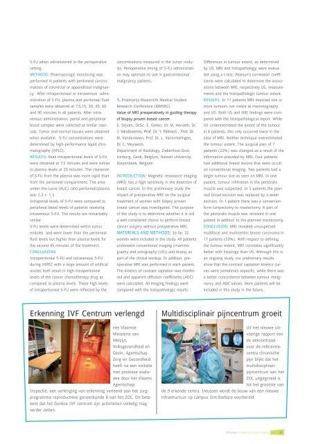
Erkenning IVF Centrum verlengd<br />
Het Vlaamse<br />
Ministerie van<br />
Welzijn,<br />
Volksgezondheid en<br />
Gezin, Agentschap<br />
Zorg en Gezondheid<br />
heeft na een visitatie<br />
met positieve evaluatie<br />
door het Vlaams<br />
Agentschap<br />
Inspectie, een verlenging van erkenning verleend aan het zorgprogramma<br />
reproductieve geneeskunde B van het ZOL. Dit betekent<br />
dat het Genkse IVF centrum zijn activiteiten volledig mag<br />
verder zetten.<br />
concentrations measured in the tumor nodules.<br />
Perioperative timing of 5-FU administration<br />
may optimize its use in gastrointestinal<br />
malignancy patients.<br />
1. Posterprijs Maastricht Medical Student<br />
Research Conference (MMSRC)<br />
Value of MRI preoperatively in guiding therapy<br />
of biopsy proven breast cancer<br />
S. Stijven, Dr.Sc. E. Gielen, Dr. M. Horvath, Dr.<br />
J. Vandevenne, Prof. Dr. Y. Palmers , Prof. Dr.<br />
M. Vandersteen, Prof. Dr. L. Vanormelingen,<br />
Dr. L. Meylaerts<br />
Department of Radiology, <strong>Ziekenhuis</strong>-<strong>Oost</strong>-<br />
<strong>Limburg</strong>, Genk, Belgium, Hasselt University,<br />
Diepenbeek, Belgium<br />
INTRODUCTION: Magnetic resonance imaging<br />
(MRI) has a high sensitivity in the detection of<br />
breast cancer. In this preliminary study the<br />
impact of preoperative MRI on the surgical<br />
treatment of women with biopsy proven<br />
breast cancer was investigated. The purpose<br />
of the study is to determine whether it is still<br />
a well-considered choice to perform breast<br />
cancer surgery without preoperative MRI.<br />
MATERIALS AND METHODS: So far, 32<br />
women were included in the study. All patients<br />
underwent conventional imaging (mammography<br />
and sonography (US)) and biopsy as<br />
part of the clinical workup. In addition, preoperative<br />
MRI was performed in each patient.<br />
The kinetics of contrast captation was monitored<br />
and apparent diffusion coefficients (ADC)<br />
were calculated. All imaging findings were<br />
compared with the histopathologic results.<br />
Differences in tumour extent, as determined<br />
by US, MRI and histopathology, were evaluated<br />
using a t-test. Pearson’s correlation coefficients<br />
were calculated to determine the associations<br />
between MRI, respectively US, measurements<br />
and the histopathologic tumour extent.<br />
RESULTS: In 17 patients MRI depicted one or<br />
more tumours not visible at mammography<br />
and US. Both US and MRI findings were compared<br />
with the histopathological report. While<br />
US underestimated the extent of the tumour<br />
in 8 patients, this only occurred twice in the<br />
case of MRI. Neither technique overestimated<br />
the tumour extent. The surgical plan of 7<br />
patients (22%) was changed as a result of the<br />
information provided by MRI. Four patients<br />
had additional breast lesions that were occult<br />
on conventional imaging. Two patients had a<br />
larger tumour size as seen on MRI. In one<br />
patient, tumour infiltration in the pectoralis<br />
muscle was suspected. In 5 patients the planned<br />
broad excision was replaced by a wider<br />
excision. In 1 patient there was a conversion<br />
form lumpectomy to mastectomy. A part of<br />
the pectoralis muscle was removed in one<br />
patient in addition to the planned mastectomy.<br />
CONCLUSION: MRI revealed unsuspected<br />
multifocal and multicentric breast carcinoma in<br />
17 patients (53%). With respect to defining<br />
the tumour extent, MRI correlates significantly<br />
better with histology than US. Although this is<br />
an ongoing study, our preliminary results<br />
show that the contrast captation kinetics curves<br />
were sometimes aspecific, while there was<br />
a better concordance between tumour malignancy<br />
and ADC values. More patients will be<br />
included in this study in the future.<br />
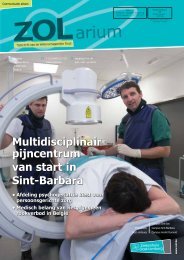
Multidisciplinair pijncentrum groeit<br />
Uit het nieuwe uitvoerige<br />
rapport van<br />
de akkoordraad<br />
voor de referentiecentra<br />
chronische<br />
pijn blijkt dat het<br />
multidisciplinair<br />
pijncentrum van het<br />
ZOL uitgegroeid is<br />
tot het grootste van<br />
de 9 erkende centra. Intussen wordt de bouw van een nieuwe<br />
infrastructuur op campus Sint-Barbara voorbereid.<br />
ZOLarium I <strong>Ziekenhuis</strong> <strong>Oost</strong>-<strong>Limburg</strong><br />
39