
Create successful ePaper yourself
Turn your PDF publications into a flip-book with our unique Google optimized e-Paper software.
12<br />
Life Science News<br />
Use <strong>of</strong> microRNAs for therapy <strong>of</strong> renal fibrosis<br />
MicroRNAs (miRNA) are small non-coding RNA molecules which bind with complementary<br />
sequences in mRNAs suppressing protein translation. They have the ability to bind to specific<br />
regions in the 3′UTR <strong>of</strong> mRNA and repress gene expression through interaction with the RNA<br />
induced silencing complex (RISC). MiRNAs are essential for renal development and homeostasis.<br />
Renal fibrosis is a result <strong>of</strong> a significant kidney injury, in which the replacement <strong>of</strong> functioning<br />
nephrons with scar tissue results in impaired renal function. Transforming growth factor<br />
β (TGFβ) is the main cytokine involved in renal damage and fibrosis here, and its main<br />
contribution to the process is to stimulate synthesis and accumulation <strong>of</strong> extracellular matrix.<br />
The feature <strong>of</strong> miRNAs to target multiple mRNAs defines that targeting a particular miRNA<br />
for therapy could stop fibrosis. MiR-21 is a miRNA very highly expressed in most tissue, has<br />
its own promoter and is independently transcribed. MiR-21 expression is closely linked to<br />
TGFβ signaling and consequently fibrosis. Many groups have shown in multiple cell types<br />
that increases in miR-21 are induced by TGFβ stimulation. This increased expression <strong>of</strong> miR-<br />
21 has been observed in multiple preclinical models <strong>of</strong> renal diseases. Treatment <strong>of</strong> diseased<br />
kidneys <strong>of</strong> knockout (genetically modified) mice with the anti-MiRNA-21 has led to reduced<br />
albuminuria, less fibrosis and inflammation, and improved survival. The severity <strong>of</strong> the renal<br />
dysfunction was reduced. This shows how a particular miRNA<br />
for therapy could have a dramatic effect on a disease process.<br />
Source: Denby, L., Baker, A.H. (2016) Targeting non-coding RNA for the therapy <strong>of</strong> renal<br />
disease. Current Opinion in Pharmacology, <strong>Volume</strong> 27, April 2016, 70–77.<br />
Science<br />
Effects <strong>of</strong> mobile phone radiation on the brain<br />
The public concern about the potential health hazards induced<br />
by radi<strong>of</strong>requency (RF) radiation has been growing with the rapid<br />
development <strong>of</strong> electrical technologies. A study was performed<br />
to investigate the effect <strong>of</strong> 1800 MHz RF radiation emitted from<br />
a mobile phone on the rat’s brain. Twenty rats were exposed to<br />
1800 MHz emitted from a mobile phone with an SAR value <strong>of</strong><br />
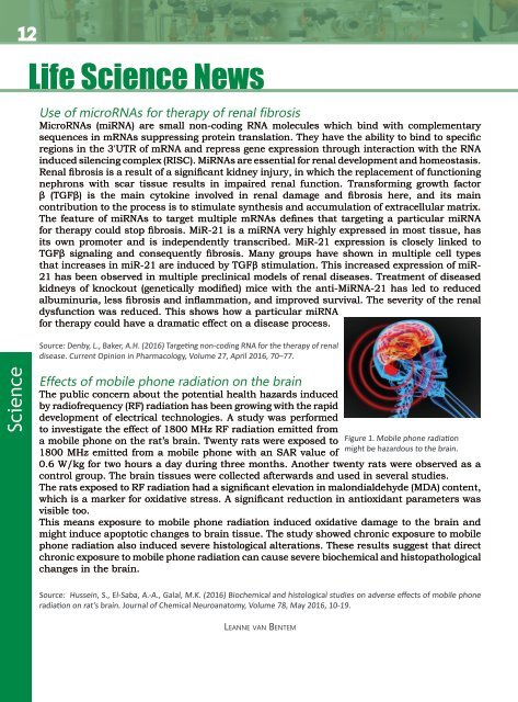
Figure 1. Mobile phone radiation<br />
might be hazardous to the brain.<br />
0.6 W/kg for two hours a day during three months. Another twenty rats were observed as a<br />
control group. The brain tissues were collected afterwards and used in several studies.<br />
The rats exposed to RF radiation had a significant elevation in malondialdehyde (MDA) content,<br />
which is a marker for oxidative stress. A significant reduction in antioxidant parameters was<br />
visible too.<br />
This means exposure to mobile phone radiation induced oxidative damage to the brain and<br />
might induce apoptotic changes to brain tissue. The study showed chronic exposure to mobile<br />
phone radiation also induced severe histological alterations. These results suggest that direct<br />
chronic exposure to mobile phone radiation can cause severe biochemical and histopathological<br />
changes in the brain.<br />
Source: Hussein, S., El-Saba, A.-A., Galal, M.K. (2016) Biochemical and histological studies on adverse effects <strong>of</strong> mobile phone<br />
radiation on rat’s brain. Journal <strong>of</strong> Chemical Neuroanatomy, <strong>Volume</strong> 78, May 2016, 10-19.<br />
Leanne van Bentem