

São Paulo (SP) - Conselho Brasileiro de Oftalmologia
São Paulo (SP) - Conselho Brasileiro de Oftalmologia
São Paulo (SP) - Conselho Brasileiro de Oftalmologia

Create successful ePaper yourself
Turn your PDF publications into a flip-book with our unique Google optimized e-Paper software.
Renesto AC, et al.<br />
A<br />
B<br />
C<br />
D<br />
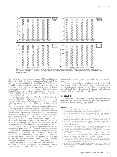
Figure 4. A) Riboflavin eyedrops group: cell-to-cell contact of epithelial cells - temporal conjunctiva. B) Riboflavin eyedrops group: nucleus-to-cytoplasm<br />
ratio - temporal conjunctiva. C) Riboflavin eyedrops group: goblet cell <strong>de</strong>nsity - temporal conjunctiva. D) Riboflavin eyedrops group: keratinization - temporal<br />
conjunctiva.<br />
junctiva. The <strong>de</strong>struction of corneal tissue by LASIK procedures may<br />
also directly or indirectly affect the population of goblet cells. These<br />
findings were observed at the 3-month follow-up. After 6 months,<br />
the goblet cells recovered (14) , which was similar to our experience.<br />
In our study, the femtosecond laser was used to create the channel<br />
ring at a <strong>de</strong>pth of 80% of the thinnest corneal thickness on the tunnel<br />
location. Thus, the creation of the channel ring by the femtosecond<br />
laser did not affect the goblet cell <strong>de</strong>nsity on the ocular surface in<br />
our group of patients.<br />
In the present study, the IC results before and after treatment<br />
were normal (gra<strong>de</strong> 0) in all corneal specimens. Although epithelial<br />
<strong>de</strong>bri<strong>de</strong>ment before corneal CXL can cause necrosis and apoptosis (15)<br />
of keratocytes at a <strong>de</strong>pth of 50 to 60 μm, any significant cytotoxic<br />
effect of CXL seems to be concentrated in the anterior part of the<br />
cornea because of the high absorption of UVA light by riboflavin. This<br />
behavior prevents the light from penetrating to <strong>de</strong>eper levels and<br />
preserves the endothelium and lens. Previous reports showed neither<br />
clinically evi<strong>de</strong>nt si<strong>de</strong> effects of CXL treatment nor persistent epithelial<br />
<strong>de</strong>fects (16) , with the exception of transient corneal e<strong>de</strong>ma and a<br />
foreign body sensation for 24-48 hours, in combination with burning<br />
and tearing that are common in photorefractive surgery. Mazzotta<br />
et al. (16) used confocal microscopy to <strong>de</strong>monstrate that the cytotoxic<br />
effects of CXL, although significant, were concentrated in the first<br />
350 μm of the stroma and did not affect the adjacent structures.<br />
In the riboflavin eyedrops group, an improvement in the cell-tocell<br />
contact of epithelial cells on superior conjunctiva was noted at the<br />
2-year follow-up. On the temporal conjunctiva, improvements in the<br />
cell-to-cell contact of epithelial cells and keratinization, and increases<br />
in the N:C ratio and goblet cell <strong>de</strong>nsity were noted. Our patients used<br />
riboflavin eyedrops for 1 month, and the aforementioned parameters<br />
improved up to the 2-year follow-up. These findings are possibly related<br />
to the more regular use of artificial tears prescribed throughout<br />
the 2-year follow-up period. These patients were followed closely and<br />
encouraged to regularly use the artificial tears. Squamous metaplasia<br />
scores may have improved in the conjunctiva because of a rise in<br />
ocular surface humidity related to the regular use of artificial tears<br />
over time.<br />
Riboflavin alone produced no cell damage, which is not surprising<br />
because riboflavin is also present in the retina, liver, and heart as<br />
an essential element in normal nutrition (17) . In a previous experimental<br />
study, riboflavin played a role in the <strong>de</strong>velopment and maintenance<br />
of the surface structures of epithelial cells (18) .<br />
CONCLUSION<br />
Cross-linking and intrastromal corneal ring segment insertion did<br />
not change the ocular surface morphology gra<strong>de</strong> in keratoconic eyes<br />
as assessed by impression cytologic analysis.<br />
REFERENCES<br />
1. Wollensak G, Spoerl E, Seiler T. Riboflavin/ultraviolet-A-induced collagen crosslinking<br />
for the treatment of keratoconus. Am J Ophthalmol. 2003;135(5):620-7.<br />
2. Tan BU, Purcell TL, Torres LF, Schanzlin DJ. New surgical approaches to the management<br />
of keratoconus and post-lasik ectasia. Trans Am Ophthalmol Soc. 2006;104:<br />
212-20.<br />
3. Miranda D, Sartori M, Francesconi C, Allemann N, Ferrara P, Campos M. Ferrara intras tromal<br />
corneal ring segments for severe keratoconus. J Refract Surg. 2003;19(6): 645-53.<br />
4. Renesto AC, Barros JN, Campos M. Impression cytologic analysis after corneal collagen<br />
cross-linking using riboflavin and ultraviolet-A light in the treatment of ke ratoconus.<br />
Cornea. 2010;29(10):1139-44.<br />
5. Torquetti L, Berbel RF, Ferrara P. Long-term follow-up of intrastromal corneal ring<br />
seg ments in keratoconus. J Cataract Refract Surg. 2009;35(10):1768-73.<br />
6. Krumeich JH, Daniel J, Knülle A. Live-epikeratophakia for keratoconus. J Cataract Re -<br />
fract Surg. 1998;24(4):456-63.<br />
7. Pinheiro MN Jr, dos Santos PM, dos Santos RC, Barros J <strong>de</strong> N, Passos LF, Cardoso Ne -<br />
to J. [Oral flaxseed oil (Linum usitatissimum) in the treatment for dry-eye Sjögren’s<br />
syndrome patients]. Arq Bras Oftalmol. 2007;70(4):649-55. Portuguese.<br />
8. Fenga C, Aragona P, Cacciola A, Ferreri F, Spatari G, Stilo A, et al. Ocular discomfort and<br />
conjunctival alterations in operating room workers. A single-institution pilot study.<br />
Int Arch Occup Environ Health. 2001;74(2):123-8.<br />
9. <strong>de</strong> Almeida SF, <strong>de</strong> Sousa LB, Vieira LA, Chiamollera MI, Barros JN. Clinic-cytologic<br />
study of conjunctivochalasis and its relation to thyroid autoimmune diseases. Cornea.<br />
2006;25(7):789-93.<br />
Arq Bras Oftalmol. 2012;75(2):101-6<br />
105


