

São Paulo (SP) - Conselho Brasileiro de Oftalmologia
São Paulo (SP) - Conselho Brasileiro de Oftalmologia
São Paulo (SP) - Conselho Brasileiro de Oftalmologia

You also want an ePaper? Increase the reach of your titles
YUMPU automatically turns print PDFs into web optimized ePapers that Google loves.
Bupivacaine and botulinum toxin to treat comitant strabismus<br />
The present study intends to analyze the effect of bupicavaine<br />
and botulinum toxin A injections in extraocular muscles with comitant<br />
strabismus, evaluating ocular motility and extraocular muscle<br />
thickness.<br />
METHODS<br />
In this study we prospectively enrolled 8 patients with comitant<br />
strabismus and either amblyopia or poor vision in one eye. These<br />
patients un<strong>de</strong>rwent injections of 2 ml of 1.5% bupivacaine in the<br />
lateral rectus (LR) if had esotropia or in the medial rectus (MR) if had<br />
exotropia and 2.5 U botulinum toxin A in the MR if had esotropia or<br />
in the LR if had exotropia. The protocol was approved by the Ethics<br />
Committee - Investigational Review Board of the Fe<strong>de</strong>ral University<br />
of São <strong>Paulo</strong>, Brazil.<br />
The criteria for enrollment in the study were: poor vision in one<br />
eye and comitant strabismus and no previous surgery or botulinum<br />
toxin A injections. All patients had visual acuity lower than 0.5 in the<br />
worst eye. Three of them had exotropia and 5 esotropia.<br />
One ophthalmologist (TSM) used EMG to localize the posterior<br />
third of the horizontal rectus muscle where the injection of 2 ml of<br />
1.5% bupivacaine was performed un<strong>de</strong>r topical anesthesia (proximetacaine).<br />
At least one third of the medication was injected along<br />
the needle trajectory while the syringe was being withdrawn. The<br />
injection was performed in the LR in case of esotropia and in the MR<br />
in case of exotropia. Botulinum toxin A was subsequently administered<br />
using Mendonça’s forceps in the contralateral horizontal rectus<br />
muscle (13) . Botulinum toxin A was diluted using 100 U ampoule and<br />
2 ml of 0.9% saline solution. Subsequently 2.5 U were injected into<br />
the MR in esotropic patients and into the LR in the exotropic ones.<br />
All patients un<strong>de</strong>rwent ocular ultrasonography (USG) of both eyes<br />
once before the procedure, and repeated 7 and 30 days after the<br />
procedure, to <strong>de</strong>termine the thickness of all extraocular muscles. All<br />
measurements were performed by the same examiner (NA) using a<br />
10-MHz transducer (Ultrascan, Alcon), and eyelid contact method<br />
(ultrasound gel was used as a coupling media), with patient’s eye<br />
aligned in primary position. One eye was examined at the time and<br />
the patient was asked to fixate on a target with that eye to ensure<br />
alignment.<br />
Patients un<strong>de</strong>rwent a comprehensive ophthalmological exam<br />
before the procedure. Clinical examination was performed after 1, 7,<br />
15, 30 days and then monthly until 180 days to <strong>de</strong>termine eye alignment,<br />
versions and the presence of complications. All the motility<br />
measurements were performed by the same examiner (LMH) using<br />
the Krimsky method or the cover/uncover method when the patient<br />
was able to fixate a target at distance.<br />
Both examiners for ocular motility exam and ultrasonography<br />
(LMH and NA) did not have access to medical records with previous<br />
measurements until all data was collected.<br />
RESULTS<br />
The eight patients who un<strong>de</strong>rwent injection of bupivacaine and<br />
botulinum Toxin A presented average age of 24.5 years (range 18 to<br />
53 years), three were male (five, female).<br />
Five presented the primary diagnosis of esotropia, four of them<br />
had infantile esotropia and amblyopia, and one had esotropia secondary<br />
to macular scar due to toxoplasmosis. The other three patients<br />
had exotropia. One of which had congenital cataract that had been<br />
operated and manifest nystagmus, other had poor vision in one eye<br />
due to a macular scar subsequent to congenital toxoplasmosis and<br />
the last one had a trauma in childhood with subsequent glaucoma<br />
and low vision.<br />
Table 1 summarizes the changes in alignment <strong>de</strong>viation during<br />
the follow-up. Four patients (2, 3, 6 and 8) <strong>de</strong>veloped a transitory<br />
vertical <strong>de</strong>viation due to the effect of the botulinum toxin A, which<br />
was recovered by day 180. Ptosis of 1 mm was present in patients<br />
1 (Figure 4), 5, 7 and 8 at the 7 th , 15 th and 30 th days of follow-up visit.<br />
Patient 2 presented a 2 mm ptosis and patient 4, 4 mm ptosis, presented<br />
at the 7 th , 15 th , 30 th and 60 th days of follow-up visit. The overall mean<br />
change in <strong>de</strong>viation was 10 prism diopters at the 180 th day visit. Patients<br />
4 (Figure 5) and 8 had an increase in <strong>de</strong>viation on day 1. All patients had<br />
the smallest angle of <strong>de</strong>viation at days 15 and 30. Patients 2, 3 and 4<br />
had a higher <strong>de</strong>viation on the 60 th day than the previous visit, with a<br />
<strong>de</strong>crease on <strong>de</strong>viation the 180 th day. Patients 5 and 6 were exclu<strong>de</strong>d<br />
due to lack of follow-up data at the 60 th and 180 th days visit.<br />
Figure 1 and table 2 show the thickness of the muscle injected<br />
with botulinum toxin type A measured by USG before the injection<br />
and after 7 and 30 days of the procedure. Figure 2 and table 3 show<br />
the thickness of the muscle injected with bupivacaine also measured<br />
by USG before the injection and after 7 and 30 days. Patient 5 un<strong>de</strong>rtook<br />
the USG exam 60 days after injection instead of 30 because of a<br />
not related clinical intervention. Figure 3 shows the muscle thickness<br />
before and after injection in each group of muscles - LR and MR.<br />
Ultrasonography showed an average increase in thickness of the<br />
muscle injected with Bupivacaine on the 7 th day of 0.43 mm. The<br />
mean increase of muscle thickness after 30 days injection of bupivacaine<br />
was equal of 1.01 mm, and after botulinum toxin A injection<br />
was equal of 0.28 mm. When comparing before and after injection,<br />
an increase in thickness of the LR injected with botulinum toxin A<br />
(0.6 mm) and with bupivacaine (1.5 mm) was observed in thickness<br />
for each group of muscles. There was no increase in thickness of the<br />
MR injected with bupivacaine, and a small increase in the botulinum<br />
toxin A MR injected group (0.1 mm).<br />
One patient (patient 5) presented a retrobulbar hemorrhage during<br />
bupivacaine injection in the MR. The complication was treated<br />
using compressive bandage of the eye with a<strong>de</strong>quate control of IOP<br />
and proptosis. After 60 days the patient showed no change in visual<br />
acuity and no optic disc abnormality at fundoscopy, but a <strong>de</strong>crease<br />
in the pupillary response with medium mydriasis.<br />
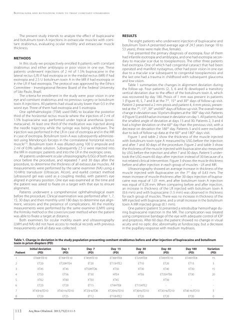
Table 1. Change in <strong>de</strong>viation in the study population presenting comitant strabismus before and after injection of bupivacaine and botulinum<br />
toxin in prism diopters (PD)<br />
Patient<br />
Initial <strong>de</strong>viation<br />
(PD)<br />
Day 1<br />
(PD)<br />
Day 7<br />
(PD)<br />
Day 15<br />
(PD)<br />
Day 30<br />
(PD)<br />
Day 60<br />
(PD)<br />
Day 180<br />
(PD)<br />
1 ET60HTE10 ET50HTE10 ET45HTE10 ET30HTE8 ET25HTE8 ET45HTE10 ET45HTE8 15<br />
2 ET20 ET20HTE4 ET20 ET15HTE2 ET10 ET20 ET15 5<br />
3 XT45 XT45 XT50HTD6 XT30 XT30 XT40 XT30 15<br />
4 ET50 ET56 ET50 HTE4 HTE6 ET35HTE6 ET30 20<br />
5 XT62 XT60 XT50 XT45 - XT56 - -<br />
6 ET20 ET20 ET15 ET4HTE8 ET15HTE2 - - -<br />
7 XT45HoTD10 XT40 HoTD10 XT25HoTD8 XT20HoTD10 XT30HoTD10 XT30 HoTD10 XT40 HoTD10 5<br />
8 ET20 ET25 ET12 ET10HTE2 ET20 ET20 ET20 0<br />
Variation<br />
(PD)<br />
112 Arq Bras Oftalmol. 2012;75(2):111-5


