

São Paulo (SP) - Conselho Brasileiro de Oftalmologia
São Paulo (SP) - Conselho Brasileiro de Oftalmologia
São Paulo (SP) - Conselho Brasileiro de Oftalmologia

Create successful ePaper yourself
Turn your PDF publications into a flip-book with our unique Google optimized e-Paper software.
Experimental mo<strong>de</strong>ls of autoimmune inflammatory ocular diseases<br />
the available mo<strong>de</strong>ls are complementary (25) . Table 1 shows the most<br />
used mo<strong>de</strong>ls for the study of autoimmune ocular inflammatory<br />
diseases.<br />
1. EXPERIMENTAL AUTOIMMUNE UVEITIS (EAU)<br />
EAU is the most used mo<strong>de</strong>l for the study of autoimmune ocular<br />
inflammation, especially in the posterior segment. It is induced by<br />
the subcutaneous injection of a single dose of soluble retinal antigen<br />
(21,26) . The EAU clinical and pathological characteristics <strong>de</strong>pend on<br />
the species and the antigen used for induction, but T-cell mediated<br />
inflammatory response is always present, mainly CD4+ cells. CD4+<br />
cells recognize antigens presented by antigen presenting cells (APC)<br />
in the context of class II main histocompatibility complex (MHC)<br />
molecules. Pro-inflammatory cytokines, such as IL-2, IFN-γ, and TNF-α<br />
are produced by the subset Th1 and are important mediators of the<br />
cellular immune response (27) .<br />
1.1 S-Antigen<br />
The S-antigen, also called S-arrestin, is a photoreceptor protein<br />
present in abundance in photoreceptors and pineal gland cells (21) .<br />
It plays a physiological role in <strong>de</strong>sensitization of the photoactivated<br />
transduction casca<strong>de</strong> by inhibiting coupling of rhodopsin to trans -<br />
ducin. In addition, S-antigen is highly antigenic and is a potent inducer<br />
of autoimmune uveoretinitis in animals.<br />
EAU is induced by the subcutaneous injection of bovine S-antigen<br />
along with the complete Freund’s adjuvant (CFA) as vehicle (28) .<br />
The inflammatory response begins between 10 days and 2 months<br />
and lasts from few days to 1 year, <strong>de</strong>pending on the species and the<br />
dose of the antigen. In Lewis rats, the inflammation is acute and selfli<br />
mited but it causes important damage to the retina and choroid<br />
within few days (29) . The disease is less severe and subacute in other<br />
rat strains, guinea pigs, and monkeys. In the latter, the di sease may<br />
present a longer course with progressive <strong>de</strong>struction of the retina (29) .<br />
Each antigen has a group of antigenic <strong>de</strong>terminants called epitopes.<br />
Epitopes are sequences of amino acids that elicit inflammatory<br />
response after recognition by the immune system. Some antigens<br />
may present various different epitopes, or the same epitopes may<br />
be present more than once in the same protein. Pepti<strong>de</strong> M is an<br />
18-amino acid component of S-antigen that has been shown to be<br />
highly uveitopathogenic in different species, such as rats, guinea pigs,<br />
and monkeys, producing inflammatory changes in eyes very similar<br />
to those induced by native S-antigen (30) . In humans, the amino acids<br />
sequence of the S-antigen corresponding to pepti<strong>de</strong> M was found to<br />
be virtually i<strong>de</strong>ntical to that of the bovine S-antigen (31) .<br />
Immunological mimicry between host and microbial proteins has<br />
been suggested as a potential mechanism in the <strong>de</strong>velopment of uveitis<br />
in humans. Cross-reactivity between anti-streptococcal monoclonal<br />
antibodies with retinal S-antigen from rods of the human eye<br />
hu man eye has been <strong>de</strong>monstrated, suggesting that both molecules<br />
share epitopes of antigenic similarity. These findings may suggest a<br />
possible association between host and microbial proteins, which may<br />
be a potential mechanism in the <strong>de</strong>velopment of human uveitis (32) .<br />
1.2 Interphotoreceptor retinoid-binding protein<br />
IRBP is a protein found primarily between the retinal pigment<br />
epi thelium (RPE) and the photoreceptor cells. It plays a role in the<br />
trans port of the retinoids, a product of vitamin A, between the retina<br />
and the RPE cells (33,34) . Uveitis is induced by subcutaneous injection<br />
of isolated bovine IRPB or part of bovine IRPB associated to per tussis<br />
toxin with CFA as vehicle (35) . IRBP is a strong uveitis inducer in rats (36) ,<br />
monkeys (37) , rabbits (38) , and mice (22) . However, it is a weak inducer in<br />
guinea pigs (39) . The clinical course and severity of EAU <strong>de</strong>pend on the<br />
antigen dosage, the animal species, and the association of pertussis<br />
toxin at the moment of induction. In Lewis rats, the acute inflammation<br />
may be severe and begin from 8 to 12 days after immunization,<br />
lasting 5 to 10 days (36) . In mice, uveitis has longer incubation and lasts<br />
longer. Mild anterior uveitis may be present and the prominent retinal<br />
findings are vasculitis, granuloma, serous <strong>de</strong>tachment, and loss of<br />
photoreceptors. Other findings inclu<strong>de</strong> sub-RPE infiltrates (clinical<br />
equivalent of Dallen-Fuchs nodules) and thickening of the choroid<br />
and ciliary body (40) .<br />
IRBP molecule has 1,264 amino acids, forming 4 repeated units<br />
of 300 amino acids each. Each unit presents an uveitogenic site. The<br />
pepti<strong>de</strong> 1117-1191 of bovine IRBP is capable of generating important<br />
uveal inflammation (41,42) . Another study suggests that the rat IRBP<br />
pepti<strong>de</strong> 273-283 also elicits severe uveitis (43) . Approximately 84% of<br />
the bovine and human IRBP present i<strong>de</strong>ntical sequence of amino<br />
acids (44,45) . Many studies using EAU mo<strong>de</strong>ls induced by IRBP have<br />
aimed the i<strong>de</strong>ntification of the exact immunogenic epitopes (46-48)<br />
and interactions with the MHC molecules. Specific IRBP epitopes<br />
ha ve already been recognized as uveitogenic in susceptible mice<br />
strains: Pepti<strong>de</strong> 1-20 for C57BL/6, 161-180 for B10.RIII, and 201-216<br />
for B10.A (49) , and recent studies revealed several additional uveitogenic<br />
epitopes of human IRBP molecule that elicit EAU in mice (48) .<br />
2. EXPERIMENTAL MELANIN PROTEIN-INDUCED UVEITIS (EMIU)<br />
Non-soluble melanin proteins isolated from pigmented bovine<br />
eye tissues can induce an uveitis mo<strong>de</strong>l named EMIU (50,51) . According<br />
to Matteson et al. (52) , this mo<strong>de</strong>l can be induced by subcutaneous<br />
injection of melanin proteins in association with Hunter adjuvant.<br />
CD4+ T cells are the primary mediators of the inflammatory res -<br />
ponse, as well as in EAU (53) , but the target tissue is the uvea, where<br />
the melanocytes are located, not the retina. Therefore, ocular inflammation<br />
is mostly evi<strong>de</strong>nt in the iris, ciliary body, and choroid. Clinical<br />
manifestations begin 10 to 14 days after induction and consists of<br />
con junctival hyperemia, corneal e<strong>de</strong>ma, anterior uveitis, iris vessels<br />
dilation, hypopyon, and synechiae (40) . The 2-week acute uveitis may<br />
present spontaneous recurrences as observed in EAU in mice (54) .<br />
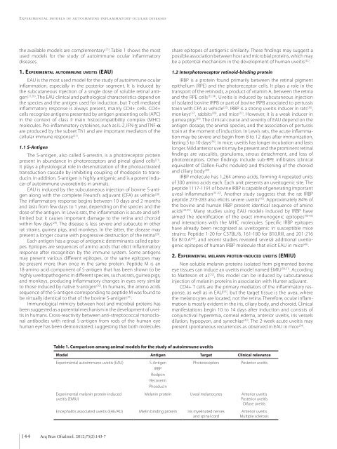
Table 1. Comparison among animal mo<strong>de</strong>ls for the study of autoimmune uveitis<br />
Mo<strong>de</strong>l Antigen Target Clinical relevance<br />
Experimental autoimmune uveitis (EAU)<br />
Experimental melanin protein-induced<br />
uveitis (EMIU)<br />
S-Antigen<br />
IRBP<br />
Rodpsin<br />
Recoverin<br />
Phosducin<br />
Photoreceptors<br />
Posterior uveitis<br />
Melanin protein Uveal melanocytes Anterior uveitis<br />
Posterior uveitis<br />
Difuse uveitis<br />
Encephalitis associated uveitis (EAE/AU) Mielin binding protein Iris myelinated nerves<br />
and spinal cord<br />
Anterior uveitis<br />
Multiple sclerosis<br />
144 Arq Bras Oftalmol. 2012;75(2):143-7


