

Respircase Cilt: 6 - Sayı: 1 Yıl: 2017

Create successful ePaper yourself
Turn your PDF publications into a flip-book with our unique Google optimized e-Paper software.
Respiratory Case Reports<br />
CASE<br />
A 64-year-old male patient who was followed for COPD<br />
for 10 years presented with a complaint of severe cough<br />
persisting even during stable periods of his condition for<br />
the past two years. During follow-up, his complaint of<br />
cough was attributed to his present condition and he had<br />
an episode of hospitalization. Being an ex-smoker for five<br />
years, he had a 50 pack/year history of smoking with<br />
purulent sputum without hemoptysis. He had no comorbidities.<br />
Pulmonary function test showed a forced vital<br />
capacity (FVC) of 2500mL (66%), a forced expiratory<br />
volume in 1 second (FEV1) of 1330mL (46%), and a<br />
FEV1/FVC ratio of 46%. Auscultation revealed diminished<br />
breath sounds with a prolonged expirium, and thin rales<br />
in the right base, while the results of other system examinations<br />
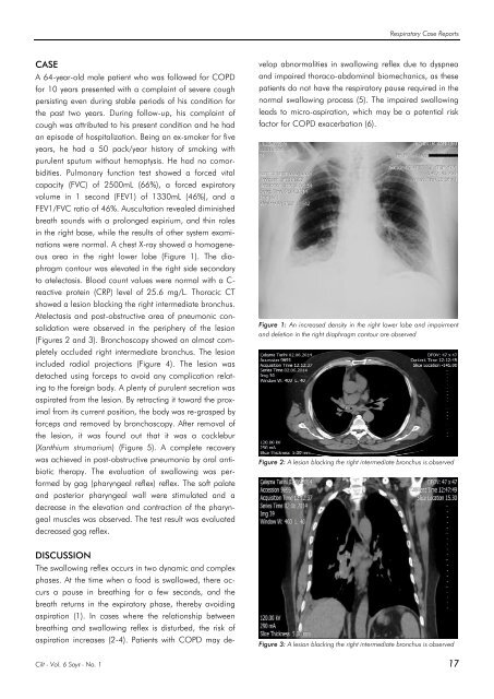
were normal. A chest X-ray showed a homogeneous<br />
area in the right lower lobe (Figure 1). The diaphragm<br />
contour was elevated in the right side secondary<br />
to atelectasis. Blood count values were normal with a C-<br />
reactive protein (CRP) level of 25.6 mg/L. Thoracic CT<br />
showed a lesion blocking the right intermediate bronchus.<br />
Atelectasis and post-obstructive area of pneumonic consolidation<br />
were observed in the periphery of the lesion<br />
(Figures 2 and 3). Bronchoscopy showed an almost completely<br />
occluded right intermediate bronchus. The lesion<br />
included radial projections (Figure 4). The lesion was<br />
detached using forceps to avoid any complication relating<br />
to the foreign body. A plenty of purulent secretion was<br />
aspirated from the lesion. By retracting it toward the proximal<br />
from its current position, the body was re-grasped by<br />
forceps and removed by bronchoscopy. After removal of<br />
the lesion, it was found out that it was a cocklebur<br />
(Xanthium strumarium) (Figure 5). A complete recovery<br />
was achieved in post-obstructive pneumonia by oral antibiotic<br />
therapy. The evaluation of swallowing was performed<br />
by gag (pharyngeal reflex) reflex. The soft palate<br />
and posterior pharyngeal wall were stimulated and a<br />
decrease in the elevation and contraction of the pharyngeal<br />
muscles was observed. The test result was evaluated<br />
decreased gag reflex.<br />
The swallowing reflex occurs in two dynamic and complex<br />
phases. At the time when a food is swallowed, there occurs<br />
a pause in breathing for a few seconds, and the<br />
breath returns in the expiratory phase, thereby avoiding<br />
aspiration (1). In cases where the relationship between<br />
breathing and swallowing reflex is disturbed, the risk of<br />
aspiration increases (2-4). Patients with COPD may develop<br />
abnormalities in swallowing reflex due to dyspnea<br />
and impaired thoraco-abdominal biomechanics, as these<br />
patients do not have the respiratory pause required in the<br />
normal swallowing process (5). The impaired swallowing<br />
leads to micro-aspiration, which may be a potential risk<br />
factor for COPD exacerbation (6).<br />
Figure 1: An increased density in the right lower lobe and impairment<br />
and deletion in the right diaphragm contour are observed<br />
Figure 2: A lesion blocking the right intermediate bronchus is observed<br />
DISCUSSION<br />
Figure 3: A lesion blocking the right intermediate bronchus is observed<br />
<strong>Cilt</strong> - Vol. 6 <strong>Sayı</strong> - No. 1 17


