

Respircase Cilt: 6 - Sayı: 1 Yıl: 2017

You also want an ePaper? Increase the reach of your titles
YUMPU automatically turns print PDFs into web optimized ePapers that Google loves.
Co-Incidence of Echinococcus Alveolaris and Echinococcus Granulosus in the Lung: A Rare Case | Akıncı Özyürek et al.<br />
treatment, and cyst excision via right thoracotomy and<br />
liver cyst excision have been planned.<br />
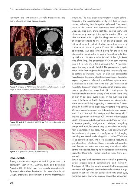
Figure 1: Imaging of PET-CT and thoracic CT: Multiple nodules in both<br />
lungs, of which some had cavitary characteristics<br />
Figure 2A and B: E. alveolaris (HEX40) (A: Cuticle membrane B: Lung<br />
parenchyma)<br />
Figure 3: E. granulosis (HEX40) (Cyst membrane)<br />
DISCUSSION<br />
Turkey is an endemic region for both E. granulosus. It is<br />
particularly seen in the Central, East, and South-East<br />
Anatolia (1). Hydatid cysts are usually asymptomatic.<br />
Symptoms depend on the size and location of the lesion.<br />
Cough, chest pain, and hemoptysis are the most frequent<br />
symptoms. The most diagnostic symptom in cystic echinococcosis<br />
is the expectoration of the cyst fluid or membranes,<br />
indicating that the cyst is perforated. The overall<br />
status of the patient may deteriorate after perforation.<br />
Dyspnea, chest pain, and anaphylaxis can be seen. Lung<br />
abscesses may develop, if the cyst is infected. Our case<br />
also presented with cough. For diagnostic work-up, it is<br />
an important finding to live in an endemic region and<br />
history of animal contact. Routine laboratory tests may<br />
not be helpful in the diagnosis. Eosinophilia in blood can<br />
be detected. Our case owned a dog for one year. No<br />
abnormality was detected in routine laboratory tests. Cyst<br />
hydatid has a tendency to be located at the right lower<br />
lobe of the lung. The percentage of CH in both liver and<br />
lung is 6 to 13% (2). In the diagnosis of CH, X-ray imaging<br />
of the lung is usually helpful. The presence of a cystic<br />
lesion in the liver supports the diagnosis. It is usually seen<br />
as solitary or multiple, round or oval well-demarcated<br />
mass lesions. In case of alveolar echinococcus, the radiological<br />
diagnosis is difficult due to different appearances.<br />
E. alveolaris is mostly seen in the liver (3). There can be<br />
metastatic lesions in other intra-abdominal organs, bone,<br />
muscle, lymph nodes, lungs, brain (4). It is diagnosed by<br />
the fine needle aspiration biopsy of the lesions in the lung<br />
or liver. In our case, cystic lesions in the liver were also<br />
present. In the cranial MRI, there was a hypodense lesion<br />
in the left frontal lobe, suggesting a metastasis of E. alveolaris.<br />
In the differential diagnosis, metastatic lung cancer,<br />
Wegener granulomatosis, and tuberculosis were considered,<br />
due to the spicular multiple nodules, some which<br />
showed cavitation in thoracic CT. Alveolar echinococcus<br />
usually shows a gradual progression and, thus, may mimic<br />
slow-progressing malignancies. Multiple, irregularly<br />
marginated, nodular lesions may be mistaken for malignant<br />
metastases. In our case, PET-CT was performed with<br />
the preliminary diagnosis of a malignancy. This imaging<br />
modality was useful in deciding which nodule TTNB was<br />
to be performed from. The TTNB supported vasculitis or<br />
granulomatous infections. Blood elements extravasated<br />
from the vascular structures in the lung parenchyma adjacent<br />
to the nodules, leading to the appearance mimicking<br />
vasculitis. Diagnostic wedge resection was, then, performed.<br />
Early diagnosis and treatment are essential in preventing<br />
serious disease-related complications and morbidity.<br />
Treatment of CH of the lung is surgical. Following surgery,<br />
treatment with systemic benzimidazole derivations is suggested.<br />
In patients with non-complicated cysts, small cysts,<br />
numerous cysts, and when surgery cannot be performed,<br />
43 www.respircase.com


