

Respircase Cilt: 6 - Sayı: 1 Yıl: 2017

Create successful ePaper yourself
Turn your PDF publications into a flip-book with our unique Google optimized e-Paper software.
RESPIRATORY CASE REPORTS<br />
Respir Case Rep <strong>2017</strong>;6(1):72-73 DOI: 10.5505/respircase.<strong>2017</strong>.80774<br />
LETTER TO EDITOR<br />
EDİTÖRE MEKTUP<br />
Relation of Floppy Eyelid Syndrome with<br />
Obstructive Sleep Apnea<br />
Gevşek Gözkapağı Sendromu ile Obstrüktif Uyku Apnesi İlişkisi<br />
To The Editor,<br />
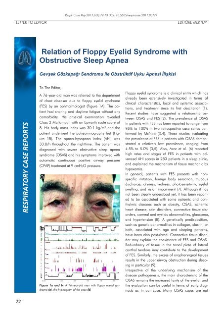
A 76-year-old man was referred to the department<br />
of chest diseases due to floppy eyelid syndrome<br />
(FES) by an ophthalmologist (Figure 1A). The patient<br />
had snoring and daytime fatigue without any<br />
comorbidity. His physical examination revealed<br />
Class 2 Mallampati with an Epworth scale score of<br />
8. His body mass index was 30.1 kg/m 2 and the<br />
patient underwent the polysomnography test (Figure<br />
1B). The apnea-hypopnea index (AHI) was<br />
33.8/h throughout the nighttime. The patient was<br />
diagnosed with severe obstructive sleep apnea<br />
syndrome (OSAS) and his symptoms improved with<br />
automatic continuous positive airway pressure<br />
(CPAP) treatment at 9 cmH 2O pressure.<br />
Figure 1a and b: A 76-year-old men with floppy eyelid syndrome<br />
(a), the hypnogram of the case (b)<br />
Floppy eyelid syndrome is a clinical entity which has<br />
already been extensively investigated in terms of<br />
clinical characteristics, local and systemic associations,<br />
and treatment since its first description (1).<br />
Recent studies have suggested a relationship between<br />
OSAS and FES (2). The prevalence of OSAS<br />
in patients with FES has been reported to range from<br />
96% to 100% in two retrospective case series performed<br />
by McNab (3,4). These studies evaluating<br />
the prevalence of FES in patients with OSAS demonstrated<br />
a relatively low prevalence, ranging from<br />
4.5% to 5.0% (3,5). Also, Acar et al. (6) reported<br />
high rates and stages of FES in patients with advanced<br />
AHI scores in 280 patients in a sleep clinic,<br />
and explained the mechanism of tissue mechanic by<br />
hypoxemia.<br />
In general, patients with FES presents with nonspecific<br />
irritation, foreign body sensation, mucous<br />
discharge, dryness, redness, photosensitivity, eyelid<br />
swelling, and vision impairment (7). Although it has<br />
not been clearly understood yet, it has been reported<br />
to be associated with some systemic and ophthalmic<br />
diseases such as obesity, OSAS, ischemic<br />
heart disease, skin disorders, connective tissue disorders,<br />
corneal and eyelids abnormalities, glaucoma,<br />
and hypertension (8). A genetically predisposition,<br />
such as genetic abnormalities in collagen, elastin, or<br />
both, associated with age and sleeping patterns,<br />
have been also postulated. Connective tissue disorder<br />
may explain the coexistence of FES and OSAS.<br />
Redundancy of tissue in the tarsal plate of lateral<br />
canthal tendons may contribute to the development<br />
of FES. Similarly, the excess of oropharyngeal tissues<br />
results in the upper airway obstruction during sleeping<br />
in particular (9).<br />
Irrespective of the underlying mechanism of the<br />
disease pathogenesis, the main characteristic of the<br />
OSAS remains the increased laxity of the eyelid, and<br />
the evaluation can be useful in terms of early diagnosis<br />
as in our case. Many OSAS cases are not<br />
72


