

Respircase Cilt: 6 - Sayı: 1 Yıl: 2017

You also want an ePaper? Increase the reach of your titles
YUMPU automatically turns print PDFs into web optimized ePapers that Google loves.
Respiratory Case Reports<br />
CASE<br />
A 29-year-old male patient was admitted to the outpatient<br />
clinic with complaints of cough and chest pain. His<br />
medical and family history revealed no indications of<br />
typical symptoms, except living in a rural area. He was<br />
smoking a pack of cigarettes/day for 10 years. His physical<br />
examination was non-specific. The laboratory examination<br />
showed that C-reactive protein level was 81 mg/L<br />
(0-5), the leukocyte count was 12,400 (3,600-10,200),<br />
the number of neutrophils was 11,200 (2000-6900),<br />
lymphocyte count was 141 (600-3400), and the monocyte<br />
count was 994 (0-900). Liver and kidney functions<br />
were normal.<br />
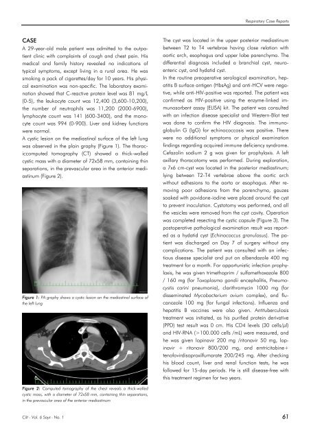
A cystic lesion on the mediastinal surface of the left lung<br />
was observed in the plain graphy (Figure 1). The thoraciccomputed<br />
tomography (CT) showed a thick-walled<br />
cystic mass with a diameter of 72x58 mm, containing thin<br />
separations, in the prevascular area in the anterior mediastinum<br />
(Figure 2).<br />
Figure 1: PA graphy shows a cystic lesion on the mediastinal surface of<br />
the left lung<br />
The cyst was located in the upper posterior mediastinum<br />
between T2 to T4 vertebrae having close relation with<br />
aortic arch, esophagus and upper lobe parenchyma. The<br />
differential diagnosis included a branchial cyst, neuroenteric<br />
cyst, and hydatid cyst.<br />
In the routine preoperative serological examination, hepatitis<br />
B surface antigen (HbsAg) and anti-HCV were negative,<br />
while anti-HIV-positive was reported. The patient was<br />
confirmed as HIV-positive using the enzyme-linked immunosorbent<br />
assay (ELISA) kit. The patient was consulted<br />
with an infection disease specialist and Western-Blot test<br />
was done to confirm the HIV diagnosis. The immunoglobulin<br />
G (IgG) for echinococcosis was positive. There<br />
were no additional symptoms or physical examination<br />
findings regarding acquired immune deficiency syndrome.<br />
Cefazolin sodium 2 g was given for prophylaxis. A left<br />
axillary thoracotomy was performed. During exploration,<br />
a 7x6 cm-cyst was located in the posterior mediastinum;<br />
lying between T2-T4 vertebrae above the aortic arch<br />
without adhesions to the aorta or esophagus. After removing<br />
poor adhesions from the parenchyma, gauzes<br />
soaked with povidone-iodine were placed around the cyst<br />
to prevent inoculation. Cystotomy was performed, and all<br />
the vesicles were removed from the cyst cavity. Operation<br />
was completed resecting the cystic capsule (Figure 3). The<br />
postoperative pathological examination result was reported<br />
as a hydatid cyst (Echinococcus granulosus). The patient<br />
was discharged on Day 7 of surgery without any<br />
complications. The patient was consulted with an infectious<br />
disease specialist and put on albendazole 400 mg<br />
treatment for a month. For opportunistic infection prophylaxis,<br />
he was given trimethoprim / sulfamethoxazole 800<br />
/ 160 mg (for Toxoplasmo gondii encephalitis, Pneumocystis<br />
carini pneumonia), clarithromycin 1000 mg (for<br />
disseminated Mycobacterium avium complex), and flucanozole<br />
100 mg (for fungal infections). Influenza and<br />
hepatitis B vaccines were also given. Antituberculosis<br />
treatment was initiated, as his purified protein derivative<br />
(PPD) test result was 0 cm. His CD4 levels (30 cells/µl)<br />
and HIV-RNA (>100.000 cells /mL) were measured, and<br />
he was given lopinavir 200 mg /ritonavir 50 mg, lopinavir<br />
+ ritonavir 800/200 mg, and emtricitabine+<br />
tenofovirdisoproxilfumarate 200/245 mg. After checking<br />
his blood count, liver and renal function tests, he was<br />
followed for 15-day periods. He is still disease-free with<br />
this treatment regimen for two years.<br />
Figure 2: Computed tomography of the chest reveals a thick-walled<br />
cystic mass, with a diameter of 72x58 mm, containing thin separations,<br />
in the prevascular area of the anterior mediastinum<br />
<strong>Cilt</strong> - Vol. 6 <strong>Sayı</strong> - No. 1 61


