

Respircase Cilt: 6 - Sayı: 1 Yıl: 2017

You also want an ePaper? Increase the reach of your titles
YUMPU automatically turns print PDFs into web optimized ePapers that Google loves.
Respiratory Case Reports<br />
CASE<br />
A 15-year-old boy presented with chest pain and dyspnea.<br />
Chest X-ray showed a homogenous mass in the right<br />
thoracic cavity. Thoracic computed tomography (CT)<br />
which was performed at another center showed a large<br />
predominantly cystic, well-defined mass (Figure 1). It had<br />
also multiple enhancing septations and irregular calcified<br />
areas. Magnetic resonance imaging (MRI) in our hospital<br />
demonstrated a lesion consisting of multiple cystic areas,<br />
the contents of which varied in intensity and the contours<br />
of which were lobulated. The lesion was located in the<br />
midline of the upper lobe of the left lung and about<br />
77x68x70 mm in size with multiple septations. No<br />
marked diffusion limitation was observed. Following the<br />
injection of the contrast medium, contrast enhancement<br />
in the cystic walls and septa was observed. There was a<br />
remarkable atelectasis in the pulmonary parenchyma<br />
around the cyst. Bone structures and soft tissues of the<br />
chest wall were normal. The ribs were also normal. Surgical<br />
excision of the mass and pulmonary parenchyma by<br />
the side of mass was performed. Gross examination revealed<br />
a cystic lesion 80x70x70 mm in size with a hemorrhaging<br />
smooth surface. There were cystic structures filled<br />
with blood and the walls of which were calcific in the<br />
surfaces of the section. Microscopically, cystic areas lined<br />
by cuboidal cells were detected, with hemorrhaging in<br />
most of them. The septa were rich in fibroblasts and giant<br />
cells (Figures 2 and 3). There were also focal areas of<br />
calcification and osteoid (Figure 4). Hemorrhaging regions,<br />
inflammatory cells, and atelectasis in the pulmonary<br />
parenchyma adhering to the mass were also present.<br />
All of these pathological findings were consistent with<br />
those of an ABC. Clinical and pathological examinations<br />
during two-year-follow-up showed no abnormality or<br />
recurrence.<br />
Figure 2: Hemorrhagic areas within a cystic space and multi-nucleate<br />
giant cells (H.E. X 200)<br />
Figure 3: Hemorrhagic areas within a cystic space and multi-nucleate<br />
giant cells (H.E. X 200)<br />
Figure 4: Osteoblastic rimming and osteoid element with aneurismal<br />
bone cyst (H.E. X 400)<br />
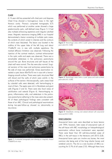
Figure 1: A computed tomography scan showing an irregularly shaped<br />
77X68X70 mm mass<br />
DISCUSSION<br />
Aneurysmal bone cysts were described as bone lesions<br />
until 1994; however, later cases of aneurysmal cysts of<br />
the soft tissue with ABC morphology on histopathological<br />
examination without bone involvement were reported.<br />
There were fewer than 20 well-documented cases of<br />
ABCs in the soft tissue in the literature until 2012 (3,8).<br />
Aneurysmal bone cysts can involve any bones, the most<br />
common site being end of the long tubular bones and<br />
vertebral column. The rib is a rare location of ABCs accounting<br />
for 2.7% of all cases (7,9). Although there have<br />
<strong>Cilt</strong> - Vol. 6 <strong>Sayı</strong> - No. 1 26


