Acta Soc. Zool. Bohem. 66: 85–97, 2002ISSN 1211-376XOn <strong>the</strong> morphology and surface ultrastructure of some parasitic nematodes(Nematoda) of birds (Aves)Denisa FRANTOVÁInstitute of Parasitology, Academy of Sciences of <strong>the</strong> <strong>Czech</strong> <strong>Republic</strong>, Branišovská 31,CZ–370 05 eské Budjovice, <strong>Czech</strong> <strong>Republic</strong>Department of Parasitology, Faculty of Biological Sciences, University of South Bohemia, Branišovská 31,CZ–370 05 eské Budjovice, <strong>Czech</strong> <strong>Republic</strong>Received February 1, 2001; accepted October 16, 2001Published June 28, 2002Abstract. The morphology of four species of avian nematodes was studied us<strong>in</strong>g scann<strong>in</strong>g electronmicroscopy (SEM): Porrocaecum depressum (Zeder, 1800) from Buteo buteo; third- and fourth-stagelarvae of Porrocaecum semiteres (Zeder, 1800) from Larus ridibundus and Turdus philomelos; Acuariaanthuris (Rudolphi, 1819) from Corvus frugilegus; Cosmocephalus obvelatus (Crepl<strong>in</strong>, 1825) from Larusridibundus. The exam<strong>in</strong>ation of <strong>the</strong> head end of adult Porrocaecum depressum and fourth-stage larvae ofP. semiteres revealed a pattern of labial papillae that is typical of ascaridoid genera. The structure of <strong>the</strong>head end of third- and fourth-stage larvae of P. semiteres seems to be identical with that of <strong>the</strong> related P.ensicaudatum, which occurs <strong>in</strong> <strong>the</strong> same species of <strong>in</strong>termediate and def<strong>in</strong>ite host. The fourth-stage larvaeof P. semiteres was redescribed. A detailed exam<strong>in</strong>ation of <strong>the</strong> oral region of Acuaria anthuris revealed teethat <strong>the</strong> anterior end of <strong>the</strong> cordons, which may serve to host tissue damage dur<strong>in</strong>g feed<strong>in</strong>g. The deirids of A.anthuris are very small, with a bicuspid tip.Morphology, surface ultrastructure, nematode, parasite, AvesINTRODUCTIONThe taxonomy of parasitic nematodes of birds (Aves) is often based on <strong>in</strong>complete descriptionsand draw<strong>in</strong>gs. Dur<strong>in</strong>g a recent study of some materials from birds (Frantová 2002) provided by <strong>the</strong>Helm<strong>in</strong>thological Collection of <strong>the</strong> Institute of Parasitology, Academy of Sciences of <strong>the</strong> <strong>Czech</strong><strong>Republic</strong> (ASCR), <strong>in</strong> eské Budjovice, morphology of four common species of nematodes wasstudied <strong>in</strong> detail, us<strong>in</strong>g light microscopy and scann<strong>in</strong>g electron microscopy (SEM): Porrocaecumdepressum, P. semiteres, Acuaria anthuris and Cosmocephalus obvelatus.Porrocaecum depressum is a common parasite of birds of prey (Falconiformes, Strigiformes)and its morphology had been described several times us<strong>in</strong>g light microscopy (see Mozgovoy1953, Hartwich 1975). P. semiteres is typical of birds of <strong>the</strong> order Charadriiformes. It frequentlyoccurs as a third- or fourth-stage larva <strong>in</strong> <strong>the</strong> digestive tract of small passer<strong>in</strong>es (especially Turdidae),but is unable to mature <strong>in</strong> <strong>the</strong>se atypical hosts (Iygis 1967). Descriptions and draw<strong>in</strong>gs ofthird-stage larvae are <strong>in</strong>cluded <strong>in</strong> <strong>the</strong> works of Mozgovoy & Bishaeva (1959), Iygis (1967) andMoravec (1971). There are few accounts of <strong>the</strong> morphology of <strong>the</strong> fourth-stage larvae (Iygis 1967).A related species, P. ensicaudatum (Zeder, 1800), also uses small passer<strong>in</strong>es (especially Turdidae)as def<strong>in</strong>itive hosts, but is also found as larvae <strong>in</strong> Charadriiformes (Iygis 1967, 1970). Consequently,larvae of P. ensicaudatum and P. semiteres can occur <strong>in</strong> <strong>the</strong> same species of avian host. They canbe dist<strong>in</strong>guished by <strong>the</strong> ratio of <strong>the</strong> stomach to <strong>the</strong> <strong>in</strong>test<strong>in</strong>al caecum length (Supryaga & Supryaga1971, Baruš et al. 1978b). In <strong>the</strong> genus Porrocaecum Railliet et Henry, 1912, only P. ensicaudatumwas exam<strong>in</strong>ed us<strong>in</strong>g SEM (Wharton 1978, Baruš et al. 1983, McNeill & Anderson 1990a, b).85
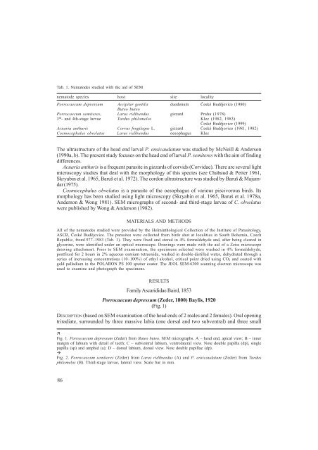
Tab. 1. Nematodes studied with <strong>the</strong> aid of SEMnematode species host site localityPorrocaecum depressum Accipiter gentilis duodenum eské Budjovice (1980)Buteo buteoPorrocaecum semiteres, Larus ridibundus gizzard Praha (1978)3 rd - and 4th-stage larvae Turdus philomelos Klec (1982, 1983)eské Budjovice (1999)Acuaria anthuris Corvus frugilegus L. gizzard eské Budjovice (1981, 1982)Cosmocephalus obvelatus Larus ridibundus oesophagus KlecThe ultrastructure of <strong>the</strong> head end larval P. ensicaudatum was studied by McNeill & Anderson(1990a, b). The present study focuses on <strong>the</strong> head end of larval P. semiteres with <strong>the</strong> aim of f<strong>in</strong>d<strong>in</strong>gdifferences.Acuaria anthuris is a frequent parasite <strong>in</strong> gizzards of corvids (Corvidae). There are several lightmicroscopy studies that deal with <strong>the</strong> morphology of this species (see Chabaud & Petter 1961,Skryab<strong>in</strong> et al. 1965, Baruš et al. 1972). The cordon ultrastructure was studied by Baruš & Majumdar(1975).Cosmocephalus obvelatus is a parasite of <strong>the</strong> oesophagus of various piscivorous birds. Itsmorphology has been studied us<strong>in</strong>g light microscopy (Skryab<strong>in</strong> et al. 1965, Baruš et al. 1978a,Anderson & Wong 1981). SEM micrographs of second- and third-stage larvae of C. obvelatuswere published by Wong & Anderson (1982).MATERIALS AND METHODSAll of <strong>the</strong> nematodes studied were provided by <strong>the</strong> Helm<strong>in</strong>thological Collection of <strong>the</strong> Institute of Parasitology,ASCR, eské Budjovice. The parasites were collected from birds shot at localities <strong>in</strong> South Bohemia, <strong>Czech</strong><strong>Republic</strong>, from1977–1983 (Tab. 1). They were fixed and stored <strong>in</strong> 4% formaldehyde and, after be<strong>in</strong>g cleared <strong>in</strong>glycer<strong>in</strong>e, were identified under an optical microscope. Draw<strong>in</strong>gs were made with <strong>the</strong> aid of a Zeiss microscopedraw<strong>in</strong>g attachment. Prior to SEM exam<strong>in</strong>ation, <strong>the</strong> specimens selected were washed <strong>in</strong> 4% formaldehyde,postfixed for 2 hours <strong>in</strong> 2% aqueous osmium tetraoxide, washed <strong>in</strong> double-distilled water, dehydrated through aseries of <strong>in</strong>creas<strong>in</strong>g concentrations (10–100%) of ethyl alcohol, critical po<strong>in</strong>t dried us<strong>in</strong>g CO2 and coated withgold palladium <strong>in</strong> <strong>the</strong> POLARON PS 100 sputter coater. The JEOL SEM-6300 scann<strong>in</strong>g electron microscope wasused to exam<strong>in</strong>e and photograph <strong>the</strong> specimens.RESULTSFamily Ascarididae Baird, 1853Porrocaecum depressum (Zeder, 1800) Baylis, 1920(Fig. 1)DESCRIPTION (based on SEM exam<strong>in</strong>ation of <strong>the</strong> head ends of 2 males and 2 females). Oral open<strong>in</strong>gtriradiate, surrounded by three massive labia (one dorsal and two subventral) and three smallFig. 1. Porrocaecum depressum (Zeder) from Buteo buteo. SEM micrographs. A – head end, apical view; B – <strong>in</strong>nermarg<strong>in</strong> of labium with detail of teeth; C – subventral labium, ventrolateral view. Note double papilla (dp), s<strong>in</strong>glepapilla (sp) and amphid (a); D – dorsal labium, dorsal view. Note double papillae (dp).Fig. 2. Porrocaecum semiteres (Zeder) from Larus ridibundus (A) and P. ensicaudatum (Zeder) from Turdusphilomelos (B). Third-stage larvae, lateral view. Scale bar <strong>in</strong> mm.86
- Page 2: Acta Soc. Zool. Bohem. 66: 81-84, 2
- Page 5: REFERENCESBERAN L. & HORSÁK M. 199
- Page 10: Tab. 2. The dimensions (mm) of thir
- Page 14 and 15: Family Acuariidae Railliet, Henry e
- Page 16 and 17: Fig. 7. Cosmocephalus obvelatus (Cr
- Page 18 and 19: A c k n o w l e d g e m e n t sThe
- Page 20 and 21: Acta Soc. Zool. Bohem. 66: 99-119,
- Page 22 and 23: Figs 1-10. Harpalus (Harpalus) atra
- Page 24 and 25: Figs 11-19. Harpalus (Harpalus) cis
- Page 26 and 27: Figs 20-27. Harpalus (Harpalus) lut
- Page 28 and 29: Figs 28-35. Harpalus (Harpalus) pic
- Page 30 and 31: Figs 36-44. Harpalus (Harpalus) sax
- Page 32 and 33: Figs 45-53. Harpalus (Harpalus) ser
- Page 34 and 35: Figs 54-61. Harpalus (Harpalus) sol
- Page 36 and 37: Figs 62-70. Harpalus (Harpalus) xan
- Page 38 and 39: Based on the study of our larval ma
- Page 40 and 41: EMDEN F. I. van 1942: A key to the
- Page 42 and 43: Acta Soc. Zool. Bohem. 66: 121-140,
- Page 44 and 45: and D. papillifera (Fuhrmann, 1908)
- Page 46 and 47: about 1 mm) with only a few eggs in
- Page 48 and 49: The number of catfish examined each
- Page 50 and 51: number (3.65) of trematodes with ma
- Page 52 and 53: cestode was distinctly associated w
- Page 54 and 55: from individual months. Tab. 2 show
- Page 56 and 57:
number (5.57) of males occurred in
- Page 58 and 59:
intermediate host, copepod, catfish
- Page 60 and 61:
CHUBB J. C. 1982: Seasonal occurren
- Page 62 and 63:
Acta Soc. Zool. Bohem. 66: 141-150,
- Page 64 and 65:
efore distal angle of discoidal tri
- Page 66 and 67:
cells between them and IR2 or MA”
- Page 68 and 69:
character is less pronounced in Oli
- Page 70 and 71:
REFERENCESBACHMAYER F. 1952: Fossil
- Page 72 and 73:
Acta Soc. Zool. Bohem. 66: 151-160,
- Page 74 and 75:
elow) and a 170×90×70 type was us
- Page 76 and 77:
the brood, first into heaps near th
- Page 78 and 79:
trophallaxis until the termination
- Page 80 and 81:
4. The queen that was accepted was