PELONews April/May 2019

You also want an ePaper? Increase the reach of your titles
YUMPU automatically turns print PDFs into web optimized ePapers that Google loves.
PELONEWS<br />
WITH FOCUS ON PRIMARY CELLS<br />
PELOBiotech Newsletter <strong>April</strong> /<strong>May</strong> <strong>2019</strong><br />
“Science without religion<br />
is lame, religion without<br />
science is blind.“<br />
-Albert Einstein, Aus meinen späten<br />
Jahren, Selbstporträt, 1936) -<br />
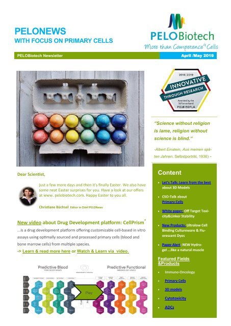
Dear Scientist,<br />
just a few more days and then it's finally Easter. We also have<br />
some neat Easter surprises for you. Have a look at our offers<br />
at www. pelobiotech.com. Happy Easter to you all.<br />
Christiane Büchsel Editor in Chief <strong>PELONews</strong><br />
New video about Drug Development platform: CellPrism ®<br />
...is a drug development platform offering customizable cell-based in vitro<br />
assays using optimally sourced and processed primary cells (blood and<br />
bone marrow cells) from multiple species.<br />
-> Learn & read more here or Watch & Learn via video.<br />
Content<br />
Let‘s Talk: Learn from the best<br />
about 3D Models<br />
CSO-Talk about<br />
Primary Cells<br />
White paper: Off Target Toxicity&Linker<br />
Stability<br />
New Products: Ultralow Cell<br />
Binding Cultureware & Fluorescent<br />
Dyes<br />
Paper Alert: NEW Hydrogel<br />
...like a natural muscle<br />
Featured Fields<br />
&Products<br />
Play<br />
<br />
<br />
<br />
<br />
<br />
Immuno-Oncology<br />
Primary Cells<br />
3D models<br />
Cytotoxicity<br />
ADCs
<strong>PELONews</strong><br />
We have talked: Optimised Cancer<br />
Treatments and EV research with 3D Models<br />
To an expert- talk about “Optimised Cancer Treatments and EV research with 3D Models”<br />
invited PELOBIotech GmbH and its Swiss partner abc biopply at the IZB Sky-Lounge end of<br />
March <strong>2019</strong>. Around 20 scientists from Bavarian biotech companies and institutes such as<br />
TUM, LMU, Helmholtz Centre in Munich and Fraunhofer in Regensburg discussed with highlevel<br />
cancer research experts Dr. med. Andreas Thomsen and Dr. Irina Nazarenko from Universitätsklinikum<br />
Freiburg about new models for tissue-specific interaction of cells and how<br />
they optimize their treatments.<br />
First Dr. Marco Leu, Co-Owner abc biopply, gave a short overview of a novel and innovative 3D microwell array in<br />
the analysis of adhesion independent micro-organoids, named 3D CoSeedis. “I am glad abc biopply was able to<br />
collaborate with Dr. Andreas Thomsen and to further develop this unique and novel technology. We are proud<br />
to have recently released the first standardizes systems it to the scientific world” says Dr. Leu. 3DCoSeedis allows<br />
the support of the growth of feeder cells in the formation of 3D cell constructs that are not in direct contact with<br />
the test cells themselves.<br />
„It’s totally cool!“<br />
Both key speakers are already working with the new system 3D CoSeedis, as in fact Dr.<br />
Thomsen invented the system himself. His lecture “Creating Unique Microenvironments<br />
in 3D Models” showed a broad range of applications and inspired the researchers on their<br />
projects on tissue-specific interaction of cells. A lot of wows he earned showing his results<br />
in comparison with standard 2d methods and the new 3D model. “It makes it so much easier”,<br />
the Freiburg radio-oncologist says. His survey “Do you enjoy colony formation assays?” performed among<br />
colleagues made the audience laugh. They knew almost everybody dislike it and now are given a possibility to<br />
work much more efficient.<br />
A lot of interest had the lecture by Dr. Irina Nazarenko: Studying Intercellular Communication by the mean of<br />
Extracellular Vesicles in 3D Models. She talked about the impact environment on production, cargo, and function<br />
of extracellular vesicles (EVs) in several cancer models. EVs mediate intercellular communication between tumor<br />
and tumor stroma, tumor and immune system, playing a pivotal role in metastases, immunosuppression, angiogenesis<br />
and other systemic effects in progressing tumor. In her experience 3DCoSeedis TM is applicable for EV isolation,<br />
analytic and functional characterization. Using the 3D microwell array more EVs/cell can be isolated in a<br />
considerably more cost- and efforts- efficient manner than 2D culture. “My co-workers and me love working with<br />
it”, Dr. Nazarenko says, as the cells are growing in more physiological environment and producing more EVs. 3D<br />
environments are likely to trigger release of smaller EVs with increased amounts of certain miRNAs and decreased<br />
amounts of their target proteins. She also emphasized that functional impact of 3D environment on EVs<br />
differs between cell lines and should be individually analyzed.<br />
„We are already switching!“<br />
Dr. Nazarenko is more than convinced by the 3D CoSeedis system. Asked whether she would<br />
switch if she had the opportunity: “Definitely, it´s genius, totally cool. So, where it makes<br />
sense, we are already switching, step by step”, she outlines. One of the participants confirms<br />
her point of view: “We are more and more searching for 3D models, also in EV research. So<br />
this system sounds very promising to us.”<br />
Peek into presentation Dr. Irina Narzarenko and download paper here.<br />
Download presentation Dr. med. Andreas Thomsen, and download paper here:<br />
Peek into presentation Dr. Marco Leu
Here some<br />
impressions of<br />
this great<br />
3D model<br />
symposium end<br />
of March.
<strong>PELONews</strong><br />
Primary Cells<br />
CSO-Talk: Why Isolation and Cultivation of PCs is not always easy<br />
by Dr. Lothar Steeb<br />
Primary cell culture is the only basis for developing alternative test systems and methods. Theoretically, if one<br />
reads the method parts of publications, primary cells can be obtained and cultured quite easily.<br />
However, anyone who has already dealt with this topic, the isolation and cultivation of primary cells from human<br />
tissues, will quickly notice that there are some challenges that are not mentioned in every methods and that these<br />
cells can only show their advantages if they are cultivated differently from permanent cell lines with special microenvironment<br />
conditions. An endothelial cell does not grow in an environment for neurons or epithelial cells and<br />
vice versa. In addition, the processing of donor tissue must always be based on infectious material, which requires<br />
stringent methodological work. Nevertheless, the advantages of primary cells are so great that it is worth working<br />
with them.<br />
Primary cells are of particular importance with regard to<br />
- metabolism<br />
- morphology<br />
- physiology<br />
- Genetic characteristics<br />
- demeanor.<br />
The most in vivo state is the one closest to the body and therefore form the link<br />
CSO: Dr Lothar Steeb<br />
between the body and ex vivo cultures. Cross-contaminations and false characterizations can also be excluded.<br />
The markers are usually unambiguous. Another disadvantage of cell lines is that they often differ genetically and<br />
phenotypically from their original tissue. In contrast, primary cells retain many of the important markers and functions<br />
observed in vivo. For example, endothelial cell lines lack various functional markers, while primary endothelial<br />
cells retain these important characteristics. In animal cells, in particular, an advantage is extremely important<br />
because it enables cross-generational and genetically modified investigations.<br />
Many years ago, PELOBiotech was one of the first suppliers of cells that recognised the increasing demands on cell<br />
culture media and the cells‘ environment represent an opportunity not only to maintain the individuality of cells,<br />
but also to increase the comparability of the results. By creating defined and standardised environmental conditions<br />
the results from different working groups are more stringent and also more reliable. They cannot be influenced<br />
by unknown factors such as serum components or raw products such as matrices or extracts.<br />
With its wide range of products, PELOBiotech offers exactly these opportunities. We are happy to help find the cells you are<br />
looking for (there are currently more than a thousand possibilities open to you), the media selection and all the tools you need<br />
for optimal primary cell cultivation. PELOBiotech stands for solutions and assistance for all questions regarding primary cell<br />
culture and supports in choosing the most efficient, cost-effective and optimized solution from our almost 30,000 products.<br />
Want to know more, click on the following links:<br />
Animal Cells, Media & Tools:<br />
Human cells,Media & Tools
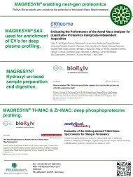
Assays&Arrays<br />
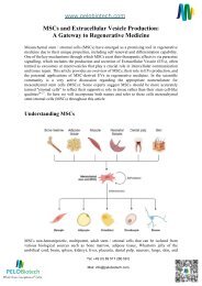
WHITE PAPER | Off-Target Toxicity and Linker Stability<br />
By Emer Clarke, CSO ReachBio Research Labs<br />
CSO Dr. Emer Clarke, PhD<br />
The Primary Cell Experts<br />
ReachBio Research Labs’ scientists use human primary cells<br />
every day in our contract services lab for high value projects for<br />
clients in the pharma and biotechnology industries. Their extensive<br />
experience in handling and using human primary cells is<br />
used in the production of our human cell products, resulting in<br />
high quality cells that you can rely on for your own important<br />
experiments. We use Cryostor CS5 serum-free cryopreservation<br />
medium to ensure high post-thaw viability and overfill each vial<br />
to ensure you always get at least the number of cells we advertise<br />
on the vial when they are thawed correctly.<br />
-> Ask us at PELOBiotech for more.<br />
F<br />
Antibody drug conjugates (ADCs) combine the benefits of both therapeutic monoclonal antibodies (mAb) and potent<br />
small molecule cytotoxic drugs. These moieties are connected through linkers that should be stable within<br />
systemic circulation, while maintaining the ability to cleave within the mAb-targeted cells to release highly specific<br />
payloads.<br />
In addition to the targeted killing of antigen-positive tumor cells, some ADCs appear to have significant off-target<br />
effects, causing neutropenia and thrombocytopenia, which have necessitated some clinical trials being terminated<br />
early. However, the success of some ADCs in treating diseases has stimulated the continued research of these<br />
constructs.<br />
ReachBio has two distinct assay platforms to assess cytotoxicity caused by ADCs<br />
In the initial platform, they can evaluate off-target toxicity using colony forming cell (CFC) assays to assess effects<br />
on erythroid, myeloid, or megakaryocyte progenitors using marrow derived from human, NHP, rat, and mouse.<br />
Our second platform addresses linker stability. They have adopted the neutrophil differentiation assay described<br />
by Zhao et al (2017), whereby neutrophils cause their own demise by releasing enzymes which cleave the payload<br />
from the antibody and thus kill developing neutrophils.<br />
Download Emer`s White paper here
<strong>PELONews</strong><br />
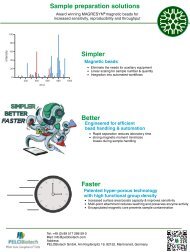
New Products<br />
Ultralow Cell Binding Cultureware<br />
HydroCell is a low cell biding cultureware. On its surface, super hydrophilic<br />
polymer was immobilized at nano-thickness. This hydrophilic character helps<br />
the formation of embryoid bodies of ES/iPS cell or Spheroid in culture.: Compared<br />
to other brands' low cell binding cultureware, HydroCell shows lower<br />
cell binding.<br />
<br />
• Super hydrophilic polymer is immobilized to the surface of the cultureware<br />
at nano-thickness.<br />
• Lowest attachment rate among similar competitive brands.<br />
• Three formats : Microplates, Dishes and Flask types.<br />
Fig.1 Overview of HydroCell surface<br />
<br />
• Formation of embryoid bodies of ES cells in culture<br />
• Formation of anchorage-independent colonies<br />
• Differentiation such as cartilage (spheroid formation)<br />
• Culture & storage of macrophage/immune cells<br />
• Screening of anti-cancer drugs (replacement for soft agar assays)<br />
<br />
Fig.2 Comparison to other brand products<br />
Mouse macrophages were incubated for 3 days in (1) HydroCell, (2) another brand's product and (3) non-treated cultureware. Only HydroCell allowed<br />
uniform suspension culture (>99% of the cells remained in suspension), simple cell dissociation and harvesting by pipetting.<br />
For 3D<br />
Cell Culture<br />
Fig. Stem cell sphere formation iPS spheres are formed on HydroCell (left) or other brand low cell binding dish.A complete sphere was surely formed on<br />
HydroCell, while adhesion was observed on other low cell binding dish. (Data provided by A. Umezawa, National Center for Child Health and Development)<br />
For more infos and prices, just call us: 0049 89 517286 59–0
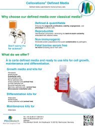
Reversible Fluorescent Dye for Nucleus<br />
NucleoSeeing is a novel live cell imaging probe which emits green fluorescence<br />
by binding to DNA specifically. NucleoSeeing can be used for not only in animal cells or tissues, but<br />
also Guard cell of Arabidopsis thaliana with high S/N ratio. Moreover, NucleoSeeing can be used as a pH sensor in<br />
nucleus. This product has been commercialized under the license from Nagoya Institute of Technology.<br />
<br />
NucleoSeeing is composed of green fluorescent dye and DNA binding tag. Under DNA free condition, Nucleoseeing<br />
is in folded conformation and no fluorescent is emitted. However, once it binds to DNA, the conformation<br />
will change andemitts green fluorescence.<br />
Ex/<br />
Em<br />
Fluoresce<br />
nce<br />
Phototoxicity<br />
Nucleu<br />
s<br />
Spe<br />
cifici<br />
ty<br />
Cell<br />
Me<br />
m.<br />
Permea<br />
bilit<br />
y<br />
Cyto-<br />
toxi-<br />
city<br />
Live Fixed<br />
NucleoS<br />
Green Low Yes Yes Low Yes Yes<br />
488/<br />
520<br />
eeing<br />
Hoech 350/<br />
st 461<br />
DAPI 350/<br />
461<br />
Company<br />
X<br />
Company<br />
Y<br />
485/<br />
498<br />
646/<br />
680<br />
Blue High Yes Yes<br />
Hig<br />
h<br />
Yes Yes<br />
Blue High Yes No N/A No Yes<br />
Green Low No Yes N/A Yes Yes<br />
Red Low Yes Yes<br />
Hig<br />
h<br />
Yes Yes<br />
<br />
Comparison table of dyes for nucleus<br />
Fig.1 Staining of nucleus in various cultured cells<br />
References<br />
1. Nakamura, A., et al., Chem. Commun., 50, 6149-6152 (2014)<br />
Hoechst tagging: a modular strategy to design synthetic fluorescent<br />
probes for live-cell nucleus imaging.<br />
2.<br />
2. Ueda, M., et al., ACS Cent. Sci., 3 (5), 462-472 (2017)<br />
Noncanonical function of a small-molecular virulence factor coronatine<br />
against plant immunity: an in vivo raman imaging approach.<br />
3 papers is equal to NucleoSeeing.<br />
3. Nakamura, A. and Tsukiji, S., Bioorg. Med. Chem. Lett., 27 (14),<br />
3127-3130 (2017)Ratiometric fluorescence imaging of nuclear pH in<br />
living cells using hoechst-tagged fluorescein. * hoeAc2FL in above<br />
More Product Information/Datasheet here.
<strong>PELONews</strong><br />
PELO<br />
Academy<br />
You like to learn from<br />
the best but missed<br />
one of our LIVE lectures?<br />
No worries.<br />
Sign up for<br />
PELOAcademy and we<br />
send you regular invitations<br />
to our events.<br />
Plus if you sign in for<br />
our past webinars you<br />
can watch the recording<br />
immediatley. You<br />
are very welcome.<br />
<strong>PELONews</strong>: Refer a friend<br />
We are convinced that we convince you with our expertise and our outstanding service.<br />
So if you are already a PELO-Fan, please recommend us to a colleague and<br />
friend – you will get a coupon and nice thank-you reward. More details? Call us now.<br />
Want to stay tuned in<br />
and informed? Then<br />
sign up for our regular<br />
Webinar Invitation<br />
Reminder. Just check<br />
the box with the infos<br />
you like to receive and<br />
sign up here.<br />
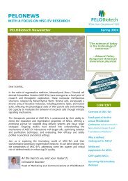
Paper Alert from our partner Noviocell<br />
New hydrogel stiffens and<br />
softens like a natural muscle<br />
„A new synthetic hydrogel that becomes up to 50 times stiffer upon heating<br />
just a few degrees has been developed by researchers at Radboud University<br />
in the Netherlands. The stiffening process is reversible, and the team believes<br />
that the hydrogel could be used in a range of new applications including tissue<br />
engineering. The controlled stiffening or softening of materials is very common<br />
in biology, playing roles in processes such as muscle contraction, tissue<br />
fibrosis, the enzymatic degradation of tissues and tumour formation. It involves<br />
a biological cell converting chemical energy into mechanical stresses,<br />
which cause the cell’s cytoskeleton to stiffen with stress. However, mimicking<br />
this ability to stiffen and soften in a synthetic material such as a hydrogel has<br />
proven very tricky to achieve.<br />
Read more here.<br />
We like your feedback – tell us what you love, don’t like so much and what you would<br />
like to get, please. Just reply Please update your subscription anytime, as we like to<br />
comply to the new GDPR guidelines.<br />
How to<br />
reach us<br />
If you need any further<br />
assistance or if you like<br />
what you see, tell us:<br />
PELOBiotech GmbH<br />
Klopferspitz 19 82152<br />
Planegg | Germany<br />
Tel.: +49 89 517286<br />
59 0<br />
info@pelobiotech.com<br />
| www.pelobiotech.com<br />
Managing Directors:<br />
Dr. Peter Frost,<br />
Dr Lothar Steeb