Physiological Basis of the Electrocardiogram
Physiological Basis of the Electrocardiogram
Physiological Basis of the Electrocardiogram

You also want an ePaper? Increase the reach of your titles
YUMPU automatically turns print PDFs into web optimized ePapers that Google loves.
20 The <strong>Physiological</strong> <strong>Basis</strong> <strong>of</strong> <strong>the</strong> <strong>Electrocardiogram</strong><br />
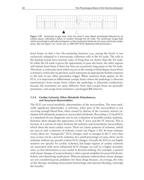
Figure 1.23 Ventricular escape beat. Note <strong>the</strong> atrial P wave (black arrowhead) followed by an<br />
evident pause, indicating a failure to conduct through <strong>the</strong> AV node. The ventricular escape beat<br />
(white arrowhead) is a fail-safe mechanism so that conduction blocks do not cause ventricular cardiac<br />
arrest. Also see Figure 1.21. (From: [2]. c○ 2004 MIT OCW. Reprinted with permission.)<br />
heart beats, so that a key life-sustaining function (e.g., pacing <strong>the</strong> heart) is not<br />
exclusively relegated to a microscopic collection cells in <strong>the</strong> SA node. The cells in<br />
<strong>the</strong> backup system have intrinsic rates <strong>of</strong> firing that are slower than <strong>the</strong> SA node.<br />
So while <strong>the</strong> SA node is given <strong>the</strong> opportunity to pace <strong>the</strong> heart, <strong>the</strong> o<strong>the</strong>r regions<br />
will initiate heart beats if <strong>the</strong>re has been an excessively long pause in <strong>the</strong> SA node.<br />
Therefore, a ventricular beat which occurs in <strong>the</strong> setting <strong>of</strong> third-degree heart block<br />
is termed a ventricular escape beat, and it represents an appropriate healthy response<br />
to <strong>the</strong> lack <strong>of</strong> any o<strong>the</strong>r pacemaker trigger. When nonsinus beats appear on <strong>the</strong><br />
ECG, it is important to differentiate ectopic beats (where <strong>the</strong> pathology is aberrant<br />
automaticity) from escape beats (where <strong>the</strong> pathology is abnormal conduction),<br />
because <strong>the</strong> treatments are quite different. Note that ectopic beats are generally<br />
premature, and escape beats terminate a prolonged RR interval.<br />
1.3.4 Cardiac Ischemia, O<strong>the</strong>r Metabolic Disturbances,<br />
and Structural Abnormalities<br />
The ECG can reveal metabolic abnormalities <strong>of</strong> <strong>the</strong> myocardium. The most medically<br />
significant abnormality is ischemia, when part <strong>of</strong> <strong>the</strong> myocardium is not<br />
receiving enough blood flow, <strong>of</strong>ten caused by disease <strong>of</strong> <strong>the</strong> coronary arteries (ischemia<br />
will ultimately progress to myocardial cell death). Recording a 12-lead ECG<br />
is a standard-<strong>of</strong>-care diagnostic test in any evaluation <strong>of</strong> possible cardiac ischemia.<br />
Ischemia <strong>of</strong>ten changes <strong>the</strong> appearance <strong>of</strong> <strong>the</strong> T wave and <strong>the</strong> ST interval. This is<br />
because <strong>of</strong> a current <strong>of</strong> injury between <strong>the</strong> ischemic and nonischemic myocardium,<br />
which alters <strong>the</strong> main cardiac vector. There are classic patterns <strong>of</strong> ischemia, which<br />
are seen in only a minority <strong>of</strong> ischemic events (see Figure 1.24). In most ischemic<br />
events, <strong>the</strong>re are “nonspecific” ECG changes, such as changes in <strong>the</strong> T wave that<br />
may or may not be caused by ischemia. In a small percentage <strong>of</strong> cases, <strong>the</strong>re can be<br />
ischemia without any grossly evident ECG changes. Overall, <strong>the</strong> ECG is not highly<br />
sensitive nor specific for cardiac ischemia, but larger regions <strong>of</strong> cardiac ischemia<br />
are associated with more substantial ECG changes (as well as a higher mortality<br />
rate), so this information is very useful in decision-making. For example, a patient<br />
with classic changes <strong>of</strong> acute ischemia is <strong>of</strong>ten a good candidate for powerful thrombolytic<br />
(clot dissolving) medication. Whereas patients without those classic changes<br />
are not considered good candidates for <strong>the</strong>se drugs because, on average, <strong>the</strong> risks<br />
<strong>of</strong> <strong>the</strong> <strong>the</strong>rapy, including intracranial hemorrhage and internal bleeding, outweigh<br />
<strong>the</strong> benefits.