Physiological Basis of the Electrocardiogram
Physiological Basis of the Electrocardiogram
Physiological Basis of the Electrocardiogram

You also want an ePaper? Increase the reach of your titles
YUMPU automatically turns print PDFs into web optimized ePapers that Google loves.
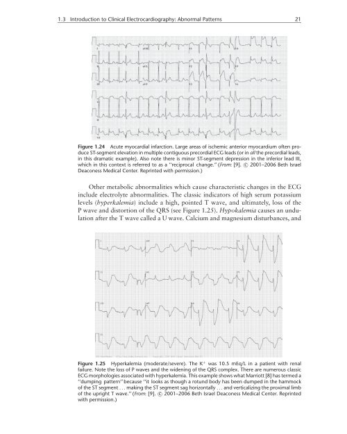
1.3 Introduction to Clinical Electrocardiography: Abnormal Patterns 21<br />
Figure 1.24 Acute myocardial infarction. Large areas <strong>of</strong> ischemic anterior myocardium <strong>of</strong>ten produce<br />
ST-segment elevation in multiple contiguous precordial ECG leads (or in all <strong>the</strong> precordial leads,<br />
in this dramatic example). Also note <strong>the</strong>re is minor ST-segment depression in <strong>the</strong> inferior lead III,<br />
which in this context is referred to as a ‘‘reciprocal change.” (From: [9]. c○ 2001–2006 Beth Israel<br />
Deaconess Medical Center. Reprinted with permission.)<br />
O<strong>the</strong>r metabolic abnormalities which cause characteristic changes in <strong>the</strong> ECG<br />
include electrolyte abnormalities. The classic indicators <strong>of</strong> high serum potassium<br />
levels (hyperkalemia) include a high, pointed T wave, and ultimately, loss <strong>of</strong> <strong>the</strong><br />
P wave and distortion <strong>of</strong> <strong>the</strong> QRS (see Figure 1.25). Hypokalemia causes an undulation<br />
after <strong>the</strong> T wave called a U wave. Calcium and magnesium disturbances, and<br />
Figure 1.25 Hyperkalemia (moderate/severe). The K + was 10.5 mEq/L in a patient with renal<br />
failure. Note <strong>the</strong> loss <strong>of</strong> P waves and <strong>the</strong> widening <strong>of</strong> <strong>the</strong> QRS complex. There are numerous classic<br />
ECG morphologies associated with hyperkalemia. This example shows what Marriott [8] has termed a<br />
‘‘dumping pattern’’ because ‘‘it looks as though a rotund body has been dumped in <strong>the</strong> hammock<br />
<strong>of</strong> <strong>the</strong> ST segment ...making <strong>the</strong> ST segment sag horizontally ...and verticalizing <strong>the</strong> proximal limb<br />
<strong>of</strong> <strong>the</strong> upright T wave.’’ (From: [9]. c○ 2001–2006 Beth Israel Deaconess Medical Center. Reprinted<br />
with permission.)