Physiological Basis of the Electrocardiogram
Physiological Basis of the Electrocardiogram
Physiological Basis of the Electrocardiogram

Create successful ePaper yourself
Turn your PDF publications into a flip-book with our unique Google optimized e-Paper software.
1.2 The Physical <strong>Basis</strong> <strong>of</strong> Electrocardiography 7<br />
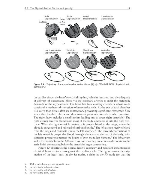
Figure 1.4 Trajectory <strong>of</strong> a normal cardiac vector. (From: [2]. c○ 2004 MIT OCW. Reprinted with<br />
permission.)<br />
<strong>the</strong> cardiac tissue, <strong>the</strong> heart’s electrical rhythm, valvular function, and <strong>the</strong> adequacy<br />
<strong>of</strong> delivery <strong>of</strong> oxygenated blood via <strong>the</strong> coronary arteries to meet <strong>the</strong> metabolic<br />
demands <strong>of</strong> <strong>the</strong> myocardium. The heart has four cavitary chambers whose walls<br />
consist <strong>of</strong> a mechanical syncytium <strong>of</strong> myocardial cells. At <strong>the</strong> exit <strong>of</strong> each chamber<br />
is a valve that closes after its contraction, preventing significant retrograde flow<br />
when <strong>the</strong> chamber relaxes and downstream pressures exceed chamber pressures.<br />
The right heart includes a small atrium leading into a larger right ventricle. 6 The<br />
right atrium receives blood from most <strong>of</strong> <strong>the</strong> body and feeds it into <strong>the</strong> right ventricle.<br />
When <strong>the</strong> right ventricle contracts, it propels blood to <strong>the</strong> lungs, where <strong>the</strong><br />
blood is oxygenated and relieved <strong>of</strong> carbon dioxide. 7 The left atrium receives blood<br />
from <strong>the</strong> lungs and conducts it into <strong>the</strong> left ventricle. 8 The forceful contractions <strong>of</strong><br />
<strong>the</strong> left ventricle propel <strong>the</strong> blood through <strong>the</strong> aorta to <strong>the</strong> rest <strong>of</strong> <strong>the</strong> body, with<br />
sufficient pressure to perfuse <strong>the</strong> brains <strong>of</strong> even <strong>the</strong> tallest humans. 9 The left atrium<br />
and left ventricle form <strong>the</strong> left heart. As noted earlier, under normal conditions <strong>the</strong><br />
atria finish contracting before <strong>the</strong> ventricles begin contracting.<br />
Figure 1.4 illustrates <strong>the</strong> normal heart’s geometry and resultant instantaneous<br />
electrical heart vectors throughout <strong>the</strong> cardiac cycle. The figure shows <strong>the</strong> origination<br />
<strong>of</strong> <strong>the</strong> heart beat (at <strong>the</strong> SA node), a delay at <strong>the</strong> AV node (so that <strong>the</strong><br />
6. With a valve known as <strong>the</strong> tricuspid valve.<br />
7. Its valve is <strong>the</strong> pulmonic valve.<br />
8. Its valve is <strong>the</strong> mitral valve.<br />
9. Its valve is <strong>the</strong> aortic valve.