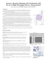
TRIzol Reagent - Invitrogen
TRIzol Reagent - Invitrogen
TRIzol Reagent - Invitrogen

Create successful ePaper yourself
Turn your PDF publications into a flip-book with our unique Google optimized e-Paper software.
This is common with skin samples. It is assumed that there is fat in these samples, and the fat micelles "try to spin to the<br />
top of the tube" during the centrifugation. In skin samples, the micelles pick up melanin pigment and cause the aqueous<br />
phase to appear colored. Fat micelles may also pick up pigment from the <strong>TRIzol</strong> itself and cause a pinkish color. If a<br />
sample is thought to contain fat, the sample homogenate in <strong>TRIzol</strong> may be centrifuged prior to addition of chloroform.<br />
The fat will appear as a clear layer at the top of the supernatant; this should be pipetted off and discarded.<br />
If a sample contains a lot of blood, the aqueous phase may appear cloudy and/or yellowish (this may be due to iron in the<br />
hemoglobin). If the centrifuge used is not cold, the organic phase will be a deeper maroon color; some of this color may<br />
come into the aqueous phase and cause it to appear orange or yellow.<br />
A pinkish aqueous phase may also be caused by over-dilution of the sample (i.e., the sample:<strong>TRIzol</strong> ratio > 1:10), as well<br />
as too much salt or protein in the sample. This can cause premature phase separation, which can be remedied by adding a<br />
bit more <strong>TRIzol</strong> to the sample. If the RNA is isolated from a pinkish aqueous phase, chances are that it will be<br />
contaminated with DNA.<br />
Precipitate At the Bottom of the tube following centrifugation after adding Chloroform (before isopropanol is added)<br />
(back to Table of Contents)<br />
(back to Protocol and Application Notes)<br />
(back to RNA Isolation)<br />
This is most likely polysaccharides or cell membranes; DNA should be in the interphase. In samples containing blood<br />
(e.g., liver), a red viscous layer may be visible on top of the pellet. This is most likely due to blood products and should<br />
not be carried over with the supernatant.<br />
Intensity of the Ribosomal RNA Bands from Prep to Prep is Inconsistent<br />
(back to Table of Contents)<br />
(back to Protocol and Application Notes)<br />
(back to RNA Isolation)<br />
Check the composition and freshness of the loading buffer. The composition should be such that the final concentration of<br />
formamide is 50% and the formamide must be fresh.<br />
No Bands around 200 bp on a Northern Blot<br />
(back to Table of Contents)<br />
(back to Protocol and Application Notes)<br />
(back to RNA Isolation)<br />
Proteoglycans co-purify with RNA in <strong>TRIzol</strong> and can be transferred onto a Northern Blot. [Schick (1995)18.4, 575]. See<br />
steps above for removing proteoglycans at the isopropanol precipitation step.<br />
A260/A280 ratio >2.0 for RNA<br />
(back to Table of Contents)<br />
(back to Protocol and Application Notes)<br />
(back to RNA Isolation)<br />
Degraded RNA can cause an increased absorbance at 260 nm.<br />
DNA Isolation with <strong>TRIzol</strong><br />
(back to Table of Contents)<br />
(back to Protocol and Application Notes)<br />
Detailed Protocol Steps for DNA Isolation with <strong>TRIzol</strong><br />
DNA Precipitation<br />
DNA Wash<br />
Redissolving the DNA<br />
Additional Notes for DNA Isolation with <strong>TRIzol</strong><br />
Alternative procedure for DNA isolation<br />
16