Ch05: Red and White Lesions of the Oral Mucosa
Ch05: Red and White Lesions of the Oral Mucosa
Ch05: Red and White Lesions of the Oral Mucosa

Create successful ePaper yourself
Turn your PDF publications into a flip-book with our unique Google optimized e-Paper software.
<strong>Red</strong> <strong>and</strong> <strong>White</strong> <strong>Lesions</strong> <strong>of</strong> <strong>the</strong> <strong>Oral</strong> <strong>Mucosa</strong> 115<br />
A<br />
B<br />
FIGURE 5-40 A, Fordyce’s granules, appearing as multiple yellowish<br />
white papules <strong>and</strong> <strong>of</strong>ten seen as aggregates in <strong>the</strong> buccal mucosa. B,<br />
Photomicrograph <strong>of</strong> Fordyce’s granules, which microscopically are identical to<br />
normal sebaceous gl<strong>and</strong>s. (Hematoxylin <strong>and</strong> eosin, ×20 original magnification)<br />
to rupture <strong>and</strong> disappear spontaneously. The eponyms<br />
“Epstein’s pearls” <strong>and</strong> “Bohn’s nodules” have both been used<br />
to describe odontogenic cysts <strong>of</strong> dental lamina origin, but<br />
<strong>the</strong>se terms are not considered to be accurate. Epstein originally<br />
described keratin-filled nodules found along <strong>the</strong> midpalatal<br />
region, probably derived from entrapped epi<strong>the</strong>lium<br />
along <strong>the</strong> lines <strong>of</strong> fusion <strong>of</strong> <strong>the</strong> palatal processes. These are considered<br />
quite rare. Bohn’s nodules are thought to be keratinfilled<br />
cysts scattered across <strong>the</strong> palate but most plentiful along<br />
<strong>the</strong> junction <strong>of</strong> <strong>the</strong> hard palate <strong>and</strong> s<strong>of</strong>t palate <strong>and</strong> are thought<br />
to be derived from palatal salivary gl<strong>and</strong>s. Bohn’s nodules<br />
probably relate to what are presently called gingival cysts <strong>of</strong> <strong>the</strong><br />
newborn. Krisover reported finding 65 examples <strong>of</strong> gingival<br />
cysts in 17 infants. 242 The incidence in Japanese infants was<br />
almost 90%, an incidence considered significantly higher than<br />
that seen in black or white newborns. 263 Palatal cysts <strong>of</strong> <strong>the</strong><br />
newborn occasionally persist into adult life <strong>and</strong> appear as<br />
peripheral odontogenic keratocysts.<br />
Features in <strong>the</strong> Adult<br />
Gingival cysts <strong>of</strong> <strong>the</strong> adult are thought to arise from dental<br />
lamina rests or from entrapment <strong>of</strong> surface epi<strong>the</strong>lium. 264,265<br />
They are most common in <strong>the</strong> canine <strong>and</strong> premolar area <strong>of</strong><br />
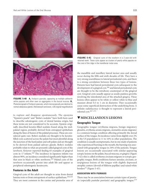
FIGURE 5-41 Gingival cysts <strong>of</strong> <strong>the</strong> newborn in a 2-year-old with<br />
retained teeth. These cysts appear as clusters <strong>of</strong> pearly white papules on<br />
<strong>the</strong> crest <strong>of</strong> <strong>the</strong> ridge in <strong>the</strong> m<strong>and</strong>ibular molar area.<br />
<strong>the</strong> m<strong>and</strong>ible <strong>and</strong> maxillary lateral incisor area <strong>and</strong> usually<br />
occur during <strong>the</strong> fifth <strong>and</strong> sixth decades <strong>of</strong> life. They have a<br />
very strong resemblance to lateral periodontal cysts, <strong>and</strong> <strong>the</strong>re<br />
is a strong correlation between <strong>the</strong>se two types <strong>of</strong> lesions.<br />
Patients have had lateral periodontal cysts subsequent to <strong>the</strong><br />
development <strong>of</strong> a gingival cyst, 266 <strong>and</strong> lateral periodontal cysts<br />
are thought to be <strong>the</strong> intrabony counterpart <strong>of</strong> <strong>the</strong> gingival<br />
cyst. Gingival cysts usually appear as sessile painless growths<br />
involving <strong>the</strong> interdental area <strong>of</strong> <strong>the</strong> attached gingiva. These<br />
lesions <strong>of</strong>ten appear to be white or yellow white to blue <strong>and</strong><br />
measure about 0.5 to 1 cm in diameter. They occasionally<br />
cause some superficial destruction <strong>of</strong> <strong>the</strong> underlying bone. A<br />
definite radiolucency is thought to represent a lateral periodontal<br />
cyst. 267–272<br />
▼ MISCELLANEOUS LESIONS<br />
Geographic Tongue<br />
Geographic tongue (ery<strong>the</strong>ma migrans, benign migratory<br />
glossitis, ery<strong>the</strong>ma areata migrans, stomatitis areata migrans)<br />
is a common benign condition affecting primarily <strong>the</strong> dorsal<br />
surface <strong>of</strong> <strong>the</strong> tongue. Its incidence varies from slightly over 2%<br />
in <strong>the</strong> US population to 11 to 16% in o<strong>the</strong>r populations. The<br />
condition is usually asymptomatic, but in one study <strong>of</strong> patients<br />
who experienced burning in <strong>the</strong> mouth, <strong>the</strong> burning was associated<br />
with geographic tongue in 24% <strong>of</strong> <strong>the</strong> patients. Tongue<br />
lesions are occasionally associated with similar-appearing<br />
ectopic lesions on <strong>the</strong> palate, buccal mucosa, or gingiva (Figure<br />
5-42); this is called ery<strong>the</strong>ma circinata migrans or ectopic geographic<br />
tongue. Both conditions feature annular, circinate, or<br />
serpiginous lesions <strong>of</strong> <strong>the</strong> tongue with slightly depressed<br />
atrophic centers (devoid <strong>of</strong> filiform papillae) <strong>and</strong> raised white<br />
borders 15 (Figure 5-43).<br />
ASSOCIATION WITH PSORIASIS<br />
There may be an association between certain types <strong>of</strong> psoriasis<br />
(especially pustular psoriasis) <strong>and</strong> geographic tongue. 273