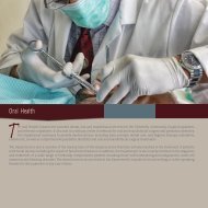
Ch05: Red and White Lesions of the Oral Mucosa
Ch05: Red and White Lesions of the Oral Mucosa
Ch05: Red and White Lesions of the Oral Mucosa

You also want an ePaper? Increase the reach of your titles
YUMPU automatically turns print PDFs into web optimized ePapers that Google loves.
<strong>Red</strong> <strong>and</strong> <strong>White</strong> <strong>Lesions</strong> <strong>of</strong> <strong>the</strong> <strong>Oral</strong> <strong>Mucosa</strong> 93<br />
A B<br />
FIGURE 5-13 A, Typical white corrugated leukoplakia in <strong>the</strong> maxillary vestibule, associated with sanguinaria use. B, M<strong>and</strong>ibular vestibular lesion in<br />
<strong>the</strong> same patient.<br />
vestibule exhibit prolonged retention <strong>of</strong> <strong>the</strong> product due to <strong>the</strong><br />
greater distance from <strong>the</strong> major salivary ducts. Histopathologically,<br />
all biopsy specimens demonstrate significant<br />
surface keratosis with a verrucoid pattern. Minimal atypical<br />
changes (including basilar hyperplasia, nuclear hyperchromatism,<br />
<strong>and</strong> increased nucleocytoplasmic ratios) limited to <strong>the</strong><br />
lower one-third <strong>of</strong> <strong>the</strong> epi<strong>the</strong>lium are noted in most specimens.<br />
More significant atypical changes have also been reported. 78<br />
TREATMENT<br />
No appropriate treatment has been established for sanguinariainduced<br />
leukoplakia. However, an initial biopsy is m<strong>and</strong>atory. If<br />
a histopathologic diagnosis <strong>of</strong> dysplasia is rendered, <strong>the</strong> condition<br />
should be treated in a fashion similar to <strong>the</strong> treatment <strong>of</strong><br />
o<strong>the</strong>r potentially premalignant processes. The less severe changes<br />
should be managed according to clinical judgment, depending on<br />
<strong>the</strong> extent <strong>and</strong> duration <strong>of</strong> <strong>the</strong> lesion. In all cases, complete discontinuation<br />
<strong>of</strong> Sanguinaria-containing products <strong>and</strong> cessation<br />
<strong>of</strong> any o<strong>the</strong>r harmful habits such as tobacco or alcohol use is<br />
m<strong>and</strong>atory. All patients should be given careful clinical followup,<br />
with a biopsy <strong>of</strong> any recurrent or worsening lesion(s).<br />
▼ INFECTIOUS WHITE LESIONS AND<br />
WHITE AND RED LESIONS<br />
<strong>Oral</strong> Hairy Leukoplakia<br />
<strong>Oral</strong> hairy leukoplakia is a corrugated white lesion that usually<br />
occurs on <strong>the</strong> lateral or ventral surfaces <strong>of</strong> <strong>the</strong> tongue in<br />
patients with severe immunodeficiency. 81,82 The most common<br />
disease associated with oral hairy leukoplakia is HIV<br />
infection. 82 <strong>Oral</strong> hairy leukoplakia is reported in about 25% <strong>of</strong><br />
adults with HIV infection but is not as common in HIVinfected<br />
children. Its prevalence reaches as high as 80% in<br />
patients with acquired immunodeficiency syndrome (AIDS). 83<br />
Epstein-Barr virus (EBV) is implicated as <strong>the</strong> causative agent<br />
in oral hairy leukoplakia. 84–86 A positive correlation with<br />
decreasing cluster designation 4 (CD4) cell counts has been<br />
established in HIV-positive patients. The presence <strong>of</strong> this<br />
lesion has been associated with <strong>the</strong> subsequent development<br />
<strong>of</strong> AIDS in a large percentage <strong>of</strong> HIV positive patients. 83 Hairy<br />
leukoplakia has also occasionally been reported in patients<br />
with o<strong>the</strong>r immunosuppressive conditions, such as patients<br />
undergoing organ transplantation <strong>and</strong> patients undergoing<br />
prolonged steroid <strong>the</strong>rapy. 87–89 Rare cases may occur in<br />
immunocompetent persons after topical steroid <strong>the</strong>rapy. 90,91<br />
TYPICAL FEATURES<br />
<strong>Oral</strong> hairy leukoplakia most commonly involves <strong>the</strong> lateral border<br />
<strong>of</strong> <strong>the</strong> tongue but may extend to <strong>the</strong> ventral or dorsal surfaces.<br />
<strong>Lesions</strong> on <strong>the</strong> tongue are usually corrugated <strong>and</strong> may have<br />
a shaggy or frayed appearance, mimicking lesions caused by<br />
tongue chewing (Figure 5-14). <strong>Oral</strong> hairy leukoplakia may also<br />
present as a plaquelike lesion <strong>and</strong> is <strong>of</strong>ten bilateral.<br />
Histopathologic examination <strong>of</strong> <strong>the</strong> epi<strong>the</strong>lium reveals severe<br />
hyperparakeratosis with an irregular surface, acanthosis with<br />
superficial edema, <strong>and</strong> numerous koilocytic cells (virally affected<br />
FIGURE 5-14 Bilateral linear leukoplakic lesions on <strong>the</strong> dorsolateral<br />
tongue, suggestive <strong>of</strong> oral hairy leukoplakia. (Courtesy <strong>of</strong> Dr. Parnell Taylor,<br />
Riverton, Wyoming)