

Buccinator myomucosal flap: clinical results and review of anatomy ...
Buccinator myomucosal flap: clinical results and review of anatomy ...
Buccinator myomucosal flap: clinical results and review of anatomy ...

Create successful ePaper yourself
Turn your PDF publications into a flip-book with our unique Google optimized e-Paper software.
184<br />
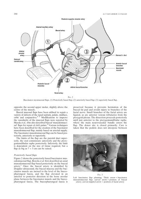
FIG. 2<br />
<strong>Buccinator</strong> <strong>myomucosal</strong> <strong>flap</strong>s. (1) Posteriorly based <strong>flap</strong>; (2) anteriorly based <strong>flap</strong>; (3) superiorly based <strong>flap</strong>.<br />
opposite the second upper molar, slightly above the<br />
centre <strong>of</strong> the muscle.<br />
Buccal mucosal <strong>flap</strong>s have been utilised to repair a<br />
variety <strong>of</strong> defects <strong>of</strong> the nasal septum, palate, midface,<br />
orbit <strong>and</strong> conjunctiva. 4–8 Modifications to improve<br />
the vascularity <strong>of</strong> the mucosal <strong>flap</strong>s were reported by<br />
Maeda et al., who also described buccal ‘musculomucosal’<br />
<strong>flap</strong>s for repair <strong>of</strong> cleft palate. 9 Various techniques<br />
have been described for the creation <strong>of</strong> the buccinator<br />
musculomucosal <strong>flap</strong>, mainly based on arterial supply.<br />
The buccinator musculomucosal <strong>flap</strong> can be based posteriorly,<br />
anteriorly or superiorly.<br />
The limits <strong>of</strong> the <strong>flap</strong> are the parotid duct superiorly,<br />
the oral commissure anteriorly <strong>and</strong> the pterygom<strong>and</strong>ibular<br />
raphe posteriorly. Inferiorly, the limit<br />
is dependent on the size <strong>of</strong> tissue required, but a<br />
<strong>flap</strong> as big as 7 5 cm can be raised.<br />
Posteriorly based <strong>flap</strong>s<br />
Figure 2 shows the posteriorly based buccinator musculomucosal<br />
<strong>flap</strong>. Bozola et al. first described an axial<br />
musculomucosal <strong>flap</strong> based posteriorly on the buccal<br />
artery. 1 Once the buccal artery is identified by<br />
Doppler ultrasound, the buccal mucosa <strong>and</strong> the buccinator<br />
muscle are incised to the level <strong>of</strong> the buccopharyngeal<br />
fascia, <strong>and</strong> the <strong>flap</strong> elevated in an<br />
anterior to posterior direction in the loose areolar<br />
plane between the buccinator muscle <strong>and</strong> the buccopharyngeal<br />
fascia. The buccopharyngeal fascia is<br />
A C VAN LIEROP, J J FAGAN<br />
preserved because it prevents herniation <strong>of</strong> the<br />
buccal fat pad <strong>and</strong> avoids injury to branches <strong>of</strong> the<br />
facial nerve. Small branches <strong>of</strong> the facial artery are<br />
ligated, as are anterior venous tributaries from the<br />
pterygoid plexus. The dissection proceeds posteriorly<br />
until just anterior to the pterygom<strong>and</strong>ibular raphe,<br />
where the main neurovascular bundle enters the<br />
<strong>flap</strong>. The donor site is closed primarily. Care is<br />
taken that the pedicle does not interpose between<br />
FIG. 3<br />
Left buccinator <strong>flap</strong> planning. Thick arrow ¼ buccinator<br />
musculomucosal <strong>flap</strong>; curved arrow ¼ position <strong>of</strong> buccal<br />
artery; curved stippled arrow ¼ position <strong>of</strong> facial artery


