Buccinator myomucosal flap: clinical results and review of anatomy ...
Buccinator myomucosal flap: clinical results and review of anatomy ...
Buccinator myomucosal flap: clinical results and review of anatomy ...
Create successful ePaper yourself
Turn your PDF publications into a flip-book with our unique Google optimized e-Paper software.
The Journal <strong>of</strong> Laryngology & Otology (2008), 122, 181–187.<br />
# 2007 JLO (1984) Limited<br />
doi:10.1017/S0022215107008353<br />
Printed in the United Kingdom<br />
First published online 22 May 2007<br />
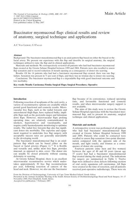
<strong>Buccinator</strong> <strong>myomucosal</strong> <strong>flap</strong>: <strong>clinical</strong> <strong>results</strong> <strong>and</strong> <strong>review</strong><br />
<strong>of</strong> <strong>anatomy</strong>, surgical technique <strong>and</strong> applications<br />
ACVAN LIEROP, JJFAGAN<br />
Main Article<br />
Abstract<br />
Background: The buccinator musculomucosal <strong>flap</strong> is an axial-pattern <strong>flap</strong> based on either the buccal or the<br />
facial artery. We present our experience with this <strong>flap</strong> <strong>and</strong> describe its surgical <strong>anatomy</strong>, the surgical<br />
techniques utilised to raise the <strong>flap</strong> <strong>and</strong> its <strong>clinical</strong> applications.<br />
Materials <strong>and</strong> methods: We retrospectively <strong>review</strong>ed all patients who had had buccinator <strong>myomucosal</strong><br />
<strong>flap</strong>s created at the Groote Schuur Hospital between 1999 <strong>and</strong> 2004. Patients were also recalled to assess<br />
<strong>flap</strong> sensation <strong>and</strong> to record reduction <strong>of</strong> mouth opening as a consequence <strong>of</strong> donor site scarring.<br />
Results: Of the 14 patients who had had a buccinator <strong>myomucosal</strong> <strong>flap</strong> created, there was one <strong>flap</strong><br />
failure. Sensation was present in 71 per cent <strong>of</strong> <strong>flap</strong>s, <strong>and</strong> there was no trismus due to donor site scarring.<br />
Conclusions: The buccinator myomycosal <strong>flap</strong> is a dependable <strong>flap</strong> with good functional outcome <strong>and</strong><br />
low morbidity.<br />
Key words: Mouth; Carcinoma; Fistula; Surgical Flaps; Surgical Procedures, Operative<br />
Introduction<br />
Following resection <strong>of</strong> neoplasms <strong>of</strong> the oral cavity, a<br />
variety <strong>of</strong> reconstructive options are available which<br />
permit good functional <strong>and</strong> cosmetic <strong>results</strong>. Microvascular<br />
free <strong>flap</strong>s, such as the radial forearm <strong>and</strong><br />
anterolateral thigh <strong>flap</strong>s, have replaced bulky, impliable<br />
<strong>flap</strong>s such as the pectoralis major <strong>and</strong> latissimus<br />
dorsi <strong>flap</strong>s. However, microvascular <strong>flap</strong>s prolong<br />
operating time, are relatively contraindicated in<br />
smokers, hypertensives <strong>and</strong> vasculopaths, <strong>and</strong><br />
require costly haemodynamic monitoring to optimise<br />
<strong>results</strong>. The radial free forearm <strong>flap</strong> also has significant<br />
donor site morbidity. The expertise <strong>and</strong> equipment<br />
required to undertake free <strong>flap</strong> surgery with<br />
acceptable success rates are generally unavailable<br />
in the developing world.<br />
The buccinator musculomucosal <strong>flap</strong> is an axialpattern<br />
<strong>flap</strong> which can be based either on the<br />
buccal or facial arteries (Figure 1). 1,2 It is flexible<br />
<strong>and</strong> versatile <strong>and</strong>, unlike most free <strong>flap</strong>s, provides<br />
mucosal, as opposed to skin, cover. The donor site<br />
can usually be closed primarily without causing<br />
deformity or scarring.<br />
At Groote Schuur Hospital, there is an excellent<br />
microvascular reconstructive service which undertakes<br />
approximately 30 free <strong>flap</strong> reconstructions<br />
per year, following resection <strong>of</strong> cancers <strong>of</strong> the<br />
head <strong>and</strong> neck. However, we also use the buccinator<br />
<strong>flap</strong> because <strong>of</strong> its convenience, reduced operating<br />
time, <strong>and</strong> favourable functional <strong>and</strong> cosmetic<br />
<strong>results</strong>, <strong>and</strong> when microvascular surgery support is<br />
not available.<br />
The aims <strong>of</strong> this study were to <strong>review</strong> the Groote<br />
Schuur Hospital experience with the buccinator <strong>myomucosal</strong><br />
<strong>flap</strong>, <strong>and</strong> to present its <strong>anatomy</strong>, surgical<br />
technique <strong>and</strong> <strong>clinical</strong> applications.<br />
Materials <strong>and</strong> methods<br />
A retrospective <strong>review</strong> was performed <strong>of</strong> all patients<br />
who had had buccinator musculomucosal <strong>flap</strong>s<br />
created at Groote Schuur Hospital between 1999<br />
<strong>and</strong> 2004. Patients who could be contacted were<br />
recalled to assess <strong>flap</strong> sensation (two-point discrimination<br />
compared with the opposite side <strong>of</strong> the<br />
mouth, <strong>and</strong> light touch), <strong>and</strong> trismus as a consequence<br />
<strong>of</strong> donor site scarring.<br />
Fourteen patients (nine men <strong>and</strong> five women)<br />
between the ages <strong>of</strong> 40 <strong>and</strong> 68 years (mean age<br />
57 years) had had a buccinator musculomucosal<br />
<strong>flap</strong> created during the study period. The indications<br />
for surgery are summarised in Table I. Twelve<br />
<strong>flap</strong>s were utilised to close defects following excision<br />
<strong>of</strong> oral cavity squamous cell carcinomas. All <strong>flap</strong>s<br />
were created at the time <strong>of</strong> resection, <strong>and</strong> all patients<br />
received post-operative radiotherapy. There were six<br />
From the Division <strong>of</strong> Otolaryngology, University <strong>of</strong> Cape Town Medical School, South Africa.<br />
Presented at the Annual Academic Meeting <strong>of</strong> the South African Society <strong>of</strong> Otorhinolaryngology Head <strong>and</strong> Neck Surgery, 14–17<br />
November 2004, Port Elizabeth, South Africa.<br />
Accepted for publication: 7 February 2007.<br />
181
182<br />
cancers <strong>of</strong> the tongue (four T2 <strong>and</strong> two T3 lesions<br />
(tumour–node–metastasis (TNM) classification)),<br />
three <strong>of</strong> the floor <strong>of</strong> the mouth (all T1), <strong>and</strong> three<br />
<strong>of</strong> the tonsil or palate (all T2).<br />
Patient number seven had had an adenoid cystic<br />
carcinoma <strong>of</strong> the hard palate, which had been<br />
treated with neutron therapy. She developed a<br />
local recurrence 10 years later, for which a wide<br />
local excision was undertaken. A buccinator musculomucosal<br />
<strong>flap</strong> was utilised to repair the palatal<br />
FIG. 1<br />
Blood supply <strong>of</strong> the buccinator muscle.<br />
TABLE I<br />
PATIENT SUMMARY<br />
defect. Patient 10 had a nasal septal perforation.<br />
He had had a cleft palate repair in infancy <strong>and</strong> subsequently<br />
sustained severe facial fractures in a<br />
motor vehicle accident. Following nasal reconstruction<br />
with rib cartilage, he had a large, symptomatic<br />
nasal septal perforation. As there was no suitable<br />
local tissue with which to repair the defect, a buccinator<br />
musculomucosal <strong>flap</strong>, based on the distal end<br />
<strong>of</strong> the facial artery, was utilised to repair the septal<br />
perforation.<br />
Patient no Age (years)/sex Tumour site Stage Base <strong>of</strong> <strong>flap</strong> Radiotherapy?<br />
1 53/F S<strong>of</strong>t palate & tonsil T 2 N 1 Posteriorly Y<br />
2 65/M Anterior floor <strong>of</strong> mouth T 1 N 1 Anteriorly Y<br />
3 56/F Anterior tongue T2 N0 Anteriorly Y<br />
4 64/M Tonsil T 2 N 1 Posteriorly Y<br />
5 61/M Lateral tongue, floor <strong>of</strong> mouth T 3 N 2 Anteriorly Y<br />
6 53/M Tongue T 2 N 2 Anteriorly Y<br />
7 60/F Palatal fistula (adenoid cystic) T 2 N 0 Posteriorly Neutrons<br />
8 63/M Tongue T 2 N 0 Anteriorly Y<br />
9 59/M Floor <strong>of</strong> mouth T 1 N 2 Anteriorly Y<br />
10 42/M Nasal septal perforation – Superiorly –<br />
11 40/M Anterior floor <strong>of</strong> mouth T 1 N 1 Anteriorly Y<br />
12 49/F Tongue, s<strong>of</strong>t palate T 2 N 0 Posteriorly Y<br />
13 51/M Anterior floor <strong>of</strong> mouth, anterolateral tongue T 3 N 1 Anteriorly Y<br />
14 68/F S<strong>of</strong>t palate, tonsil, retromolar trigone T 2 N 1 Posteriorly Y<br />
No ¼ number; F ¼ female; M ¼ male; T ¼ tumour; N ¼ node; Y ¼ yes<br />
A C VAN LIEROP, J J FAGAN
BUCCINATOR MYOMUCOSAL FLAP 183<br />
Results<br />
The outcome <strong>of</strong> the buccinator <strong>myomucosal</strong> <strong>flap</strong>s<br />
was assessed retrospectively by chart <strong>review</strong> <strong>and</strong> by<br />
recall <strong>of</strong> those patients who could be contacted<br />
(Table II).<br />
Complete <strong>flap</strong> failure occurred only in patient<br />
seven (7 per cent), who had had palatal repair following<br />
neutron therapy. Scarring <strong>and</strong> vascular injury to<br />
the recipient site from the neutron therapy was a<br />
likely contributing factor to failure. One patient<br />
had suffered very minimal distal <strong>flap</strong> necrosis for<br />
which no additional surgery was needed, as the<br />
wound had healed by secondary intention with a<br />
good functional outcome. There was one case <strong>of</strong><br />
partial <strong>flap</strong> dehiscence, which had healed with no<br />
further intervention. Donor sites were closed primarily<br />
with absorbable sutures. There was one case <strong>of</strong><br />
dehiscence <strong>of</strong> the donor site closure, which had<br />
healed by secondary intention. No other <strong>flap</strong> complications<br />
were noted.<br />
Only seven patients could be recalled to assess<br />
long-term <strong>results</strong> regarding <strong>flap</strong> sensation <strong>and</strong><br />
trismus (Table II). Both light touch sensation <strong>and</strong><br />
two-point discrimination were present, although<br />
reduced, in 71 per cent ( five <strong>of</strong> seven) <strong>of</strong> the <strong>flap</strong>s.<br />
The average two-point discrimination <strong>of</strong> the <strong>flap</strong><br />
was 21.6 mm, compared with 12.8 mm for normal<br />
buccal mucosa. No patient had trismus as a result<br />
<strong>of</strong> scarring at the donor site.<br />
Discussion<br />
The buccinator muscle is a thin, quadrilateral cheek<br />
muscle (Figure 1). It originates from the outer surfaces<br />
<strong>of</strong> the alveolar processes <strong>of</strong> the maxilla <strong>and</strong><br />
m<strong>and</strong>ible, overlying the three molar teeth. Posteriorly,<br />
it arises from the pterygom<strong>and</strong>ibular raphe<br />
<strong>and</strong> inserts anteriorly into the orbicularis oris<br />
muscle. Laterally, it is related to the ramus <strong>of</strong> the<br />
m<strong>and</strong>ible, the masseter <strong>and</strong> medial pterygoid<br />
muscles, the buccal fat pad <strong>and</strong> the buccopharyngeal<br />
fascia. Medially, it is covered by the submucosa<br />
<strong>and</strong> mucosa <strong>of</strong> the cheek. It is part <strong>of</strong> the<br />
TABLE II<br />
OUTCOME OF BUCCINATOR MYOMUCOSAL FLAPS<br />
pharyngeal-buccal-orbicularis sphincter system <strong>and</strong><br />
functions to facilitate whistling, sucking, propelling<br />
food during mastication <strong>and</strong> voiding the buccal cavity.<br />
The buccal, facial <strong>and</strong> posterosuperior alveolar<br />
arteries make up the main blood supply to the<br />
muscle (Figures 1, 2 <strong>and</strong> 3). The buccal artery is a<br />
branch <strong>of</strong> the internal maxillary artery <strong>and</strong> supplies<br />
the posterior half <strong>of</strong> the muscle. It courses anteroinferiorly<br />
under the lateral pterygoid muscle to reach<br />
the posterior half <strong>of</strong> the muscle, where it anastomoses<br />
with the posterior buccal branch <strong>of</strong> the facial<br />
artery. 3 The facial artery hooks around the lower<br />
border <strong>of</strong> the m<strong>and</strong>ible at the anterior edge <strong>of</strong> the<br />
masseter muscle, <strong>and</strong> supplies numerous branches<br />
to the buccinator muscle (Figure 1), the largest <strong>of</strong><br />
which is the posterior buccal, which supplies the posterior<br />
half <strong>of</strong> the muscle. The facial artery gives <strong>of</strong>f<br />
one to three inferior buccal branches to supply the<br />
inferior half <strong>of</strong> the muscle, <strong>and</strong> then continues in<br />
an anterosuperior direction to give <strong>of</strong>f three to five<br />
small, anterior buccal branches to the anterior half<br />
<strong>of</strong> the muscle. The posterosuperior alveolar artery,<br />
a branch <strong>of</strong> the internal maxillary artery, provides<br />
two small branches to the muscle, which enter the<br />
muscle posterosuperiorly, <strong>and</strong> the infraorbital<br />
artery gives <strong>of</strong>f a few small branches which enter it<br />
anterosuperiorly. All these arteries form an extensive<br />
vascular anastomosis on the lateral surface <strong>of</strong><br />
the muscle <strong>and</strong> within its fibres.<br />
Venous drainage occurs via the pterygoid plexus<br />
(<strong>and</strong> internal maxillary vein). This lies posterior,<br />
superior <strong>and</strong> superficial to the buccinator <strong>and</strong><br />
drains into the buccal vein via the deep facial vein.<br />
Anteriorly, the deep facial vein drains into the<br />
facial vein proper.<br />
Sensory innervation is via the long buccal nerve, a<br />
branch <strong>of</strong> the maxillary nerve, which courses with the<br />
buccal branch <strong>of</strong> the internal maxillary artery. The<br />
motor supply to the buccinator muscle is the temporal<br />
<strong>and</strong> cervical divisions <strong>of</strong> the facial nerve,<br />
which form a plexus near the buccal fat pad.<br />
The parotid duct has an important anatomical<br />
relationship to the muscle <strong>and</strong> pierces the buccinator<br />
Patient no Complications 2-point discrimination (mm) Light touch Mouth opening (cm)<br />
Flap Opposite side<br />
1 – NT NT NT NT<br />
2 – 25 15 þ 3.5<br />
3 – NT NT NT NT<br />
4 – NT NT NT NT<br />
5 – 19 10 þ 4<br />
6 – NT NT NT NT<br />
7 Complete <strong>flap</strong> failure NT NT NT NT<br />
8 Partial <strong>flap</strong> failure 25 15 þ 5<br />
9 – NT NT NT NT<br />
10 – NT NT NT NT<br />
11 Donor site breakdown 20 10 þ 3.5<br />
12 Partial <strong>flap</strong> dehiscence No sensation No sensation – 2<br />
13 – No sensation No sensation – 3.5<br />
14 – 19 14 þ 2<br />
No ¼ number; NT ¼ not tested
184<br />
FIG. 2<br />
<strong>Buccinator</strong> <strong>myomucosal</strong> <strong>flap</strong>s. (1) Posteriorly based <strong>flap</strong>; (2) anteriorly based <strong>flap</strong>; (3) superiorly based <strong>flap</strong>.<br />
opposite the second upper molar, slightly above the<br />
centre <strong>of</strong> the muscle.<br />
Buccal mucosal <strong>flap</strong>s have been utilised to repair a<br />
variety <strong>of</strong> defects <strong>of</strong> the nasal septum, palate, midface,<br />
orbit <strong>and</strong> conjunctiva. 4–8 Modifications to improve<br />
the vascularity <strong>of</strong> the mucosal <strong>flap</strong>s were reported by<br />
Maeda et al., who also described buccal ‘musculomucosal’<br />
<strong>flap</strong>s for repair <strong>of</strong> cleft palate. 9 Various techniques<br />
have been described for the creation <strong>of</strong> the buccinator<br />
musculomucosal <strong>flap</strong>, mainly based on arterial supply.<br />
The buccinator musculomucosal <strong>flap</strong> can be based posteriorly,<br />
anteriorly or superiorly.<br />
The limits <strong>of</strong> the <strong>flap</strong> are the parotid duct superiorly,<br />
the oral commissure anteriorly <strong>and</strong> the pterygom<strong>and</strong>ibular<br />
raphe posteriorly. Inferiorly, the limit<br />
is dependent on the size <strong>of</strong> tissue required, but a<br />
<strong>flap</strong> as big as 7 5 cm can be raised.<br />
Posteriorly based <strong>flap</strong>s<br />
Figure 2 shows the posteriorly based buccinator musculomucosal<br />
<strong>flap</strong>. Bozola et al. first described an axial<br />
musculomucosal <strong>flap</strong> based posteriorly on the buccal<br />
artery. 1 Once the buccal artery is identified by<br />
Doppler ultrasound, the buccal mucosa <strong>and</strong> the buccinator<br />
muscle are incised to the level <strong>of</strong> the buccopharyngeal<br />
fascia, <strong>and</strong> the <strong>flap</strong> elevated in an<br />
anterior to posterior direction in the loose areolar<br />
plane between the buccinator muscle <strong>and</strong> the buccopharyngeal<br />
fascia. The buccopharyngeal fascia is<br />
A C VAN LIEROP, J J FAGAN<br />
preserved because it prevents herniation <strong>of</strong> the<br />
buccal fat pad <strong>and</strong> avoids injury to branches <strong>of</strong> the<br />
facial nerve. Small branches <strong>of</strong> the facial artery are<br />
ligated, as are anterior venous tributaries from the<br />
pterygoid plexus. The dissection proceeds posteriorly<br />
until just anterior to the pterygom<strong>and</strong>ibular raphe,<br />
where the main neurovascular bundle enters the<br />
<strong>flap</strong>. The donor site is closed primarily. Care is<br />
taken that the pedicle does not interpose between<br />
FIG. 3<br />
Left buccinator <strong>flap</strong> planning. Thick arrow ¼ buccinator<br />
musculomucosal <strong>flap</strong>; curved arrow ¼ position <strong>of</strong> buccal<br />
artery; curved stippled arrow ¼ position <strong>of</strong> facial artery
BUCCINATOR MYOMUCOSAL FLAP 185<br />
the molar teeth, as this may interfere with mastication.<br />
Relevant molars might need to be extracted or<br />
an isl<strong>and</strong> <strong>flap</strong> created; alternatively, the vascular<br />
pedicle may be divided after a delay <strong>of</strong> a few weeks.<br />
Modifications <strong>of</strong> this procedure include isolation<br />
<strong>of</strong> the pedicle to create an isl<strong>and</strong> <strong>flap</strong>, in order to<br />
facilitate rotation, 10 <strong>and</strong> creation <strong>of</strong> a ‘buccinator<br />
<strong>myomucosal</strong> neurovascular isl<strong>and</strong> pedicle <strong>flap</strong>’<br />
based on the buccal artery, the buccal venous<br />
plexus <strong>and</strong> nerves innervating the muscle. The<br />
mucosa at the posterior end <strong>of</strong> the <strong>flap</strong> is divided<br />
from the underlying muscle <strong>and</strong> freed <strong>of</strong> its insertion<br />
from the pterygom<strong>and</strong>ibular raphe. 3 Then the<br />
<strong>flap</strong> is passed through a short tunnel under the<br />
pterygom<strong>and</strong>ibular ligament.<br />
Anteriorly based <strong>flap</strong><br />
The anteriorly based <strong>flap</strong> (Figure 2) is based anteroinferiorly<br />
on the inferior buccal branches <strong>of</strong> the<br />
facial artery, 2,11,12 the main trunk <strong>of</strong> which may be<br />
identified with a Doppler probe to establish its position.<br />
The mucosa <strong>and</strong> the buccinator muscle are<br />
incised superiorly <strong>and</strong> the facial artery <strong>and</strong> vein<br />
ligated. The dissection continues in a plane lateral<br />
to the vessels, as the <strong>flap</strong> is raised from front to<br />
back while branches <strong>of</strong> the facial artery are ligated<br />
(Figures 4 <strong>and</strong> 5).<br />
Superiorly based <strong>flap</strong><br />
Zhao et al. described the superiorly based ‘buccinator<br />
<strong>myomucosal</strong> reversed-flow arterial isl<strong>and</strong> <strong>flap</strong>’<br />
(Figure 2). 3 It is based on the distal end <strong>of</strong> the<br />
facial artery <strong>and</strong> its anterior buccal branches. The<br />
course <strong>of</strong> the artery is outlined by Doppler ultrasound,<br />
<strong>and</strong> dissection starts at the inferior margin<br />
<strong>of</strong> the <strong>flap</strong> with incision <strong>of</strong> the mucosa <strong>and</strong> buccinator<br />
muscle. The facial artery is ligated inferiorly <strong>and</strong> the<br />
<strong>flap</strong> is elevated in a superior direction. The arc <strong>of</strong><br />
rotation is centred between the oral commissure<br />
<strong>and</strong> medial canthus.<br />
FIG. 4<br />
Left anteriorly based buccinator <strong>flap</strong> elevated <strong>and</strong> ready for<br />
insertion in anterior floor <strong>of</strong> mouth defect (arrow).<br />
FIG. 5<br />
Left anteriorly based buccinator <strong>flap</strong> inserted in floor <strong>of</strong> mouth<br />
defect (arrow).<br />
The buccinator <strong>flap</strong> has proved to be very reliable.<br />
Our only <strong>clinical</strong>ly significant failure was in a patient<br />
who had previously received neutron therapy; this<br />
failure was probably related to a compromised microvascular<br />
blood supply, either to the <strong>flap</strong> or the recipient<br />
site.<br />
Light touch sensation <strong>and</strong> two-point discrimination<br />
were intact in 71 per cent <strong>of</strong> patients, although<br />
sensation was reduced. However, even reduced sensation<br />
is advantageous in terms <strong>of</strong> oral function,<br />
when compared with insensate <strong>flap</strong>s. Recovery <strong>of</strong><br />
sensation within the early post-operative period has<br />
been reported ( following creation <strong>of</strong> anteriorly <strong>and</strong><br />
posteriorly based buccinator <strong>flap</strong>s for tongue reconstruction).<br />
When a <strong>flap</strong> is posteriorly based, such<br />
sensory recovery can be attributed to the fact that<br />
the buccal nerve is included in the <strong>flap</strong>. 10,11 We<br />
have also noted very early post-operative recovery<br />
<strong>of</strong> sensation.<br />
FIG. 6<br />
Donor site defect closed primarily following raising <strong>of</strong><br />
anteriorly based buccinator <strong>flap</strong> (arrows).
186<br />
All donor sites could be closed primarily<br />
(Figure 6). Even when harvesting a large <strong>flap</strong>,<br />
primary closure could usually be achieved due to<br />
the mobility <strong>and</strong> elasticity <strong>of</strong> the remaining buccal<br />
mucosa <strong>and</strong> muscle. Should the wound dehisce, it<br />
heals with minimal scarring, as was shown by the<br />
absence <strong>of</strong> trismus in our patients.<br />
. The buccinator <strong>myomucosal</strong> <strong>flap</strong> is an<br />
axial-pattern <strong>flap</strong> based on the buccal or facial<br />
artery, <strong>and</strong> is used in the reconstruction <strong>of</strong><br />
small <strong>and</strong> moderately sized defects <strong>of</strong> the oral<br />
cavity <strong>and</strong> oropharynx<br />
. The authors report use <strong>of</strong> this <strong>flap</strong>, presenting<br />
the <strong>flap</strong> <strong>anatomy</strong>, the surgical technique<br />
utilised in elevatation, <strong>and</strong> the complications<br />
<strong>and</strong> applications <strong>of</strong> its use in reconstruction<br />
. The study included 14 patients, <strong>of</strong> whom seven<br />
were <strong>review</strong>ed to assess <strong>flap</strong> sensation <strong>and</strong><br />
overall morbidity<br />
. Seventy-one per cent <strong>of</strong> patients had sensation<br />
in the <strong>flap</strong>, albeit reduced compared with the<br />
opposite side<br />
. Primary closure <strong>of</strong> the donor site was achieved<br />
in all cases, <strong>and</strong> the complication rate in raising<br />
the <strong>flap</strong> was low, with only one <strong>flap</strong> failure<br />
. The buccinator <strong>myomucosal</strong> <strong>flap</strong> was found to<br />
be a reliable <strong>flap</strong> with good functional<br />
outcome <strong>and</strong> low morbidity<br />
The buccinator musculomucosal <strong>flap</strong> is a versatile<br />
<strong>flap</strong> that can be used to reconstruct a variety <strong>of</strong><br />
defects. It is remarkably elastic <strong>and</strong> malleable, <strong>and</strong><br />
can be stretched to conform to complexly shaped<br />
defects. The procedures we performed are summarised<br />
in Table I. The arc <strong>of</strong> rotation <strong>of</strong> posteriorly<br />
based <strong>flap</strong>s will allow it to reach velar, palatine <strong>and</strong><br />
lateral pharyngeal sites. We have used it to reconstruct<br />
defects <strong>of</strong> the tonsil bed, s<strong>of</strong>t <strong>and</strong> hard palate, tonsillolingual<br />
sulcus, <strong>and</strong> retromolar trigone. It has also been<br />
used in primary cleft palate repair. 1 We used the anteriorly<br />
based buccinator musculomucosal <strong>flap</strong> to reconstruct<br />
the lip, <strong>and</strong> for repair <strong>of</strong> floor <strong>of</strong> mouth defects<br />
<strong>and</strong> reconstruction <strong>of</strong> the anterior <strong>and</strong> lateral tongue.<br />
It is particularly useful for anterior floor <strong>of</strong> mouth<br />
<strong>and</strong> tongue defects as it provides a normal mucosal<br />
covering <strong>and</strong> preserves mobility <strong>of</strong> the anterior<br />
tongue. It has also been described for closure <strong>of</strong><br />
oro-antral <strong>and</strong> palatal fistulae, <strong>and</strong> for alveolar reconstruction.<br />
2 The superiorly based buccinator musculomucosal<br />
<strong>flap</strong> has been described as a means <strong>of</strong><br />
closing defects in the anterior hard palate, alveolus,<br />
maxillary antrum, nasal cavity, upper lip <strong>and</strong> lower<br />
lip, as well as the orbit. 3,11,13 We employed this technique<br />
to close a large nasal septal perforation.<br />
The buccinator musculomucosal <strong>flap</strong> has distinct<br />
advantages over free <strong>flap</strong> reconstruction. It is<br />
ideally suited to surgical units that lack expertise in<br />
A C VAN LIEROP, J J FAGAN<br />
microvascular free tissue transfer. Even in units<br />
that have this option, the considerable advantages<br />
<strong>of</strong> this form <strong>of</strong> reconstruction make it a good<br />
choice for many oral cavity <strong>and</strong> oropharyngeal<br />
defects. The operating time needed to raise <strong>and</strong><br />
inset the buccinator musculomucosal <strong>flap</strong> is 45<br />
minutes to one hour, which is considerably less<br />
than that required for both free <strong>flap</strong>s <strong>and</strong> distant<br />
axial <strong>flap</strong>s. <strong>Buccinator</strong> musculomucosal <strong>flap</strong>s also<br />
require a shorter hospital stay <strong>and</strong> are associated<br />
with low donor site morbidity. The buccinator musculomucosal<br />
<strong>flap</strong> is technically simple <strong>and</strong> does not<br />
require microvascular expertise. The <strong>flap</strong> has great<br />
versatility, as is evident from the variety <strong>of</strong> defects<br />
reconstructed. It is also very useful in patients with<br />
vascular disease, in whom there may be an increased<br />
risk <strong>of</strong> free <strong>flap</strong> failure. It is ideally suited for reconstruction<br />
following resection <strong>of</strong> T 1,T 2 <strong>and</strong> smaller T 3<br />
oral cavity <strong>and</strong> oropharyngeal lesions, at the time <strong>of</strong><br />
excision <strong>of</strong> the primary tumour. However, it is not<br />
suited for larger defects for which more bulk is<br />
required; in these cases, distant axial or free <strong>flap</strong>s<br />
would be better options.<br />
Conclusions<br />
The buccinator <strong>myomucosal</strong> <strong>flap</strong> is a versatile <strong>flap</strong><br />
useful for reconstruction <strong>of</strong> a variety <strong>of</strong> small <strong>and</strong><br />
moderately sized defects <strong>of</strong> the oral cavity <strong>and</strong> oropharynx.<br />
It has a well described <strong>and</strong> constant blood<br />
supply, which makes it a dependable <strong>flap</strong> with little<br />
risk <strong>of</strong> <strong>flap</strong> failure. It has significant advantages<br />
over many other forms <strong>of</strong> oral reconstruction, including:<br />
technical simplicity; short operating time; lack <strong>of</strong><br />
external scars; minimal donor site morbidity; donor<br />
<strong>and</strong> recipient sites in the same operative field; replacement<br />
<strong>of</strong> mucosa with mucosa; sensory innervation,<br />
which aids oral rehabilitation, mastication <strong>and</strong><br />
speech; <strong>and</strong> <strong>flap</strong> reliability. Limitations include the<br />
amount <strong>of</strong> tissue available for reconstruction, <strong>and</strong><br />
the fact that the anteriorly based <strong>flap</strong> cannot be<br />
used if the facial artery has been taken at neck<br />
dissection.<br />
References<br />
1 Bozola A, Gasques J, Carriquiry C, Cordosa de Oliveira M.<br />
The buccinator musculomucosal <strong>flap</strong>: anatomic study <strong>and</strong><br />
<strong>clinical</strong> application. Plast Reconstr Surg 1989;84:250–7<br />
2 Carstens MH, St<strong>of</strong>man GM, Hurwitz DJ, Futrell JW,<br />
Patterson GT, Sotereanos G. The buccinator <strong>myomucosal</strong><br />
isl<strong>and</strong> pedicle <strong>flap</strong>: anatomic study <strong>and</strong> case report.<br />
Plast Reconstr Surg 1991;88:39–50<br />
3 Zhao Z, Li S, Yan Y, Li Y, Yang M, Mu L et al. New buccinator<br />
<strong>myomucosal</strong> isl<strong>and</strong> <strong>flap</strong>: anatomic study <strong>and</strong> <strong>clinical</strong><br />
application. Plast Reconstr Surg 1999;104:55–64<br />
4 Tezel E. Buccal mucosal <strong>flap</strong>s: a <strong>review</strong>. Plast Reconstr Surg<br />
2002;109:735–41<br />
5 Tipton JB. Closure <strong>of</strong> large septal perforations with labialbuccal<br />
<strong>flap</strong>. Plast Reconstr Surg 1970;46:514–15<br />
6 Jackson IT. Closure <strong>of</strong> secondary palatal fistulae with<br />
intra-oral tissue <strong>and</strong> bone grafting. Br J Plast Surg 1972;<br />
25:93–105<br />
7 Kaplan EN. S<strong>of</strong>t palate repair by levator muscle reconstruction<br />
<strong>and</strong> a buccal mucosal <strong>flap</strong>. Plast Reconstr Surg<br />
1975;56:129–36<br />
8 Rayner CR. Oral mucosal <strong>flap</strong>s in midfacial reconstruction.<br />
Br J Plast Surg 1984;37:43–7
BUCCINATOR MYOMUCOSAL FLAP 187<br />
9 Maeda K, Ojimi H, Utsugi R, Ando S. A T-shaped musculomucosal<br />
buccal <strong>flap</strong> method for cleft palate surgery. Plast<br />
Reconstr Surg 1987;79:888–96<br />
10 Licamelli CR, Dolan R. <strong>Buccinator</strong> musculomucosal <strong>flap</strong>.<br />
Arch Otolaryngol Head Neck Surg 1998;124:69–72<br />
11 Zhao Z, Zhang Z, Li Y, Li S, Xiao S, Fan X et al. The buccinator<br />
musculomucosal isl<strong>and</strong> <strong>flap</strong> for partial tongue<br />
reconstruction. J Am Coll Surg 2003;196:753–60<br />
12 St<strong>of</strong>man GM, Carstens MH, Berman PD, Arena S,<br />
Sotereanos GC. Reconstruction <strong>of</strong> the floor <strong>of</strong> mouth by<br />
means <strong>of</strong> an anteriorly based buccinator <strong>myomucosal</strong><br />
isl<strong>and</strong> <strong>flap</strong>. Laryngoscope 1995;105:90–6<br />
13 Dupoirieux L, Plane C, Penneau M. Anatomical basis<br />
<strong>and</strong> <strong>results</strong> <strong>of</strong> the facial artery musculomucosal <strong>flap</strong> for<br />
oral reconstruction. Br J Oral Maxill<strong>of</strong>ac Surg 1999;37:25–8<br />
Address for correspondence:<br />
Pr<strong>of</strong>essor J J Fagan,<br />
Division <strong>of</strong> Otolaryngology,<br />
Groote Schuur Hospital,<br />
Observatory 7925,<br />
Cape Town, South Africa.<br />
Fax: (0)21 4488865<br />
E-mail: antonvl@worldonline.co.za<br />
Pr<strong>of</strong>essor J J Fagan takes responsibility for the integrity<br />
<strong>of</strong> the content <strong>of</strong> the paper.<br />
Competing interests: None declared


