

Thyroidectomy - Vula - University of Cape Town
Thyroidectomy - Vula - University of Cape Town
Thyroidectomy - Vula - University of Cape Town

Create successful ePaper yourself
Turn your PDF publications into a flip-book with our unique Google optimized e-Paper software.
OPEN ACCESS ATLAS OF OTOLARYNGOLOGY, HEAD &<br />
NECK OPERATIVE SURGERY<br />
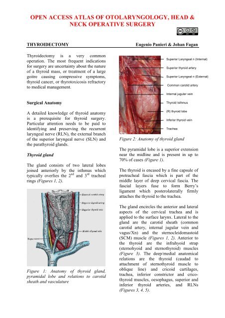
THYROIDECTOMY Eugenio Panieri & Johan Fagan<br />
<strong>Thyroidectomy</strong> is a very common<br />
operation. The most frequent indications<br />
for surgery are uncertainty about the nature<br />
<strong>of</strong> a thyroid mass, or treatment <strong>of</strong> a large<br />
goitre causing compressive symptoms,<br />
thyroid cancer, or thyrotoxicosis refractory<br />
to medical management.<br />
Surgical Anatomy<br />
A detailed knowledge <strong>of</strong> thyroid anatomy<br />
is a prerequisite for thyroid surgery.<br />
Particular attention needs to be paid to<br />
identifying and preserving the recurrent<br />
laryngeal nerve (RLN), the external branch<br />
<strong>of</strong> the superior laryngeal nerve (SLN) and<br />
the parathyroid glands.<br />
Thyroid gland<br />
The gland consists <strong>of</strong> two lateral lobes<br />
joined anteriorly by the isthmus which<br />
typically overlies the 2 nd and 3 rd tracheal<br />
rings (Figures 1, 2).<br />
Figure 1: Anatomy <strong>of</strong> thyroid gland,<br />
pyramidal lobe and relations to carotid<br />
sheath and vasculature<br />
Figure 2: Anatomy <strong>of</strong> thyroid gland<br />
Superior Laryngeal n (Internal)<br />
Superior thyroid artery<br />
Superior Laryngeal n (External)<br />
Common carotid artery<br />
Internal jugular vein<br />
Thyroid Isthmus<br />
(R) thyroid lobe<br />
Inferior thyroid vein<br />
Trachea<br />
RLN<br />
The pyramidal lobe is a superior extension<br />
near the midline and is Common present carotid<br />
in up to<br />
70% <strong>of</strong> cases (Figure 1).<br />
The thyroid is encased by a fine capsule <strong>of</strong><br />
pretracheal fascia which is part <strong>of</strong> the<br />
middle layer <strong>of</strong> deep cervical fascia. The<br />
fascial layers fuse to form Berry’s<br />
ligament which posterolaterally firmly<br />
attaches the thyroid to the trachea.<br />
The gland encircles the anterior and lateral<br />
aspects <strong>of</strong> the cervical trachea and is<br />
applied to the surface larynx. Lateral to the<br />
gland are the carotid sheath (common<br />
carotid artery, internal jugular vein and<br />
vagus/Xn) and the sternocleidomastoid<br />
(SCM) muscle (Figures 1, 2). Anterior to<br />
the thyroid are the infrahyoid strap<br />
(sternohyoid and sternothyroid) muscles<br />
(Figure 3). The deep/medial anatomical<br />
relations are the thyroid (caudad to<br />
attachment <strong>of</strong> sternothyroid muscle to<br />
oblique line) and cricoid cartilages,<br />
trachea, inferior constrictor and cricothyroid<br />
muscles, oesophagus, superior and<br />
inferior thyroid arteries, and RLNs<br />
(Figures 3, 4, 5).
Figure 3: The superficial relations <strong>of</strong> the<br />
thyroid are the infrahyoid strap muscles<br />
(sternohyoid and sternothyroid) and SCM<br />
Figure 4: Structures deep to thyroid gland:<br />
Note the oblique line to which the<br />
sternothyroid muscle inserts and which<br />
defines the anterosuperior limit <strong>of</strong> the<br />
thyroid (Wikipedia)<br />
The thyroid gland weighs 15-25g. The<br />
thyroid lobes are normally cone-shaped<br />
and measure approximately 5cm in length<br />
and 2-3cms in width in both transverse and<br />
anteroposterior dimensions.<br />
The Tubercle <strong>of</strong> Zukerkandl is a pyramidal<br />
enlargement <strong>of</strong> the lateral edge <strong>of</strong> the<br />
thyroid lobe that stems from the fusion <strong>of</strong><br />
the lateral and medial thyroid anlages<br />
(Figure 6). It is recognisable in up to 75%<br />
<strong>of</strong> thyroids. It is anatomically closely<br />
related to the RLN, the inferior thyroid<br />
artery, Berry's ligament and the superior<br />
Ext Carotid artery<br />
Sup thyroid art<br />
Inf constrictor<br />
Cricothyroid<br />
Parathyroids<br />
Inf thyroid art<br />
RLNs<br />
Oesophagus<br />
Thyrocervical<br />
trunk<br />
Figure 5: Posterior view <strong>of</strong> the thyroid<br />
gland demonstrating the deep / medial<br />
anatomical relations, the RLNs and the<br />
superior and inferior thyroid arteries<br />
parathyroid gland. The tubercle usually<br />
projects lateral to the RLN. Elevating the<br />
tubercle allows the RLN to be readily<br />
located. Less commonly the RLN courses<br />
lateral to an enlarged tubercle; this places<br />
the nerve at risk <strong>of</strong> injury. The superior<br />
parathyroid gland, also derived from the 4 th<br />
branchial cleft, is commonly located close<br />
to and cephalad to the tubercle.<br />
Figure 6: Tubercle <strong>of</strong> Zuckerkandl (TZ)<br />
and its relationship to the superior<br />
parathyroid gland and RLN<br />
TZ<br />
Superior parathyroid<br />
RLN<br />
2
Blood supply<br />
The arterial supply is based on the<br />
superior thyroid (STA) and inferior thyroid<br />
(ITA) arteries. Occasionally the thyroidea<br />
ima artery is encountered inferiorly but is<br />
seldom <strong>of</strong> surgical relevance. It arises from<br />
the innominate artery or aortic arch and<br />
ascends along the front <strong>of</strong> the trachea.<br />
The superior thyroid artery (STA) is the<br />
first branch <strong>of</strong> the external carotid artery<br />
(Figures 2, 5, 7). It courses over the<br />
external surface <strong>of</strong> the inferior constrictor<br />
muscle <strong>of</strong> the larynx, entering the gland<br />
posteromedially just below the highest<br />
point <strong>of</strong> the upper pole where it usually is<br />
located superficial to the external branch <strong>of</strong><br />
the SLN (Figure 2). Its branches communicate<br />
with the ITA and cross to the<br />
contralateral thyroid lobe via the thyroid<br />
isthmus.<br />
STA<br />
ITA<br />
Thyrocervical<br />
Subclavian a<br />
Figure 7: Superior thyroid artery (STA),<br />
subclavian artery, thyrocervical trunk and<br />
inferior thyroid artery (ITA)<br />
The inferior thyroid artery (ITA) is a<br />
branch <strong>of</strong> the thyrocervical trunk which<br />
originates from the subclavian artery<br />
(Figures 5, 7). It courses superiorly along<br />
the surface <strong>of</strong> the anterior scalene muscle<br />
before it turns medially behind the carotid<br />
sheath from where it reaches the inferior<br />
pole <strong>of</strong> the thyroid (Figure 5). It provides<br />
blood supply to the thyroid, upper<br />
oesophagus and trachea, and is the sole<br />
arterial supply to all the parathyroid<br />
glands, both superior and inferior. The<br />
relationship <strong>of</strong> the ITA and RLN is<br />
reviewed later.<br />
Venous drainage is quite variable and<br />
occurs via a capsular network <strong>of</strong> thinwalled,<br />
freely intercommunicating veins<br />
which drain through the superior thyroid<br />
veins (adjacent to the STA), the inferior<br />
thyroid veins (exit the inferior pole), and<br />
the middle thyroid vein(s), which course<br />
laterally to drain directly into the internal<br />
jugular vein (Figure 1). The middle<br />
thyroid vein is surgically most relevant; it<br />
is encountered early during thyroid<br />
mobilisation, and failure to secure it causes<br />
bothersome bleeding.<br />
Lymphatic drainage parallels the venous<br />
drainage and occurs to the lateral deep<br />
cervical and pre- and paratracheal lymph<br />
nodes (Figure 8). Understanding the<br />
pattern <strong>of</strong> nodal drainage is particularly<br />
important in managing patients with<br />
thyroid cancer since the cervicocentral<br />
compartment is most commonly involved<br />
in metastatic thyroid cancer.<br />
Figure 8: Posterior view <strong>of</strong> the course <strong>of</strong><br />
the lymphatics and RLNs<br />
3
Recurrent Laryngeal Nerve (RLN)<br />
During thyroid surgery, identification and<br />
preservation <strong>of</strong> the RLN and all <strong>of</strong> its<br />
divisions is essential to minimise<br />
morbidity. The RLN innervates all the<br />
intrinsic muscles <strong>of</strong> the larynx except the<br />
cricothyroid muscle (SLN) and provides<br />
sensory innervation to the larynx. Even<br />
minor neuropraxia may cause dysphonia;<br />
irreversible injury confers permanent<br />
hoarseness. The reported incidence <strong>of</strong> RLN<br />
injury during thyroidectomy is 0 - 28% and<br />
is the most common reason for medicolegal<br />
claims following thyroidectomy.<br />
The RLNs originate from the Xn. After<br />
circling around the subclavian artery<br />
(right) and aortic arch (left) the RLNs<br />
ascend superiorly and medially toward the<br />
tracheoesophageal groove (Figures 8, 9).<br />
The right RLN enters the root <strong>of</strong> the neck<br />
from a more lateral direction. Its course is<br />
less predictable than that <strong>of</strong> the left RLN.<br />
The RLNs enter the larynx deep to the<br />
inferior constrictor muscles and posterior<br />
to the cricothyroid joint.<br />
Xns<br />
RLNs<br />
Subclavian arteries<br />
Aortic arch<br />
Figure 9: Typical anatomical course <strong>of</strong><br />
RLNs (Non-recurrent RLN in red)<br />
The RLN may be non-recurrent in<br />
approximately 0.6% <strong>of</strong> patients i.e. does<br />
not pass around the subclavian artery, but<br />
branches from the Xn higher in the neck,<br />
passing directly to the larynx close to the<br />
superior thyroid vessels (Figure 9). This<br />
aberration almost always occurs on the<br />
right side and is associated with a<br />
retroesophageal subclavian artery.<br />
Knowledge <strong>of</strong> the anatomical relationships<br />
<strong>of</strong> the RLN to the tracheoesophageal<br />
groove, ligament <strong>of</strong> Berry, and ITA is<br />
essential. The course <strong>of</strong> the RLN with<br />
respect to the ITA is quite variable. Most<br />
commonly it crosses behind the branches<br />
<strong>of</strong> the artery, more predictably so on the<br />
left. However, the nerve may pass deep to,<br />
superficial to, or between the terminal<br />
branches <strong>of</strong> the ITA. Up to twenty<br />
anatomical variations have been described.<br />
In Figure 10 the RLN is seen to pass<br />
anterior to the artery.<br />
Figure 10: RLN passing over the inferior<br />
thyroid artery (right neck, thyroid reflected<br />
medially)<br />
The majority <strong>of</strong> RLNs are located within<br />
3mm <strong>of</strong> Berry’s ligament; rarely the nerve<br />
is embedded in it, and more commonly lies<br />
laterally to it.<br />
Classically, the RLN is identified intraoperatively<br />
in Simon’s triangle, which is<br />
formed by the common carotid artery<br />
4
laterally, the oesophagus medially, and the<br />
ITA superiorly (Figure 11).<br />
Figure 11: RLN crossing Simon’s triangle<br />
formed by trachea, inferior thyroid artery<br />
(ITA) and common carotid artery (right<br />
neck, thyroid reflected medially)<br />
The Tubercle <strong>of</strong> Zukerkandl may also be<br />
used as an anatomical landmark to identify<br />
the nerve (Figure 6). The RLN generally<br />
courses between this structure and the<br />
trachea. However, this relationship can<br />
vary with enlargement <strong>of</strong> the tuberculum<br />
thereby placing the nerve at risk during<br />
exploration.<br />
Superior Laryngeal Nerve (SLN)<br />
Trachea<br />
RLN<br />
ITA<br />
Carotid<br />
The SLN is a branch <strong>of</strong> the Xn and has<br />
both an external and internal branch<br />
(Figures 2, 12). The internal branch is<br />
situated above and outside the normal field<br />
<strong>of</strong> dissection; it is sensory and enters the<br />
larynx through the thyrohyoid membrane.<br />
The external branch innervates the<br />
cricothyroid muscle, a tensor <strong>of</strong> the vocal<br />
cord. Injury to the SLN causes hoarseness,<br />
decreased pitch and/or volume, and voice<br />
fatigue. These voice changes are more<br />
subtle than those relating to a RLN injury,<br />
and are frequently underestimated and not<br />
reported. The external branch <strong>of</strong> the SLN<br />
is at risk because <strong>of</strong> its close proximity to<br />
the STA (Figures 12, 13). Understanding<br />
its relationship to the upper pole <strong>of</strong> the<br />
thyroid and the STA is crucial to<br />
preserving its integrity.<br />
Figure 12: Anatomical relations <strong>of</strong><br />
internal and external branches <strong>of</strong> right<br />
SLN to superior thyroid artery and to<br />
superior pole <strong>of</strong> thyroid<br />
The usual configuration is that the nerve is<br />
located behind the STA, proximal to its<br />
entry into the superior pole <strong>of</strong> the thyroid.<br />
The relationships <strong>of</strong> the nerve to the<br />
superior pole and STA are however<br />
extremely variable. Variations include the<br />
nerve passing between the branches <strong>of</strong> the<br />
STA as it enters the superior pole <strong>of</strong> the<br />
thyroid gland; in such cases it is<br />
particularly vulnerable to injury.<br />
Figure 13: Note close proximity <strong>of</strong> external<br />
branch <strong>of</strong> SLN to STA and thyroid vein and<br />
to superior pole <strong>of</strong> thyroid gland<br />
Parathyroid glands<br />
XIIn<br />
SLN (internal)<br />
SLN (external)<br />
STA<br />
There are typically four parathyroid<br />
glands; however, supernumerary glands<br />
have been reported. The parathyroid glands<br />
Sup pole thyroid<br />
SLN Ext branch<br />
STA<br />
Thyroid<br />
STVs<br />
5
are generally symmetrically located in the<br />
neck. Their characteristic golden colour<br />
varies from yellow to reddish brown, and<br />
permits them to be distinguished from the<br />
pale-yellow colour <strong>of</strong> adjacent lymph<br />
nodes, thymus, mediastinal fat, and the<br />
dark-red thyroid parenchyma. They are<br />
usually oval and measure 3–8 mm. The<br />
ITA is the predominant vascular supply to<br />
both the upper and lower parathyroids.<br />
Consequently dividing the trunk <strong>of</strong> the ITA<br />
is discouraged as it places all the<br />
parathyroids on that side at risk <strong>of</strong><br />
ischaemic injury.<br />
The superior parathyroid glands originate<br />
from the 4 th pharyngeal pouch and attach<br />
to the posterior surface <strong>of</strong> the caudally<br />
migrating thyroid. They have a much<br />
shorter migration distance compared to the<br />
inferior parathyroid glands; this accounts<br />
for their more predictable location. They<br />
are embryologically and anatomically<br />
closely related to the Tubercle <strong>of</strong><br />
Zuckerkandl, and are usually located at the<br />
level <strong>of</strong> the upper two-thirds <strong>of</strong> the thyroid,<br />
in a posterior position, about 1cm above<br />
the crossing point <strong>of</strong> the RLN and inferior<br />
thyroid artery (Figure 6). Ectopic positions<br />
<strong>of</strong> the superior parathyroid glands such as<br />
in the posterior neck, retropharyngeal and<br />
retroesophageal spaces and intrathyroidally<br />
are very uncommon (1%).<br />
The dorsal wing <strong>of</strong> the 3 rd pharyngeal<br />
pouch gives rise to the inferior<br />
parathyroid glands. They join the thymus<br />
as it travels caudally and medially to its<br />
final position in the mediastinum. This<br />
accounts for the fact that they are usually<br />
found in a plane ventral to that <strong>of</strong> the<br />
superior parathyroid glands, and that<br />
ectopic inferior parathyroid glands can be<br />
found anywhere along this large area <strong>of</strong><br />
descent up to the superior border <strong>of</strong> the<br />
pericardium. Their commonest location is<br />
between the lower pole <strong>of</strong> the thyroid and<br />
isthmus, equally commonly on the anterior<br />
or the posterolateral surfaces <strong>of</strong> the lower<br />
pole <strong>of</strong> the thyroid (42%, Wang et al); or in<br />
the lower neck in proximity to the thymus<br />
(39%). Other locations are: lateral to the<br />
thyroid or within the carotid sheath (15%),<br />
within the mediastinal thymic tissue and<br />
the pericardium (2%).<br />
If the RLN’s course is viewed in a coronal<br />
plane, then the superior parathyroid gland<br />
is located deep (dorsal) and the inferior<br />
parathyroid superficial (ventral) to the<br />
plane <strong>of</strong> the nerve (Figures 14a, b).<br />
Figure 14a: The superior parathyroid<br />
gland lies deep (dorsal) and the inferior<br />
parathyroid superficial (ventral) to a<br />
coronal plane along course <strong>of</strong> RLN<br />
Inferior PT<br />
Superior PT<br />
RLN<br />
Figure 14b: The superior parathyroid<br />
gland lies deep (dorsal) and the inferior<br />
parathyroid superficial (ventral) to a<br />
coronal plane along the course <strong>of</strong> the RLN<br />
6
Types <strong>of</strong> thyroidectomy<br />
Thyroid lobectomy: Either lobe is<br />
removed, usually with a small segment <strong>of</strong><br />
the thyroid isthmus; the contralateral lobe<br />
is left undisturbed. It is most commonly<br />
performed as a diagnostic procedure for a<br />
thyroid nodule <strong>of</strong> uncertain nature. It may<br />
be a sufficient for cure in some cases <strong>of</strong><br />
thyroid carcinoma with favourable<br />
prognostic criteria.<br />
Subtotal thyroidectomy: 90-95% <strong>of</strong><br />
thyroid tissue is removed bilaterally,<br />
leaving a small (1x2cm) thyroid remnant in<br />
situ overlying the RLN. This operation has<br />
slowly lost favour as it is by its very nature<br />
inexact, is prone to recurrence <strong>of</strong> the<br />
thyroid pathology, and in expert hands<br />
does not result in lower rates <strong>of</strong> RLN injury<br />
when compared to total thyroidectomy.<br />
Total thyroidectomy: Both right and left<br />
lobes, isthmus and pyramidal lobe (when<br />
present) are removed; no macroscopic<br />
thyroid tissue is left in situ. This is the<br />
procedure <strong>of</strong> choice for the treatment <strong>of</strong><br />
thyroid carcinoma and is commonly<br />
performed for a MNG with compressive<br />
symptoms, or for thyrotoxicosis.<br />
Subtotal vs. total thyroidectomy for<br />
differentiated thyroid carcinoma<br />
Bilateral RLN injury causing airway<br />
compromise and hypoparathyroidism<br />
causing hypocalcaemia in situations where<br />
monitoring serum calcium and treating<br />
hypocalcaemia with calcium and Vitamin<br />
D are not possible may have fatal<br />
consequences. Regardless <strong>of</strong> surgical<br />
expertise, the complication rates rise with<br />
the extent <strong>of</strong> resection. Unilateral thyroid<br />
lobectomy rarely causes RLN injury and<br />
almost never causes significant<br />
hypoparathyroidism. Subtotal thyroidectomy<br />
preserves the blood supply to the<br />
ipsilateral parathyroid glands and reduces<br />
the risk <strong>of</strong> hypocalcaemia. Total thyroidectomy<br />
is however associated with both<br />
increased short- and long-term morbidity<br />
relating to RLN paralysis and hypocalcaemia,<br />
particularly in an occasional<br />
thyroid surgeon’s hands. Short-term<br />
complication rates for total thyroidectomy<br />
occur in 10-40% <strong>of</strong> patients; long-term<br />
complications (mainly hypoparathyroidism)<br />
occur in 5-20%. Most thyroidectomies<br />
are done in general hospitals by<br />
surgeons not specialising in endocrine<br />
surgery; complication rates have been<br />
reported to correlate with the number <strong>of</strong><br />
thyroidectomies done. In the absence <strong>of</strong><br />
convincing evidence that total thyroidectomy<br />
confers survival benefit in favourable<br />
differentiated thyroid cancers (especially<br />
when I 131 therapy is not available), coupled<br />
with the morbidity and mortality <strong>of</strong> total<br />
thyroidectomy, the occasional thyroid<br />
surgeon or the surgeon practising in a<br />
setting where calcium monitoring and<br />
replacement are suboptimal may therefore<br />
elect rather to perform thyroid lobectomy<br />
or subtotal thyroidectomy for differentiated<br />
thyroid cancer.<br />
Pre-operative evaluation<br />
Ultrasonography (US) permits accurate<br />
distinction between the common thyroid<br />
pathologies and is the imaging technique<br />
<strong>of</strong> choice for a thyroid mass. Neoplasms<br />
typically cause focal enlargement within a<br />
normal gland (“solitary nodule”). Features<br />
strongly suggestive <strong>of</strong> thyroid carcinoma<br />
are hypoechogenicity, increased and<br />
haphazard vascularity patterns within the<br />
lesion, microcalcifications, irregular margins,<br />
elevated height-to-width ratio, and<br />
regional lymphadenopathy. A multinodular<br />
goitre (MNG) typically shows multiple<br />
hyper- or isoechoic nodules, some cystic<br />
changes and coarse macrocalcifications<br />
involving both thyroid lobes.<br />
7
Focal thyroid masses or suspicious<br />
lymphadenopathy should be investigated<br />
by fine needle aspiration cytology.<br />
All patients with thyroid complaints must<br />
undergo thyroid function tests as clinical<br />
manifestations <strong>of</strong> thyrotoxicosis or<br />
hypothyroidism are notoriously unreliable.<br />
Thyrotoxicosis must first be controlled<br />
medically before surgical intervention.<br />
Failure to do so may precipitate a thyroid<br />
storm.<br />
CT scans are helpful in selected cases,<br />
particularly with a MNG with a suspected<br />
retrosternal component (Figure 15), or<br />
when uncertainty exists about the extent <strong>of</strong><br />
tracheal compression (Figure 16).<br />
Figure 15: Coronal CT scan demonstrating<br />
retrosternal extension<br />
http://chestatlas.com/gallery/Thyroid/HUGE goitre CT<br />
Figure 16: Tracheal compression on CT<br />
scan<br />
Thyroid uptake scans may be requested in<br />
cases <strong>of</strong> thyroid enlargement with<br />
thyrotoxicosis, but are not routinely done<br />
as they seldom add more information to<br />
that available from the US.<br />
Laryngoscopy: It is medico-legally prudent<br />
to document vocal cord function prior to<br />
thyroid surgery; it is essential in patients<br />
with symptoms <strong>of</strong> dysphonia.<br />
Preoperative consent<br />
Scar: The incision is generally well<br />
concealed if made within a natural skin<br />
crease, but tends to descend with ageing.<br />
Airway obstruction/wound haematoma:<br />
1% <strong>of</strong> thyroidectomy patients develop<br />
stridor postoperatively, either due to<br />
airway oedema or a haematoma.<br />
Voice changes: It is essential for the<br />
patient to have a clear understanding <strong>of</strong> the<br />
risk <strong>of</strong> postoperative voice change. While<br />
most are subtle and recover fully,<br />
approximately 1% <strong>of</strong> patients will have<br />
permanent hoarseness. The risk is highest<br />
for patients having surgery for carcinoma,<br />
large retrosternal goitres, and with repeat<br />
surgery.<br />
Hypoparathyroidism: Transient postoperative<br />
hypocalcaemia occurs in about 20% <strong>of</strong><br />
total thyroidectomy patients. Permanent<br />
hypocalcaemia occurs following 1-5% <strong>of</strong><br />
total thyroidectomies.<br />
Hypothyroidism: Hypothyroidism occurs<br />
uncommonly (5%) with thyroid lobectomy.<br />
It is common practice to routinely<br />
check TSH levels approximately 6-8<br />
weeks after surgery to identify such cases<br />
before it manifests clinically. It is selfevident<br />
that a patient will be hypothyroid<br />
following total thyroidectomy. The clinical<br />
effects only become apparent once the pre-<br />
8
existing thyroid hormone levels drop; this<br />
generally becomes evident 3-4 weeks<br />
following surgery. Thyroxine replacement<br />
therapy is routinely instituted immediately<br />
postoperatively to prevent hypothyroidism.<br />
The exception is if total thyroidectomy has<br />
been performed for a well-differentiated<br />
carcinoma and I 131 therapy is envisaged; a<br />
hypothyroid state is deliberately induced in<br />
such patients until the I 131 therapy has been<br />
administered.<br />
Anaesthesia, positioning and draping<br />
General anaesthesia with endotracheal<br />
intubation<br />
Prophylactic antibiotics are not<br />
indicated<br />
Neck slightly hyperextended by<br />
placing a bolster between the scapulae<br />
Head stabilised on a head ring<br />
Table tilted to 30º anti-Trendelenberg<br />
position to reduce venous engorgement<br />
Head is free-draped to allow turning <strong>of</strong><br />
the head<br />
Surgical technique<br />
Skin incision (Figure 17): A curvilinear<br />
incision is placed in a skin crease two<br />
fingerbreadths above the sternal notch<br />
between the medial borders <strong>of</strong> the<br />
sternocleidomastoid muscles.<br />
Figure 17: Curvilinear skin incision two<br />
fingerbreadths above the sternal notch<br />
Placing the incision too low causes an<br />
unsightly low scar over the heads <strong>of</strong> the<br />
clavicles when the extended neck is<br />
returned to its normal position. The width<br />
<strong>of</strong> the incision may need to be extended for<br />
large goitres or for a lateral lymph node<br />
dissection.<br />
Subplatysmal flaps: Subcutaneous fat and<br />
platysma are divided, and a subplatysmal<br />
dissection plane is developed superiorly<br />
(platysma is <strong>of</strong>ten absent in the midline)<br />
remaining superficial to the anterior<br />
jugular veins, up to the level <strong>of</strong> the thyroid<br />
cartilage above, and the sternal notch<br />
below (Figure 18).<br />
Figure 18: Subplatysmal flaps elevated<br />
AJV<br />
s<br />
Figure 19: Subplatysmal skin flaps held<br />
with Jowell’s retractor. Note anterior<br />
jugular veins (AJVs)<br />
9
The skin flaps are secured to a fixed<br />
retractor (e.g. Jowell’s retractor) to expose<br />
the thyroid region for the remainder <strong>of</strong><br />
operation (Figure 19).<br />
Separating strap muscles and exposing<br />
the anterior surface <strong>of</strong> thyroid: The fascia<br />
between the sternohyoid and sternothyroid<br />
muscles is divided along the midline with<br />
diathermy or scissors (Figure 20). This is<br />
an avascular plane, though care must be<br />
taken not to injure small veins occasionally<br />
crossing between the anterior jugular<br />
veins, particularly inferiorly. The<br />
infrahyoid (sternohyoid, sternothyroid and<br />
omohyoid) strap muscles are retracted<br />
laterally with a right-angled retractor. With<br />
massive goitres the strap muscles may be<br />
divided to improve access.<br />
Figure 20: Fascia between sternohyoid<br />
and sternothyroid muscles divided to<br />
expose thyroid gland<br />
It is usual at this stage for the surgeon to<br />
move to the side <strong>of</strong> the table opposite to the<br />
thyroid lobe to be resected.<br />
Medially rotating the thyroid: Using<br />
gentle digital retraction the surgeon rotates<br />
the thyroid gland medially (Figure 21).<br />
Dividing the middle thyroid vein(s)<br />
(Figure 21): The first important vascular<br />
structure to come into view is the middle<br />
thyroid vein(s), which is tightly stretched<br />
by medial rotation <strong>of</strong> the gland. It is<br />
divided between haemostats and ligated<br />
with a 3/0 tie. This permits further<br />
mobilisation <strong>of</strong> the gland and delivery <strong>of</strong><br />
the bulk <strong>of</strong> the lobe into the wound.<br />
Figure 21: Medial rotation <strong>of</strong> (R) thyroid<br />
lobe exposes the middle thyroid vein<br />
Figure 22: Dividing the middle thyroid<br />
vein<br />
Dividing the STA (Figure 23): The<br />
retractors are repositioned to allow full<br />
visualisation <strong>of</strong> the superior pole <strong>of</strong> the<br />
thyroid. This brings the STA into view.<br />
The author does not routinely identify the<br />
external branch <strong>of</strong> the SLN, but simply<br />
takes great care to divide the artery as<br />
close to the thyroid parenchyma as<br />
possible so as to avoid injury to nerve. The<br />
superior arterial pedicle is double ligated<br />
with 2/0 or 3/0 tie.<br />
10
Figure 23: Dividing STA pedicle below<br />
external branches <strong>of</strong> SLN<br />
Identifying superior parathyroid gland<br />
(Figure 24, 25): Full mobilization and<br />
anterior delivery <strong>of</strong> the superior pole <strong>of</strong> the<br />
thyroid brings the region <strong>of</strong> the superior<br />
parathyroid gland into direct view. The<br />
superior parathyroid gland is normally<br />
located at the level <strong>of</strong> the upper two-thirds<br />
<strong>of</strong> the thyroid, in a posterior position and<br />
closely related to the Tubercle <strong>of</strong><br />
Zuckerkandl, and about 1 cm above the<br />
crossing point <strong>of</strong> the recurrent laryngeal<br />
nerve and inferior thyroid artery.<br />
Figure 24: Position <strong>of</strong> superior parathyroid<br />
relative to Tubercle <strong>of</strong> Zuckerkandl<br />
(TZ), RLN and STA<br />
If the RLN’s course is viewed in a coronal<br />
plane, then the superior parathyroid gland<br />
lies deep (dorsal) to the plane <strong>of</strong> the nerve<br />
(Figures 14a, b). It is has a characteristic<br />
rich orange/yellow colour (Figure 25). The<br />
(occasional) parathyroid surgeon may find<br />
the parathyroids difficult to identify<br />
especially if there has been bleeding the<br />
surgical field, so care must be taken to<br />
TZ<br />
Sup parathyroid<br />
Crossing point<br />
<strong>of</strong> RLN & STA<br />
ensure meticulous haemostasis. The gland<br />
must remain in situ with blood supply<br />
intact. This is best achieved by carefully<br />
dissecting it <strong>of</strong>f the posterior aspect <strong>of</strong> the<br />
thyroid gland, and using short bursts <strong>of</strong><br />
bipolar cautery to control bleeding.<br />
Inferior PT<br />
Superior PT<br />
RLN<br />
Figure 25: Superior and inferior<br />
parathyroids (PT)<br />
Dividing inferior thyroid veins (Figure<br />
26): The retractors are again repositioned<br />
to expose the lower neck and the inferior<br />
thyroid vein(s). The veins are divided, and<br />
ligated. This exposes the trachea and<br />
permits full delivery <strong>of</strong> the thyroid gland<br />
outside the wound.<br />
Figure 26: Inferior thyroid vein being<br />
divided<br />
Identifying inferior parathyroid gland:<br />
The inferior parathyroid glands are<br />
normally located between the lower pole <strong>of</strong><br />
the thyroid and the isthmus, most<br />
commonly on the anterior or the<br />
posterolateral surface <strong>of</strong> the lower pole <strong>of</strong><br />
the thyroid (42%, Wang et al), or located<br />
in the lower neck in proximity to the<br />
thymus (39%).<br />
11
If the RLN’s course is viewed in a coronal<br />
plane then the inferior parathyroid is<br />
superficial (ventral) to the plane <strong>of</strong> the<br />
nerve (Figures 14a, b). The inferior gland<br />
may now become visible on the inferior<br />
aspect <strong>of</strong> the lower pole <strong>of</strong> the thyroid or<br />
within the thyrothymic ligament (Figure<br />
25). Care must be taken to preserve it in<br />
situ and to avoid damaging its ITA blood<br />
supply.<br />
Identifying the RLN: The thyroid is<br />
rotated medially; lateral retraction is<br />
applied to the carotid artery and jugular<br />
vein. The RLN is located by carefully<br />
dissecting/teasing apart the tissues in<br />
Simon’s triangle which is formed by the<br />
common carotid artery laterally, the<br />
oesophagus medially, and the inferior<br />
thyroid artery superiorly (Figure 11).<br />
Others favour finding the nerve at its point<br />
<strong>of</strong> entry into the larynx approx. 0.5cm<br />
caudad to the inferior cornu <strong>of</strong> the thyroid<br />
cartilage. The nerve must remain<br />
undisturbed and in situ i.e. is not<br />
skeletonised or handled.<br />
Pericapsular dissection <strong>of</strong> branches <strong>of</strong><br />
ITA: It is best to individually dividing and<br />
ligating (3/0 ties) all the branches <strong>of</strong> the<br />
ITA at the capsule <strong>of</strong> the thyroid so as to<br />
reduce the risk <strong>of</strong> handling the RLN.<br />
Avoid all forms <strong>of</strong> cautery to avoid<br />
thermal injury to the nerve.<br />
Dividing Ligament <strong>of</strong> Berry (Figure 26):<br />
The posteromedial aspect <strong>of</strong> the thyroid<br />
gland is attached to the side <strong>of</strong> the cricoid<br />
cartilage and to the 1 st and 2 nd tracheal<br />
rings by the posterior suspensory ligament/<br />
Ligament <strong>of</strong> Berry. The RLN is in close<br />
proximity (
thyroid bed and brought out through a<br />
laterally placed skin puncture<br />
The strap muscles are approximated for<br />
70% <strong>of</strong> their length, and the platysma<br />
is closed with interrupted absorbable<br />
3/0 sutures<br />
A subcuticular skin closure is achieved<br />
with an absorbable mon<strong>of</strong>ilament<br />
suture<br />
A light dressing is applied<br />
Postoperative care<br />
The patient is monitored overnight for<br />
bleeding and airway obstruction<br />
The intravenous line is removed and a<br />
normal diet is taken as tolerated<br />
If a drain has been placed it is removed<br />
when drainage is
apparent depending on the resting position<br />
<strong>of</strong> the vocal fold. Bilateral RLN paralysis<br />
usually manifests immediately following<br />
extubation with stridor or airway<br />
obstruction. Should the patient be unable<br />
to maintain an adequate airway then<br />
emergency tracheostomy or cricothyroidotomy<br />
is indicated. Subsequent management<br />
depends on the surgeon’s knowledge <strong>of</strong><br />
whether the RLNs were seen to be intact<br />
and hence the likelihood <strong>of</strong> vocal fold<br />
function to recover. Options might include<br />
a watchful waiting approach for up to a<br />
year or CO2 laser cordotomy/arytenoidectomy.<br />
Continuous electrophysiologic monitoring<br />
<strong>of</strong> the RLN during thyroid surgery:<br />
Recent studies have shown that<br />
intraoperative monitoring can assist with<br />
finding the RLN, but some pitfalls limit its<br />
usefulness: there is no consensus about<br />
which types <strong>of</strong> electrodes should be used<br />
for EMG registration which is the best<br />
method for recording nerve action, or<br />
which EMG parameters should be selected<br />
as predictive <strong>of</strong> postoperative vocal cord<br />
dysfunction. The technology is not widely<br />
available, and most endocrine surgeons<br />
achieve equivalent RLN morbidity rates<br />
without it.<br />
Tracheomalacia: This is characterized by<br />
flaccidity <strong>of</strong> the tracheal cartilages which<br />
in turn causes tracheal wall collapse. It is<br />
thought that a longstanding goiter can act<br />
as an external support structure for the<br />
trachea and predispose to secondary<br />
tracheomalacia. <strong>Thyroidectomy</strong> unmasks<br />
tracheomalacia causing respiratory<br />
obstruction. In clinical practice this is an<br />
uncommon cause <strong>of</strong> airway obstruction<br />
after thyroidectomy.<br />
Thyroid specific haemostatic devices<br />
(Figure 28): The last decade has seen the<br />
introduction <strong>of</strong> thyroid specific<br />
haemostatic devices (Ultrasonic scissors/<br />
Harmonic Scalpel and Ligasure Device),<br />
which achieve safe haemostasis and avoid<br />
the need for multiple ligatures.<br />
Figure 28: Harmonic Scalpel<br />
A number <strong>of</strong> randomised trials have shown<br />
equivalence between the commercially<br />
available products, and a significant<br />
reduction in operating time without an<br />
increase in complications when compared<br />
to standard thyroidectomy technique. The<br />
author uses the Harmonic Scalpel as a<br />
means <strong>of</strong> sealing and transecting vessels<br />
and to reduce surgical operating time.<br />
Minimally Invasive Thyroid Surgery: A<br />
number <strong>of</strong> techniques have evolved in an<br />
attempt to reduce the extent <strong>of</strong> skin<br />
incisions and bring the putative benefits <strong>of</strong><br />
minimally invasive techniques to thyroid<br />
surgery. Minimally invasive thyroidectomy<br />
can be performed via a limited 2-3cm neck<br />
incision with the visual assistance <strong>of</strong> an<br />
endoscope, specially designed retractors<br />
and a harmonic scalpel. An alternative<br />
approach is to place incisions for 3-4 ports<br />
in the axilla and periareolar regions to<br />
avoid a neck scar altogether. The clinical<br />
benefits are marginal at best, but they will<br />
continue to be driven by patient demand<br />
and the industry. Only patients with small<br />
thyroid nodules are suitable for such a<br />
surgical approach.<br />
14
Useful References<br />
1. Mohebati A, Shaha AR. Anatomy <strong>of</strong><br />
thyroid and parathyroid glands and<br />
neurovascular relations. Clin Anat.<br />
2012;25(1):19-31<br />
2. Bliss RD, Gauger PG, Delbridge LW.<br />
Surgeon's Approach to the Thyroid<br />
Gland: Surgical Anatomy and the<br />
Importance <strong>of</strong> Technique. World J<br />
Surg. 2000;24(8):891-7<br />
3. Wang C. The anatomic basis <strong>of</strong><br />
parathyroid surgery. Ann Surg. 1976;<br />
183:271–5<br />
1 st Author<br />
Eugenio Panieri MBChB, FCS<br />
Head: Oncology / Endocrine Surgery Unit<br />
Associate Pr<strong>of</strong>essor<br />
Division <strong>of</strong> General Surgery<br />
<strong>University</strong> <strong>of</strong> <strong>Cape</strong> <strong>Town</strong><br />
<strong>Cape</strong> <strong>Town</strong><br />
South Africa<br />
eugenio.panieri@uct.ac.za<br />
2 nd Author and Editor<br />
Johan Fagan MBChB, FCORL, MMed<br />
Pr<strong>of</strong>essor and Chairman<br />
Division <strong>of</strong> Otolaryngology<br />
<strong>University</strong> <strong>of</strong> <strong>Cape</strong> <strong>Town</strong><br />
<strong>Cape</strong> <strong>Town</strong><br />
South Africa<br />
johannes.fagan@uct.ac.za<br />
THE OPEN ACCESS ATLAS OF<br />
OTOLARYNGOLOGY, HEAD &<br />
NECK OPERATIVE SURGERY<br />
www.entdev.uct.ac.za<br />
The Open Access Atlas <strong>of</strong> Otolaryngology, Head &<br />
Neck Operative Surgery by Johan Fagan (Editor)<br />
johannes.fagan@uct.ac.za is licensed under a Creative<br />
Commons Attribution - Non-Commercial 3.0 Unported<br />
License<br />
15


