Norma Basalis OBJECTIVES At the end of this lecture, students will ...
Norma Basalis OBJECTIVES At the end of this lecture, students will ...
Norma Basalis OBJECTIVES At the end of this lecture, students will ...
Create successful ePaper yourself
Turn your PDF publications into a flip-book with our unique Google optimized e-Paper software.
<strong>Norma</strong> <strong>Basalis</strong><br />
<strong>OBJECTIVES</strong><br />
<strong>At</strong> <strong>the</strong> <strong>end</strong> <strong>of</strong> <strong>this</strong> <strong>lecture</strong>, <strong>students</strong> <strong>will</strong> be able to:-<br />
• Bones forming <strong>the</strong> base <strong>of</strong> skull<br />
• Narrate <strong>the</strong> details <strong>of</strong> anterior, middle and posterior part <strong>of</strong> base <strong>of</strong><br />
skull<br />
• Identify different foramina and structures passing through <strong>the</strong>m at<br />
<strong>the</strong> base<br />
• Explain <strong>the</strong> attachments and relations <strong>of</strong> base <strong>of</strong> skull<br />
• Formed by<br />
Inferior Aspect <strong>of</strong> Skull<br />
– Palatine processes <strong>of</strong> maxilla<br />
– Horizontal plate <strong>of</strong> palatine bones<br />
– Vomer<br />
– Pterygoid processes, inferior surfaces <strong>of</strong> great wings, spinous<br />
processes & part <strong>of</strong> body <strong>of</strong> sphenoid<br />
– Inferior surfaces <strong>of</strong> squamae & petrous portions <strong>of</strong> temporals<br />
– Inferior surface <strong>of</strong> occipital bone<br />
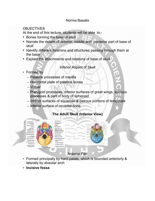
The Adult Skull (Inferior View)<br />
Anterior Part<br />
• Formed principally by hard palate, which is bounded anteriorly &<br />
laterally by alveolar arch<br />
• Incisive fossa
– Lateral incisive foramina (<strong>of</strong> Stenson) which continue as incisive<br />
canals & transmit terminal branches <strong>of</strong> greater palatine vessels<br />
& nasopalatine nerves from nasal cavity<br />
– Median incisive foramina (<strong>of</strong> Scarpa) transmit nasopalatine<br />
nerves when present<br />
• Depressions for palatine glands<br />
• Cruciate suture<br />
palatine process<br />
palatine bone<br />
vomer bone<br />
temporal bone<br />
external occipital<br />
protuberance<br />
Ventral Skull<br />
Ventral Skull<br />
sphenoid bone<br />
Palatine Process<br />
styloid process<br />
mastoid process<br />
occipital bone<br />
• The palatine process <strong>of</strong> <strong>the</strong> maxilla (palatal process), thick and<br />
strong, is horizontal and projects medialward from <strong>the</strong> nasal<br />
surface <strong>of</strong> <strong>the</strong> bone.
• It forms a considerable part <strong>of</strong> <strong>the</strong> floor <strong>of</strong> <strong>the</strong> nose and <strong>the</strong><br />
ro<strong>of</strong> <strong>of</strong> <strong>the</strong> mouth and is much thicker in front than behind.<br />
Incisive Foramen<br />
• When <strong>the</strong> two maxillæ are articulated, a funnel-shaped<br />
opening, <strong>the</strong> incisive foramen, is seen in <strong>the</strong> middle line,<br />
immediately behind <strong>the</strong> incisor teeth.<br />
Anterior Part<br />
• Greater palatine foramen<br />
– For transmission <strong>of</strong> greater palatine vessels & nerve, which<br />
desc<strong>end</strong> in greater palatine canal from pterygopalatine fossa<br />
• Lesser palatine foramina<br />
– Pyramidal process <strong>of</strong> palatine bone, perforated by one or more<br />
foramina through which course lesser palatine vessels & nerve<br />
to s<strong>of</strong>t palate<br />
– Transverse ridge for attachment <strong>of</strong> t<strong>end</strong>inous expansion <strong>of</strong><br />
tensor veli palatini muscle<br />
• Posterior nasal spine, on which attaches musculus uvulae
Middle Part<br />
• Commences just behind hard palate & ext<strong>end</strong>s to level <strong>of</strong> anterior<br />
border <strong>of</strong> foramen magnum.<br />
• Choanae<br />
• Medial pterygoid plate<br />
– Scaphoid fossa for origin <strong>of</strong> tensor veli palatini muscle<br />
– Pterygoid hamulus, around which t<strong>end</strong>on <strong>of</strong> tensor veli palatini<br />
muscle turns<br />
• Lateral pterygoid plate<br />
– Its lateral surface affords attachment to pterygoideus lateralis<br />
muscle<br />
• Pharyngeal tubercle<br />
– Near center <strong>of</strong> basilar portion <strong>of</strong> occipital bone<br />
– For attachment <strong>of</strong> fibrous raphe <strong>of</strong> pharynx<br />
– Depressions on each side for insertions <strong>of</strong> rectus capitis anterior<br />
& longus capitis<br />
• Foramen ovale<br />
– <strong>At</strong> base <strong>of</strong> lateral pterygoid plate<br />
– Through which passes mandibular nerve, accessory meningeal<br />
artery, & lesser petrosal nerve<br />
• Sphenoidal spine<br />
– Lateral to foramen ovale<br />
– <strong>At</strong>tachment to sphenomandibular ligament & tensor veli palatini<br />
• Foramen spinosum<br />
– Posterior & somewhat lateral to foramen ovale<br />
– Transmits middle meningeal vessels & small meningeal branch<br />
<strong>of</strong> mandibular nerve<br />
• Mandibular fossa<br />
– Lateral to sphenoidal spine
– Divided into 2 parts by petrotympanic fissure<br />
– Anterior portion - concave, smooth, & bounded in front by<br />
articular tubercle - articulates with condyle <strong>of</strong> mandible.<br />
– Posterior portion is rough & bounded behind by tympanic part<br />
<strong>of</strong> temporal bone.<br />
Middle Part<br />
• Foramen lacerum<br />
– <strong>At</strong> base <strong>of</strong> medial pterygoid plate in dried skull<br />
– Irregular in shape & variable in size<br />
– Not complete foramen in intact body, because its inferior part is<br />
covered over by fibrocartilaginous plate, across superior (inner<br />
or cerebral) surface <strong>of</strong> which passes internal carotid artery.<br />
– Boundary<br />
- In front by great wing <strong>of</strong> sphenoid<br />
- Behind by apex <strong>of</strong> petrous portion <strong>of</strong> temporal bone<br />
- Medially by body <strong>of</strong> sphenoid & basilar portion <strong>of</strong> occipital bone<br />
• Carotid canal<br />
– Inferior surface <strong>of</strong> petrous temporal bone is pierced by round<br />
opening.<br />
– Internal carotid artery, coursing within canal, immediately takes<br />
right angle turn to reach side <strong>of</strong> foramen lacerum.<br />
• Quadrilateral surface<br />
– Rough surface near apex <strong>of</strong> petrous portion <strong>of</strong> temporal bone,<br />
lateral to which is orifice or entrance <strong>of</strong> carotid canal.<br />
– <strong>At</strong>tachment to levator veli palatini<br />
•<br />
• Sulcus tubae auditivae<br />
– Lateral to foramen lacerum, between petrous part <strong>of</strong> temporal &<br />
great wing <strong>of</strong> sphenoid<br />
– Lodges cartilaginous part <strong>of</strong> auditory tube which is continuous<br />
with bony part within temporal bone<br />
– Petrosphenoidal fissure at bottom <strong>of</strong> <strong>this</strong> sulcus
Posterior Part<br />
• Formed principally by occipital bone<br />
• Mastoid process<br />
– Mastoid notch on medial side <strong>of</strong> each process, for posterior belly<br />
<strong>of</strong> digastricus<br />
– Occipital groove medial to mastoid notch, for occipital artery<br />
• Styloid process<br />
– Medial & slightly anterior to mastoid processes<br />
• Stylomastoid foramen<br />
– <strong>At</strong> base <strong>of</strong> styloid process<br />
– Facial nerve exits toward side <strong>of</strong> face, & stylomastoid artery<br />
enters to tympanic cavity.<br />
•<br />
• Jugular foramen<br />
– Medial to styloid process & posterior to carotid canal<br />
– Anterior compartment – inferior petrosal sinus<br />
– Intermediate – glossopharyngeal, vagus, & accessory nerves<br />
– Posterior – sigmoid sinus which leads to internal jugular vein, &<br />
some meningeal branches from occipital & asc<strong>end</strong>ing<br />
pharyngeal arteries<br />
• Petro-occipital fissure<br />
– Ext<strong>end</strong>ing anteriorly from jugular foramen to foramen lacerum<br />
carotid<br />
canal<br />
jugular<br />
foramen<br />
foramen magnum<br />
Occipital bone<br />
occipital<br />
condyle<br />
Occipital bone
Posterior Part<br />
• Foramen magnum<br />
– Posterior to basilar portion <strong>of</strong> occipital bone<br />
– Transmit<br />
- Medulla oblongata & its membranes<br />
- Accessory nerves<br />
- Vertebral arteries<br />
- Anterior & posterior spinal arteries<br />
- Ligaments connecting occipital bone with axis<br />
Occipital condyles<br />
– By which foramen magnum is bounded laterally<br />
– On medial surfaces <strong>of</strong> which attach alar ligaments<br />
– Articulate with superior articular surfaces overlying lateral<br />
masses <strong>of</strong> atlas<br />
• Jugular process<br />
– Lateral to each occipital condyle<br />
– <strong>At</strong>tachment for rectus capitis lateralis muscle & lateral atlantooccipital<br />
ligament<br />
• Hypoglossal canal<br />
– Courses forward & laterally from inner aspect <strong>of</strong> occipital bone<br />
within cranium just above foramen magnum to opening that<br />
perforates occipital bone externally at lateral part <strong>of</strong> base <strong>of</strong><br />
occipital condyle<br />
– Transmits hypoglossal nerve & a branch <strong>of</strong> posterior meningeal<br />
artery<br />
• Condyloid fossa<br />
– Posterior to each occipital condyle<br />
– Perforated on one or both sides by condyloid canal, for<br />
transmission <strong>of</strong> a vein from sigmoid sinus to vertebral veins in<br />
upper cervical region<br />
• External occipital crest<br />
• External occipital protuberance<br />
• Superior & inferior nuchal lines<br />
Thankyou


