

Access to Parapharyngeal Space - Vula - University of Cape Town
Access to Parapharyngeal Space - Vula - University of Cape Town
Access to Parapharyngeal Space - Vula - University of Cape Town

Create successful ePaper yourself
Turn your PDF publications into a flip-book with our unique Google optimized e-Paper software.
Prestyloid tumours displace the PPS fat<br />
anteromedially (Figures 4, 5).<br />
Figure 4: Direction <strong>of</strong> displacement <strong>of</strong> fat<br />
as seen on CT or MRI with prestyloid PPS<br />
mass (light blue)<br />
Figure 5: Pleomorphic adenoma <strong>of</strong> prestyloid<br />
PPS displacing fat anteromedially<br />
Examples <strong>of</strong> masses commonly<br />
encountered in the prestyloid PPS include<br />
pleomorphic adenoma (Figures 3, 5),<br />
medial extension <strong>of</strong> a deep lobe parotid<br />
tumour (Figure 6) and a ranula that has<br />
tracked in<strong>to</strong> the PPS (Figure 7).<br />
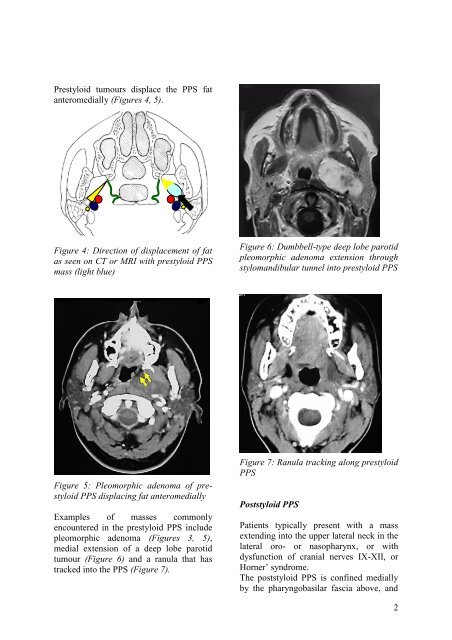
Figure 6: Dumbbell-type deep lobe parotid<br />
pleomorphic adenoma extension through<br />
stylomandibular tunnel in<strong>to</strong> prestyloid PPS<br />
Figure 7: Ranula tracking along prestyloid<br />
PPS<br />
Poststyloid PPS<br />
Patients typically present with a mass<br />
extending in<strong>to</strong> the upper lateral neck in the<br />
lateral oro- or nasopharynx, or with<br />
dysfunction <strong>of</strong> cranial nerves IX-XII, or<br />
Horner’ syndrome.<br />
The poststyloid PPS is confined medially<br />
by the pharyngobasilar fascia above, and<br />
2


