

Access to Parapharyngeal Space - Vula - University of Cape Town
Access to Parapharyngeal Space - Vula - University of Cape Town
Access to Parapharyngeal Space - Vula - University of Cape Town

Create successful ePaper yourself
Turn your PDF publications into a flip-book with our unique Google optimized e-Paper software.
estricted by the vertical ramus <strong>of</strong> the<br />
mandible, the parotid gland, the facial<br />
nerve and the styloid process with its<br />
muscular and ligamen<strong>to</strong>us attachments.<br />
Masses in the poststyloid space are mostly<br />
benign but unlike tumours <strong>of</strong> the prestyloid<br />
space, are generally tethered <strong>to</strong>/arise from<br />
major nerves and vessels. Resection<br />
requires good exposure <strong>of</strong> the mass and the<br />
major vessels and nerves via transcervical<br />
and/or transparotid approach. Rarely a<br />
mandibulo<strong>to</strong>my <strong>of</strong> the vertical ramus <strong>of</strong> the<br />
mandible is required for additional<br />
exposure. Patients should be cautioned<br />
about the sequelae <strong>of</strong> vascular and lower<br />
cranial nerve injury, as well sympathetic<br />
trunk injury causing Horner’s and “1 st<br />
Bite” syndromes. Preoperative<br />
embolisation <strong>of</strong> paragangliomas reduces<br />
intraoperative bleeding.<br />
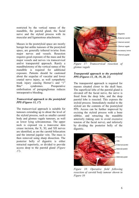
Transcervical approach <strong>to</strong> the poststyloid<br />
PPS (Figures 11, 17)<br />
The transcervical approach is suitable for<br />
tumours extending up <strong>to</strong> about the level <strong>of</strong><br />
the styloid process, such as smaller carotid<br />
body and glomus vagale tumours, as well<br />
as lower lying schwannomas. The upper<br />
neck is exposed via a transverse skin<br />
crease incision, the X, XI, and XII nerves<br />
are identified, as are the carotid bifurcation<br />
and the internal jugular vein. The mass is<br />
then removed using sharp dissection. The<br />
posterior belly <strong>of</strong> digastric is either<br />
retracted superiorly, or divided <strong>to</strong> provide<br />
access deep <strong>to</strong> the parotid gland (Figure<br />
17).<br />
Digastric<br />
Figure 17: Transcervical resection <strong>of</strong><br />
glomus vagale<br />
Transparotid approach <strong>to</strong> the poststyloid<br />
PPS (Figures 11, 18, 19, 20, 21)<br />
The transparotid approach is required for<br />
masses situated closer <strong>to</strong> the skull base.<br />
The superficial lobe <strong>of</strong> the parotid gland is<br />
elevated <strong>of</strong>f the facial nerve, the nerve is<br />
freed from the deep lobe, and the deep<br />
parotid lobe is resected. This exposes the<br />
styloid process. Immediately medial <strong>to</strong> the<br />
styloid are the contents <strong>of</strong> the poststyloid<br />
PPS. <strong>Access</strong> can be further improved by<br />
excising the styloid process with a bone<br />
nibbler, and retracting the mandible<br />
anteriorly (taking care <strong>to</strong> avoid excessive<br />
tension <strong>of</strong> the facial nerve), and inferiorly<br />
by dividing the posterior belly <strong>of</strong> the<br />
digastric.<br />
Figure 18: Operative field following<br />
resection <strong>of</strong> carotid body tumour shown in<br />
Figure 10<br />
External carotid<br />
Internal carotid<br />
Glomus vagale<br />
<strong>Access</strong>ory nerve<br />
Internal jugular<br />
6


