Overview of Gastric Pathology: Non-Neoplastic Diseases
Overview of Gastric Pathology: Non-Neoplastic Diseases
Overview of Gastric Pathology: Non-Neoplastic Diseases

Create successful ePaper yourself
Turn your PDF publications into a flip-book with our unique Google optimized e-Paper software.
Harvard-MIT Division <strong>of</strong> Health Sciences and Technology<br />
HST.121: Gastroenterology, Fall 2005<br />
Instructors: Dr. Jonathan Glickman<br />
<strong>Overview</strong> <strong>of</strong> <strong>Gastric</strong> <strong>Pathology</strong>:<br />
<strong>Non</strong>-<strong>Neoplastic</strong> <strong>Diseases</strong>
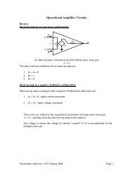
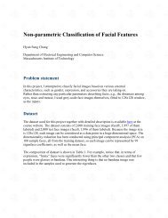
Structural Units <strong>of</strong> the Normal <strong>Gastric</strong><br />
Antral-Type<br />
Mucosa<br />
Fundic-Type<br />
Figure by MIT OCW
<strong>Non</strong>-<strong>Neoplastic</strong> <strong>Diseases</strong> <strong>of</strong> the Stomach<br />
• Developmental abnormalities<br />
• Chronic gastritis<br />
• Acute gastritis<br />
• <strong>Gastric</strong> ulcers<br />
• Mucosal hypertrophy<br />
• Infections<br />
• Vascular disorders<br />
• Systemic disorders
Patterns <strong>of</strong> Injury<br />
• Acute Injury:<br />
– Edema, congestion, and hemorrhage<br />
– Acute inflammation (neutrophils and eosinophils)<br />
– Erosions and ulcers<br />
• Chronic Injury:<br />
– Chronic inflammation (lymphocytes and plasma<br />
cells)<br />
– Lymphoid aggregates and follicles<br />
– Atrophy <strong>of</strong> specialized glands<br />
– Metaplasia (intestinal, pyloric, and pancreatic)<br />
• Repair Reactions:<br />
– Regenerative activity<br />
– Foveolar hyperplasia<br />
– Granulation tissue
Working Classification <strong>of</strong> Gastritis<br />
• Acute (erosive, hemorrhagic)<br />
• Chronic:<br />
– H. pylori gastritis<br />
– Atrophic gastritis<br />
• Type A or autoimmune or diffuse body<br />
• Type B or multi-focal or environmental<br />
– Eosinophilic gastritis (gastroenteritis)<br />
– Lymphocytic gastritis<br />
– Granulomatous gastritis<br />
• Infections<br />
• Chemical “gastropathies”<br />
• Bile reflux<br />
• NSAIDS<br />
• Alcohol
Gastritis- etiologic classification<br />
• Acute (erosive) gastritis<br />
– trauma, chemical injury, ischemia<br />
• Helicobacter-associated gastritis<br />
• <strong>Non</strong>-Helicobacter infectious gastritis<br />
• Immune-mediated- autoimmune, GVHD<br />
• Lymphocytic gastritis<br />
• Allergic (eosinophilic) gastritis<br />
• Crohn’s disease<br />
• Other- chemical, collagenous
Helicobacter Pylori Gastritis<br />
• Typical histopathology is characterized by:<br />
– Chronic active antral gastritis, with or without<br />
– Chronic active superficial gastritis in the corpus<br />
• Lymphoplasmacytic inflammation in the lamina propria<br />
• Neutrophils in the lamina propria and gastric pits<br />
• Lymphoid aggregates and follicles<br />
– Characteristic bacilli, primarily in the foveolar mucus<br />
• Histology may also include:<br />
– Increased intraepithelial lymphocytes in the antrum<br />
– Eosinophilic infiltrate
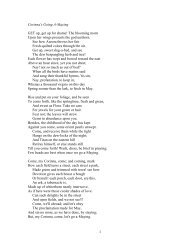
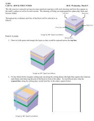
HIGH LEVEL OF ACID PRODUCTION<br />
H.pylori<br />
Normal <strong>Gastric</strong> Mucosa<br />
Acute H.pylori<br />
Infection<br />
H pylori- Natural history<br />
Chronic<br />
H.pylori<br />
Infection<br />
LOW LEVEL OF ACID PRODUCTION<br />
CHILDHOOD<br />
Antral-predominant<br />
gastritis<br />
<strong>Non</strong>atrophic<br />
pangastritis<br />
Corpus-predominant<br />
atrophic gastritis<br />
Duodenal ulcer<br />
<strong>Gastric</strong> ulcer<br />
Intestinal metaplasia<br />
Dysplasia<br />
MALT lymphoma<br />
Asymptomatic<br />
H.pylori<br />
infection<br />
<strong>Gastric</strong> cancer<br />
ADVANCED AGE<br />
Image by MIT OCW.
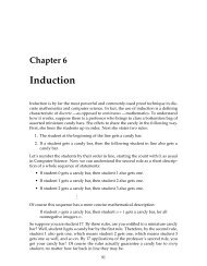
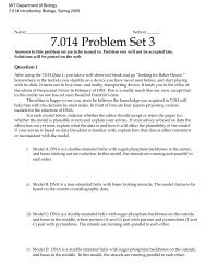
Distributions <strong>of</strong> gastritis<br />
Antral ( Type B) Fundic Gland ( Type A)<br />
Pangastritis ( Type AB)<br />
Image by MIT OCW
Autoimmune/Type A/Diffuse Atrophic<br />
Gastritis<br />
• An autoimmune autosomal dominant disease with anti-parietal cell or antiintrinsic<br />
factor autoantibodies<br />
• Histopathology is characterized by:<br />
– Chronic inflammation<br />
– Gland atrophy<br />
– Loss <strong>of</strong> parietal cells<br />
– Pyloric and intestinal metaplasia<br />
• Specific targeting <strong>of</strong> the parietal cells leads to:<br />
– Disease limited to the corpus and the fundus<br />
– Achlorohydria du to the loss <strong>of</strong> parietal cells<br />
– Pernicious anemia due to the loss <strong>of</strong> intrinsic factor<br />
– Hypergastrinemia due to the loss <strong>of</strong> gastric acid production<br />
– Endocrine cell hyperplasia and neoplasia due to<br />
1999 K. Badizadegan<br />
hypergastrinemia
Environmental/Type B/Multifocal Atrophic<br />
Gastritis<br />
• Heterogeneous disease due to chronic H. pylori gastritis,<br />
dietary factors, etc.<br />
• Disease most commonly involves the antrum and/or antrumcorpus<br />
junction, but may be seen anywhere in the stomach<br />
• Histopathology is characterized by:<br />
– Chronic inflammation<br />
– Gland atrophy<br />
– Intestinal metaplasia<br />
– Pylori metaplasia (with involvement <strong>of</strong> the corpus)<br />
– Patchy and/or focal involvement<br />
• Identified as the precancerous lesion in 95% <strong>of</strong> early gastric<br />
adenocarcinomas in Japan
“Chemical” Gastropathy<br />
• The final common pathway <strong>of</strong> mucosal damage due to chemicals, drugs,<br />
or bile reflux, characterized by any combination <strong>of</strong>:<br />
– Mucosal edema, congestion, and hemorrhage<br />
– Foveolar hyperplasia<br />
– Foveolar mucin depletion<br />
– Regenerative changes<br />
– Microscopic mucosal erosions<br />
– Increased smooth muscle fibers in the lamina propria<br />
– Relative paucity <strong>of</strong> inflammation<br />
• Alcohol, NSAIDS, and other drugs produce a similar pattern <strong>of</strong> injury
Infections
Eosinophilic Gastritis<br />
• Eosinophilic gastritis is typically part <strong>of</strong> eosinophilic gastroenteritis, which<br />
may take one <strong>of</strong> three forms:<br />
– Mucosal (bleeding, protein loss, malabsorption)<br />
– Mural (mass lesion)<br />
– Serosal (ascites)<br />
• The mucosal form <strong>of</strong> allergic gastroenteritis accounts for the majority <strong>of</strong><br />
cases, is typically “allergic” in nature, and commonly involves the gastric<br />
antrum<br />
• To establish a diagnosis <strong>of</strong> eosinophils/allergic gastroenteritis, eosinophils<br />
must be the predominant cell type, and other possible conditions must be<br />
excluded:<br />
– IBD<br />
– Reflux (esophagitis)<br />
– Parasitic infections<br />
– Vasculitis<br />
– Drug reaction<br />
– Chronic granulomatous disease<br />
– . . .
Lymphocytic Gastritis<br />
• Histopathology:<br />
– Increased foveolar intraepithelial T lymphocytes (>3 per<br />
10)<br />
– Variable degree <strong>of</strong> lymphoplasmacytic inflammation in<br />
the lamina propria<br />
– Involvement <strong>of</strong> the corpus with or without antral<br />
involvement<br />
• Approximately 80% <strong>of</strong> cases diagnosed endoscopically as chronic<br />
erosive (varioliform) gastritis meet the histological diagnostic criteria for<br />
lymphocytic gastritis<br />
• Approximately 20% <strong>of</strong> cases diagnosed histologically as lymphocytic<br />
gastritis have gross thickening <strong>of</strong> the mucosa<br />
• ? Association with H. pylori<br />
• ? Association with protein losing gastropathy<br />
• Approximately 60% <strong>of</strong> patients with active celiac disease have<br />
increased intraepithelial lymphocytes in the antrum
Granulomatous Gastritis<br />
• Crohn’s disease<br />
• Sarcoidosis<br />
• Infections:<br />
– Mycobacteria<br />
– Histoplasma<br />
• Foreign materials<br />
• Isolated granulomatous gastritis<br />
• And possibly:<br />
– Lymphoma<br />
– Malakoplakia<br />
– Whipple’s disease<br />
– Chronic granulomatous disease
Acute Gastritis<br />
• Acute infectious gastritis<br />
• Acute hemorrhagic gastritis<br />
– Stress, medications, alcohol, ischemia, . .<br />
.<br />
• Acute Stress Ulcer Disease<br />
– Cushing’s ulcer (CNS damage)<br />
– Curling’s ulcer (burn trauma)<br />
– Develops 1-2 weeks post-insult<br />
– Multifocal ulcers, typically in the body<br />
(contrast with PUD)
Developmental and Structural<br />
Abnormalities<br />
• <strong>Gastric</strong> atresia (membranes >> complete segmental defects)<br />
• Microgastria (arrested foregut development)<br />
• <strong>Gastric</strong> diverticula:<br />
– 75% are juxtacardial (on the posterior wall <strong>of</strong> the cardia)<br />
• <strong>Gastric</strong> duplication “cysts”<br />
• <strong>Gastric</strong> outlet obstruction:<br />
– Infantile hypertrophic pyloric stenosis<br />
• Heterotopias:<br />
– <strong>Gastric</strong> corpus mucosa (inlet patch, duodenal, Meckel’s,<br />
rectal)<br />
– Pancreatic tissue (gastric and duodenal wall and<br />
submucosa)<br />
– Brunner glands
Vascular Disorders<br />
• Congestive gastropathy and varices<br />
• <strong>Gastric</strong> antral vascular ectasis (GAVE)<br />
• Hereditary Hemorrhagic Telangiectasia<br />
(Osler-Weber-Rendu disease)<br />
• Sporadic telangiectasias<br />
• Caliber-persistent artery (Dieulafoy ulcer)<br />
• Arterio-venous malformations<br />
• Vasculitis<br />
• Atheroembolic disease<br />
• Amyloid vasculopathy
<strong>Gastric</strong> Mucosal Hypertrophy<br />
• Congenital hypertrophy <strong>of</strong> the rugae<br />
• Mucosal hypertrophy due to parietal cell hyperplasia<br />
– Zollinger-Ellison Syndrome<br />
• Mucosal hypertrophy due to foveolar hyperplasia<br />
– Menetrier’s Disease<br />
• Mucosal thickening (not hypertrophy) secondary to an infiltrative process
Menetrier’s Disease<br />
• Hyperplasia <strong>of</strong> the surface foveolar zone<br />
• Overproduction <strong>of</strong> mucus results in protein-losing enteropathy<br />
• Chronic disease in adults with a possible increase in the risk <strong>of</strong><br />
gastric cancer<br />
• Self-limited disease in children typically following to a viral<br />
infection
Zollinger-Ellison Syndrome<br />
• Hyperplasia <strong>of</strong> the parietal cells due to increased gastrin production<br />
• Source <strong>of</strong> gastrin may be:<br />
– A pancreatic islet cell tumor (90%)<br />
– A proximal duodenal tumor (7%)<br />
– Antral G-cell hyperplasia (3%)<br />
• Maximal stimulation <strong>of</strong> parietal cells leads to excessive acid production,<br />
resulting in multiple peptic ulcers <strong>of</strong> the stomach and the duodenum
• <strong>Non</strong>-neoplastic<br />
– Hyperplastic polyp<br />
– Fundic gland polyp<br />
<strong>Gastric</strong> polyps<br />
– Others (hamartomatous, etc.)<br />
• <strong>Neoplastic</strong><br />
– Adenoma<br />
– Carcinoma