Split hand/split foot malformation associated with sensorineural ...
Split hand/split foot malformation associated with sensorineural ...
Split hand/split foot malformation associated with sensorineural ...
Create successful ePaper yourself
Turn your PDF publications into a flip-book with our unique Google optimized e-Paper software.
Letters 405<br />
J Med Genet<br />
2001;38:405–409<br />
Department of<br />
Paediatrics, University<br />
Hospital of Innsbruck,<br />
Austria<br />
E Haberlandt<br />
H Fischer<br />
P Heinz-Erian<br />
T Müller<br />
Institute of Medical<br />
Biology and Human<br />
Genetics, University of<br />
Innsbruck,<br />
Schöpfstrasse 41, 6020<br />
Innsbruck, Austria<br />
J Löffler<br />
G Utermann<br />
A R Janecke<br />
Departments of<br />
Hearing, Speech and<br />
Voice Disorders/ENT,<br />
University Hospital of<br />
Innsbruck, Austria<br />
A Hirst-Stadlmann<br />
Department of<br />
Orthopaedics,<br />
University Hospital of<br />
Innsbruck, Austria<br />
B Stöckl<br />
Institute of MR<br />
Imaging and<br />
Spectroscopy,<br />
University Hospital of<br />
Innsbruck, Austria<br />
W Judmaier<br />
Department of<br />
Otolaryngology,<br />
University of Iowa<br />
Hospitals, Iowa City,<br />
USA<br />
R J H Smith<br />
Correspondence to:<br />
Dr Janecke,<br />
Andreas.Janecke@uibk.ac.at<br />
26 Finnilä S, Hassinen IE, Majamaa K. Restriction fragment<br />
analysis as a source of error in detection of heteroplasmic<br />
mtDNA mutations. Mutat Res 1999;406:109-14.<br />
27 Richards MB, Macaulay VA, Bandelt HJ, Sykes BC. Phylogeography<br />
of mitochondrial DNA in Western Europe. Ann<br />
Hum Genet 1998;62:241-60.<br />
28 Tanno Y, Okuizumi K, Tsuji S. mtDNA polymorphisms in<br />
Japanese sporadic Alzheimer’s disease. Neurobiol Aging<br />
1998;19(suppl):S47-51.<br />
29 Macaulay V, Richards M, Hickey E, Vega E, Cruciani F,<br />
Guida V, Scozzari R, Bonné-Tamir B, Sykes B, Torroni A.<br />
The emerging tree of west Eurasian mtDNAs: a synthesis<br />
of control-region sequences and RFLPs. Am J Hum Genet<br />
1999;64:232-49.<br />
30 Johns DR. Seminars in medicine of the Beth Israel Hospital,<br />
Boston. Mitochondrial DNA and disease. N Engl J Med<br />
1995;333:638-44.<br />
<strong>Split</strong> <strong>hand</strong>/<strong>split</strong> <strong>foot</strong> <strong>malformation</strong> <strong>associated</strong> <strong>with</strong><br />
<strong>sensorineural</strong> deafness, inner and middle ear<br />
<strong>malformation</strong>, hypodontia, congenital vertical<br />
talus, and deletion of eight microsatellite markers<br />
in 7q21.1-q21.3<br />
Edda Haberlandt, Judith LöZer, Almut Hirst-Stadlmann, Bernd Stöckl, Werner Judmaier,<br />
Helmut Fischer, Peter Heinz-Erian, Thomas Müller, Gerd Utermann, Richard J H Smith,<br />
Andreas R Janecke<br />
EDITOR—The <strong>split</strong> <strong>hand</strong>/<strong>split</strong> <strong>foot</strong> <strong>malformation</strong><br />
(SHFM, MIM 183600) is a central reduction<br />
defect of the <strong>hand</strong>s and feet and occurs<br />
both as an isolated <strong>malformation</strong> and as part of<br />
several syndromes including the EEC syndrome<br />
(MIM 129900). We report on a2year<br />
old boy <strong>with</strong> SHFM <strong>associated</strong> <strong>with</strong> features of<br />
ectodermal hypoplasia, a submucous cleft palate,<br />
congenital vertical talus, <strong>malformation</strong>s of<br />
the middle ear, profound <strong>sensorineural</strong> hearing<br />
loss resulting from Mondini dysplasia, and a de<br />
novo deletion of the paternal chromosome<br />
7q21.1-q21.3. This patient <strong>with</strong> syndromic<br />
SHFM represents a case of atypical EEC<br />
syndrome, but also displays abnormalities<br />
previously not <strong>associated</strong> <strong>with</strong> SHFM or EEC<br />
syndrome.<br />
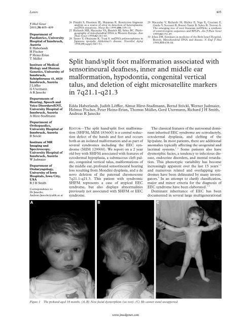
Figure 1 The proband aged 18 months. (A, B) Note facial dysmorphism (see text). (C) He cannot stand unsupported.<br />
www.jmedgenet.com<br />
The classical features of the autosomal dominant<br />
inherited EEC syndrome are ectrodactyly,<br />
ectodermal dysplasia, and clefting of the<br />
lip/palate. In most patients, there are additional<br />
anomalies typically aVecting the urogenital and<br />
lacrimal systems. 12 Some patients also have<br />
dysmorphic facies, a tendency to infectious disease,<br />
endocrine disorders, and mental retardation.<br />
This phenotypic variability has become<br />
increasingly apparent over the last 15 years 34<br />
and numerous related and overlapping syndromes<br />
have been delineated by many investigators.<br />
5 In an attempt to clarify classification,<br />
major and minor criteria for the diagnosis of<br />
EEC syndrome have been elaborated. 34<br />
Dominant inheritance of EEC has been<br />
documented in several large multigenerational
406 Letters<br />
Figure 2 Right <strong>foot</strong> of the patient. (A) Ectrodactyly (<strong>split</strong> <strong>foot</strong> <strong>malformation</strong>) <strong>with</strong> apparent absence of the 2nd toe and<br />
syndactyly of toes 3 to 5. (B) Radiograph showing syndactyly of the first and second metatarsals and absence of the second<br />
phalanges and <strong>malformation</strong> of the third to fifth phalanges.(C) Ectrodactyly and pes planovalgus (severe talus verticalis<br />
deformity).<br />
families. 6 At least 15 patients have been<br />
reported to have cytogenetic abnormalities of<br />
chromosome 7q21.2-7q22.1, including nine<br />
patients <strong>with</strong> interstitial deletions. 7–9 In addition,<br />
mutations in the gene encoding the transactivation<br />
factor p63 on chromosome 3q27<br />
have been identified in familial and sporadic<br />
cases of EEC syndrome. 10 A third locus was<br />
mapped to chromosome 19q, 11 further delineating<br />
the genetic heterogeneity of this syndrome.<br />
The reason for the phenotypic<br />
heterogeneity in EEC syndrome patients <strong>with</strong><br />
7q abnormalities is unclear but may relate to<br />
the size of the deletion.<br />
Case report<br />
Our patient is the fifth child of healthy, consanguineous,<br />
fourth cousin, Austrian parents. The<br />
father and the mother were 41 and 36 years,<br />
respectively, at the time of his birth. His four<br />
sibs are healthy. He was born after an uneventful<br />
pregnancy in the 41st week of gestation and<br />
weighed 2840 g (10th centile), was 48 cm long<br />
(10th centile), and had a head circumference of<br />
31.5 cm (10th centile). Ectrodactyly of the<br />
right <strong>foot</strong> was noted and transient evoked<br />
otoacoustic emission screening indicated hearing<br />
impairment. Further examinations were at<br />
first declined by the mother. At 15 months of<br />
asc<br />
co<br />
va es<br />
co<br />
Figure 3 Inner ear of the patient. A 3D reconstruction of a coronal MRI scan shows Mondini type <strong>malformation</strong> on both<br />
sides. (A) Right ear: overall dilated and plump structures of the inner ear. asc denotes the anterior semicircular canal, va the<br />
vestibular aqueduct <strong>with</strong> saccule and utricle, and co the cochlea showing a reduced number of coils. (B) Left ear: a large<br />
endolymphatic sac is shown (es). (C) Schema of the normal inner ear. 1. Anterior semicircular canal. 2. Membranous<br />
ampulla (MA) of the anterior semicircular canal. 3. MA of the lateral semicircular canal. 4. Saccule. 5. Cochlear canal. 6.<br />
Helicotrema. 7. Lateral semicircular canal. 8. Posterior semicircular canal. 9. MA of the posterior semicircular canal. 10.<br />
Vestibular window. 11. Cochlear window. 12. Scala vestibuli. 13. Scala tympani. 14. Utricule.<br />
www.jmedgenet.com<br />
asc
Letters 407<br />
A<br />
q21.1<br />
q21.3<br />
7 der(7)<br />
Figure 4 Cytogenetic and molecular findings. (A) High resolution cytogenetic analysis of both chromosomes 7 of the<br />
patient. The deletion is indicated by the arrow. (B) Preliminary analysis of microsatellite markers from chromosome 7q in<br />
the family of the patient. The deleted interval spans at least 8.9 cM on the paternal chromosome flanked by microsatellite<br />
markers D7S2443 and D7S2480. Data regarding microsatellite mapping are compiled from Dib et al 12 and Crackower et<br />
al. 13 14 The arrow indicates the position of the gene mutated in Pendred syndrome.<br />
age, he was referred to the hospital because of<br />
failure to thrive (weight 7200 g, below the 3rd<br />
centile; length 70 cm, below the 3rd centile;<br />
head circumference 43 cm, below the 3rd centile).<br />
Physical examination showed arched eyebrows,<br />
a small triangular nose <strong>with</strong> a depressed<br />
nasal bridge, and ears <strong>with</strong> overfolded helices<br />
and attached earlobes (fig 1). He also had<br />
hypertelorism, a large biparietal diameter,<br />
hypopigmented retina, micrognathia, a submucous<br />
cleft palate, carious primary teeth and<br />
hypodontia, sparse, light hair, pale skin,<br />
cryptorchidism, and bilateral severe congenital<br />
vertical talus, in addition to the previously<br />
noted ectrodactyly of the right <strong>foot</strong> (fig 2). CT<br />
and MRI scans showed Mondini dysplasia of<br />
the inner ear (fig 3) and cochlear implanting<br />
showed fixation of the ossicular chain. Audiometric<br />
examinations were consistent <strong>with</strong> these<br />
findings and showed conductive and profound<br />
<strong>sensorineural</strong> hearing loss. Laboratory investigations<br />
showed partial deficiency of growth<br />
hormone secretion. Mental and psychomotor<br />
developmental delay was noted.<br />
On GTG banding, we observed an interstitial<br />
deletion of chromosome 7 confined to the<br />
interval q21.1-q21.3 (fig 4A); parental karyotypes<br />
were normal. To delineate this deletion<br />
further, we used 21 chromosome 7q microsatellite<br />
markers to reconstruct parental and<br />
www.jmedgenet.com<br />
De novo<br />
deletion of<br />
about 8.9<br />
to 17.0 cM<br />
PDS gene<br />
B<br />
D7S2506<br />
D7S663<br />
D7S2455<br />
D7S634<br />
D7S2443<br />
D7S524<br />
D7S492<br />
D7S2410<br />
D7S657<br />
D7S2482<br />
D7S527<br />
D7S1812<br />
D7S821<br />
D7S2539<br />
D7S479<br />
D7S491<br />
D7S1796<br />
D7S2480<br />
D7S647<br />
D7S501<br />
D7S692<br />
3 2<br />
3 2<br />
3 1<br />
1 2<br />
2 2<br />
2 1<br />
2 2<br />
1 1<br />
1 1<br />
1 3<br />
2 1<br />
3 3<br />
2 2<br />
3 3<br />
1 2<br />
3 3<br />
2 3<br />
1 2<br />
2 2<br />
1 1<br />
3 2<br />
F<br />
D7S2506<br />
D7S663<br />
D7S2455<br />
D7S634<br />
D7S2443<br />
D7S524<br />
D7S492<br />
D7S2410<br />
D7S657<br />
D7S2482<br />
D7S527<br />
D7S1812<br />
D7S821<br />
D7S2539<br />
D7S479<br />
D7S491<br />
D7S1796<br />
D7S2480<br />
D7S647<br />
D7S501<br />
D7S692<br />
1 4<br />
4 1<br />
2 4<br />
2 3<br />
1 3<br />
2 3<br />
1 2<br />
1 1<br />
3 2<br />
2 3<br />
3 2<br />
2 1<br />
1 3<br />
2 1<br />
3 2<br />
2 1<br />
2 1<br />
2 2<br />
1 2<br />
2 3<br />
1 3<br />
SL<br />
3 1<br />
3 4<br />
3 2<br />
? 2<br />
2 1<br />
? 2<br />
- 1<br />
? 1<br />
- 3<br />
- 2<br />
? 2<br />
- 2<br />
- 1<br />
- 2<br />
- 3<br />
- 2<br />
? 2<br />
1 2<br />
2 1<br />
1 2<br />
3 1<br />
Distance<br />
Mb<br />
cM<br />
8.5<br />
10.3<br />
2.8<br />
0.7<br />
3.7<br />
3.1<br />
3.5<br />
1.5<br />
2.8<br />
1.3<br />
0.5<br />
7.8<br />
0.8<br />
patient haplotypes and found that for the eight<br />
markers flanked by D7S2443 and D7S2480,<br />
the patient had a deletion of the paternal allele<br />
(fig 4B). Two markers <strong>with</strong>in the interval<br />
(D7S2410 and D7S527) were uninformative,<br />
as were two flanking markers (D7S524 and<br />
D7S1796). These data define a deletion of 8.9<br />
to 17 cM , which includes the critical interval<br />
of
408 Letters<br />
association that has not previously been reported<br />
<strong>with</strong> either SHFM or a chromosome 7<br />
aberration. Mondini dysplasia is characterised<br />
by bony and membranous anomalies of the<br />
inner ear exhibiting a wide range of morphological<br />
and functional abnormalities. Typically,<br />
the cochlea is flat, the cochleal duct is short, the<br />
auditory and vestibular sense organs and nerves<br />
are immature, the vestibule is large, the semicircular<br />
canals are wide, small, or missing, and the<br />
endolymphatic sac is dilated. The anomaly can<br />
be unilateral or bilateral and occurs in isolation<br />
or in association <strong>with</strong> anomalies in other<br />
organs. 16 Familial examples of Mondini dysplasia<br />
generally represent examples of Pendred<br />
syndrome, an autosomal recessive disorder in<br />
which congenital <strong>sensorineural</strong> hearing impairment<br />
and goitre cosegregate.<br />
Because Pendred syndrome is caused by<br />
mutations in PDS, a gene that maps to<br />
chromosome 7q31, the simultaneous occurrence<br />
of atypical EEC syndrome and Pendred<br />
syndrome in our patient seemed an attractive<br />
possibility to explain the rare combination of<br />
physical findings. Although molecular analysis<br />
in our patient appears to place the distal<br />
breakpoint of the deletion about 9 cM centromeric<br />
to PDS, we cannot exclude a more complex<br />
chromosomal rearrangement. Assuming<br />
that the paternal copy of PDS could have been<br />
deleted, we completed a mutation screen for<br />
maternally inherited PDS allele variants. We<br />
were unable to identify any mutations by<br />
SSCP and direct sequencing as described previously<br />
17 and therefore could not establish a<br />
causal connection between the observed<br />
Mondini dysplasia and the chromosomal<br />
aberration. We also excluded an independent<br />
cause of the <strong>sensorineural</strong> hearing impairment,<br />
by sequencing the coding region and<br />
exon 1 of GJB2. 18 Mutations in this gene are<br />
the most common cause of autosomal recessive<br />
non-syndromic deafness.<br />
The simultaneous occurrence of an inner ear<br />
<strong>malformation</strong> and SHFM has rarely been<br />
reported. Berndorfer 19 noted absence of pinnae<br />
and lack of inner ears in one patient. Autosomal<br />
dominant ectrodactyly and deafness in a father<br />
and son were reported by Tolmie et al. 20 Both<br />
patients had CT verified cochlear abnormalities,<br />
which may have been consistent <strong>with</strong> Mondini<br />
dysplasia. There was no mention of any<br />
chromosomal anomaly. Moreover, <strong>sensorineural</strong><br />
hearing impairment has rarely been reported in<br />
syndromic SHFM. 21–26 In two of these cases,<br />
however, an apparently balanced translocation<br />
involving chromosome 7q was found to cosegre-<br />
gate <strong>with</strong> the disease.<br />
25 26<br />
Conductive hearing loss is observed in<br />
14-44% of cases of EEC syndrome, most commonly<br />
reflecting Eustachian tube dysfunction<br />
in association <strong>with</strong> the palatal clefting,<br />
although ossicular <strong>malformation</strong>s have been<br />
described. 27<br />
Of 10 patients <strong>with</strong> ectrodactyly in association<br />
<strong>with</strong> a deletion of 7q21-q22, microcephaly and<br />
general growth impairment have been reported<br />
in eight cases (80% 79 ) compared to only 2% and<br />
1%, respectively, in a survey of 230 patients <strong>with</strong><br />
EEC syndrome. 4 While adenohypophyseal<br />
www.jmedgenet.com<br />
3 4<br />
dysfunction in two sets of sibs has been reported<br />
in EEC syndrome, 28 29 partial growth hormone<br />
deficiency was identified in our patient as the<br />
aetiology of the growth retardation. However,<br />
growth retardation and microcephaly might<br />
also delineate a subtype of the EEC syndrome<br />
related to chromosomal aberrations involving<br />
chromosome 7q21-q22. We accordingly suggest<br />
initiating chromosomal and molecular<br />
investigations of this chromosomal region when<br />
growth retardation and microcephaly is present<br />
in patients <strong>with</strong> SHFM. Short stature, as well as<br />
low birth weight, abnormal skull shape, and ear<br />
<strong>malformation</strong>s were common findings among<br />
patients <strong>with</strong> proximal/intermediate deletions<br />
or rearrangements of chromosome 7q, <strong>with</strong> and<br />
<strong>with</strong>out SHFM. 8<br />
We believe that a specific pattern of facial<br />
anomalies characterises patients <strong>with</strong> aberrations<br />
of chromosome 7q21-q22. The facial<br />
phenotype consists of arched eyebrows, a<br />
small, triangular shaped nose <strong>with</strong> a depressed<br />
nasal bridge, abnormal ears <strong>with</strong> overfolded<br />
helices and attached earlobes, a large biparietal<br />
diameter, hypertelorism, and micrognathia. It<br />
was present in our patient and in at least six<br />
published case reports.<br />
9 30–34<br />
The <strong>split</strong> <strong>foot</strong> <strong>malformation</strong> in our patient<br />
was right sided, as has been mostly observed in<br />
cases of unilateral involvement of either the<br />
upper or lower limbs. 26 The presence of<br />
bilateral congenital vertical talus could not be<br />
explained by aplasia of the anterior calcaneus<br />
<strong>with</strong> loss of talar support or by a spinal defect,<br />
though the <strong>split</strong> <strong>foot</strong> <strong>malformation</strong> complicates<br />
the anomaly of the talus. Bilateral congenital<br />
vertical talus is an otherwise rare disorder, and<br />
to our knowledge has not been reported in the<br />
EEC syndrome or in related conditions.<br />
Our report suggests that patients <strong>with</strong><br />
syndromic SHFM should be examined for the<br />
findings we describe, and that the molecular<br />
analysis should include karyotyping and complementary<br />
studies to establish whether the<br />
critical interval of
Letters 409<br />
J Med Genet<br />
2001;38:409–411<br />
Division of<br />
Immunology/Allergy,<br />
The Infection,<br />
Immunity, Injury &<br />
Repair Program,<br />
Research Institute,<br />
Department of<br />
Pediatrics, The<br />
Hospital for Sick<br />
Children and the<br />
University of Toronto,<br />
Toronto, Canada<br />
E Grunebaum<br />
E Arpaia<br />
C M Roifman<br />
Department of<br />
Genetics, The Hospital<br />
for Sick Children and<br />
the University of<br />
Toronto, Toronto,<br />
Canada<br />
J J MacKenzie<br />
J Fitzpatrick<br />
PNRay<br />
Correspondence to:<br />
Dr Roifman, Infection,<br />
Immunity, Injury & Repair<br />
Program, Research Institute,<br />
The Hospital for Sick<br />
Children, 555 University<br />
Avenue, Toronto, Ontario<br />
M5G 1X8, Canada,<br />
chaim.roifman@sickkids.ca<br />
8 McElveen C, Carvajal MV, Moscatello D, Towner J, Lacassie<br />
Y. Ectrodactyly and proximal/intermediate interstitial<br />
deletion 7q. Am J Med Genet 1995;56:1-5.<br />
9 Marinoni JC, Stevenson RE, Evans JP, Geshuri D, Phelan<br />
MC, Schwartz CE. <strong>Split</strong> <strong>foot</strong> and developmental retardation<br />
<strong>associated</strong> <strong>with</strong> a deletion of three microsatellite markers<br />
in 7q21.2-q22.1. Clin Genet 1995;47:90-5.<br />
10 Celli J, Duijf P, Hamel BC, Bamshad M, Kramer B, Smits<br />
AP, Newbury-Ecob R, Hennekam RC, Van Buggenhout G,<br />
van Haeringen A, Woods CG, van Essen AJ, de Waal R,<br />
Vriend G, Haber DA, Yang A, McKeon F, Brunner HG,<br />
van Bokhoven H. Heterozygous germline mutations in the<br />
p53 homolog p63 are the cause of EEC syndrome. Cell<br />
1999;99:143-53.<br />
11 O’Quinn JR, Hennekam RCM, Jorde LB, Bamshad M.<br />
Syndromic ectrodactyly <strong>with</strong> severe limb, ectodermal, urogenital,<br />
and palatal defects maps to chromosome 19. Am J<br />
Hum Genet 1998;62:130-5.<br />
12 Dib C, Faure S, Fizames C, Samson D, Drouot N, Vignal A,<br />
Millasseau P, Marc S, Hazan J, Seboun E, Lathrop M,<br />
Gyapay G, Morissette J, Weissenbach J. A comprehensive<br />
genetic map of the human genome based on 5,264 microsatellites.<br />
Nature 1996;380:152-4.<br />
13 Crackower MA, Scherer SW, Rommens JM, Hui CC,<br />
Poorkaj P, Soder S, Cobben JM, Hudgins L, Evans JP, Tsui<br />
LC. Characterization of the <strong>split</strong> <strong>hand</strong>/<strong>split</strong> <strong>foot</strong> <strong>malformation</strong><br />
locus SHFM1 at 7q21.3-q22.1 and analysis of a candidate<br />
gene for its expression during limb development.<br />
Hum Mol Genet 1996;5:571-9.<br />
14 Crackower MA, Sinasac DS, Xia J, Motoyama J, Prochazka<br />
M, Rommens JM, Scherer SW, Tsui LC. Cloning and characterization<br />
of two cytoplasmic dynein intermediate chain<br />
genes in mouse and human. Genomics 1999;55:257-67.<br />
15 Akita S, Kuratomi H, Abe K, Harada N, Mukae N, Niikawa<br />
N. EC syndrome in a girl <strong>with</strong> paracentric inversion<br />
(7)(q22.1;q36.3). Clin Dysmorphol 1993;2:62-7.<br />
16 Ormerod FC. The pathology of congenital deafness. J<br />
Laryngol 1960;74:919-50.<br />
17 Van Hauwe P, Everett LA, Coucke P, Scott DA, Kraft ML,<br />
Ris-Stalpers C, Bolder C, Otten B, de Vijlder JJ, Dietrich<br />
NL, Ramesh A, Srisailapathy SC, Parving A, Cremers CW,<br />
Willems PJ, Smith RJ, Green ED, Van Camp G. Two<br />
frequent missense mutations in Pendred syndrome. Hum<br />
Mol Genet 1998;7:1099-104.<br />
18 Scott DA, Kraft ML, Carmi R, Ramesh A, Elbedour K, Yairi<br />
Y, Srisailapathy CR, Rosengren SS, Markham AF, Mueller<br />
RF, Lench NJ, Van Camp G, Smith RJ, SheYeld VC. Identification<br />
of mutations in the connexin 26 gene that cause<br />
autosomal recessive nonsyndromic hearing loss. Hum<br />
Mutat 1998;11:387-94.<br />
19 Berndorfer A. Gesichtsspalten gemeinsam mit Hand- und<br />
Fuspalten. Z Orthopad 1970;107:344-54.<br />
20 Tolmie J, Geddes NK, Knight S, Fredricks B. Autosomal<br />
dominant ectrodactyly and deafness. 5th Manchester Birth<br />
Defects Conference, 13-16 October 1992.<br />
21 Birch-Jensen A. Congenital deformities of the upper extremities.<br />
Copenhagen: Ejnar Munksgaards Forlag, 1949:19.<br />
22 Wildervanck LS. Perceptive deafness <strong>associated</strong> <strong>with</strong> <strong>split</strong><strong>hand</strong><br />
and <strong>foot</strong>, a new syndrome? Acta Genet 1963;13:161-9.<br />
23 Fraser GR. The causes of profound deafness in childhood.<br />
Baltimore: Johns Hopkins University Press, 1976.<br />
24 Anneren G, Andersson T, Lindgren PG, Kjartansson S.<br />
Ectrodactyly-ectodermal dysplasia-clefting syndrome<br />
(EEC): the clinical variation and prenatal diagnosis. Clin<br />
Genet 1991;40:257-62.<br />
25 Hasegawa T, Hasegawa Y, Asamura S, Nagai T, Tsuchiya Y,<br />
Ninomiya M, Fukushima Y. EEC syndrome (ectrodactyly,<br />
ectodermal dysplasia and cleft lip/palate) <strong>with</strong> a balanced<br />
reciprocal translocation between 7q11.21 and 9p12 (or<br />
7p11.2 and 9q12) in three generations. Clin Genet 1991;40:<br />
202-6.<br />
26 Genuardi M, Pomponi MG, Sammito V, Bellussi A, Zollino<br />
M, Neri G. <strong>Split</strong> <strong>hand</strong>/<strong>split</strong> <strong>foot</strong> anomaly in a family segregating<br />
a balanced translocation <strong>with</strong> breakpoint on 7q22.1.<br />
Am J Med Genet 1993;47:823-31.<br />
27 Robinson GC, Wildervanck LS, Chiang TP. Ectrodactyly,<br />
ectodermal dysplasia, and cleft lip-palate syndrome. Its<br />
association <strong>with</strong> conductive hearing loss. J Pediatr 1973;82:<br />
107-9.<br />
28 Knudtzon J, Aarskog D. Growth hormone deficiency <strong>associated</strong><br />
<strong>with</strong> the ectrodactyly-ectodermal dysplasia-clefting<br />
syndrome and isolated absent septum pellucidum. Pediatrics<br />
1987;79:410-12.<br />
29 Gershoni-Baruch R, Goldscher D, Hochberg Z.<br />
Ectrodactyly-ectodermal dysplasia-clefting syndrome and<br />
hypothalamo-pituitary insuYciency. Am J Med Genet<br />
1997;68:168-72.<br />
30 Young RS, Weaver DD, Kukolich MK, Heerema NA, Palmer<br />
CG, Kawira EL, Bender HA. Terminal and interstitial<br />
deletions of the long arm of chromosome 7: a review <strong>with</strong><br />
five new cases. Am J Med Genet 1984;17:437-50.<br />
31 Fryns JP, Kleczkowska A, Van den Berghe H. Moderate mental<br />
retardation and mild dysmorphic syndrome in proximal<br />
7q interstitial deletion. Ann Genet 1987;30:111-12.<br />
32 Tajara EH, Varella-Garcia M, Gusson AC. Interstitial longarm<br />
deletion of chromosome 7 and ectrodactyly. Am J Med<br />
Genet 1989;32:192-4.<br />
33 Rivera H, Sanchez-Corona J, Burgos-Fuentes VR,<br />
Melendez-Ruiz MJ. Deletion of 7q22 and ectrodactyly.<br />
Genet Couns 1991;2:27-31.<br />
34 Sharland M, Patton MA, Hill L. Ectrodactyly of <strong>hand</strong>s and<br />
feet in a child <strong>with</strong> a complex translocation including<br />
7q21.2. Am J Med Genet 1991;39:413-14.<br />
A missense mutation in the SEDL gene results in<br />
delayed onset of X linked spondyloepiphyseal<br />
dysplasia in a large pedigree<br />
E Grunebaum, E Arpaia, J J MacKenzie, J Fitzpatrick, P N Ray, C M Roifman<br />
EDITOR—Spondyloepiphyseal dysplasia (SED)<br />
is a rare osteochondroplasia, characterised by<br />
disproportionate short stature <strong>with</strong> a short<br />
neck and trunk and barrel chest. The pelvis<br />
tends to be narrow and deep, the femoral neck<br />
short, and the femoral head flattened. Mild to<br />
moderate epiphyseal dysplasia of the large<br />
joints may also be seen. The latter may lead to<br />
premature secondary osteoarthritis <strong>with</strong> significant<br />
morbidity. 1 SED may occur sporadically;<br />
however, in many cases the family<br />
history indicates an inherited condition. In<br />
some of these pedigrees, the inheritance<br />
pattern seems autosomal dominant, while in<br />
others it is consistent <strong>with</strong> autosomal recessive<br />
or X linked recessive. 2<br />
Recently, mutations in the gene designated<br />
SEDL, located on Xp22, were identified as the<br />
cause of X linked spondyloephiphyseal dysplasia<br />
tarda in three families. 3 We have previously<br />
described a large kindred of British descent<br />
www.jmedgenet.com<br />
spanning four generations aVected by SED. 1<br />
Briefly, 14 males between the ages of 10 and<br />
77 years were aVected, <strong>with</strong> early adolescence<br />
development of progressive decline in growth<br />
rate accompanied by short stature, short<br />
trunk, and barrel chest. Although some of<br />
them had to limit their activities because of hip<br />
or back limitation of movement or pain, many<br />
continued <strong>with</strong> normal activity and were able<br />
to perform in the work place <strong>with</strong>out impairment<br />
of function. There was no indication of<br />
other abnormalities previously reported in<br />
association <strong>with</strong> SED, such as mental retardation,<br />
2 immune abnormalities and retinopathy, 4<br />
cardiac dysfunction, 5 or hypogonadotrophic<br />
hypogonadism. 6 The female carriers in this<br />
pedigree had normal height. Although some of<br />
the females suVered from occasional mild back<br />
or hip pain, it did not aVect their daily activity,<br />
nor was there objective radiological evidence<br />
of spinal or joint involvement compatible <strong>with</strong>


