Split hand/split foot malformation associated with sensorineural ...
Split hand/split foot malformation associated with sensorineural ...
Split hand/split foot malformation associated with sensorineural ...

You also want an ePaper? Increase the reach of your titles
YUMPU automatically turns print PDFs into web optimized ePapers that Google loves.
406 Letters<br />
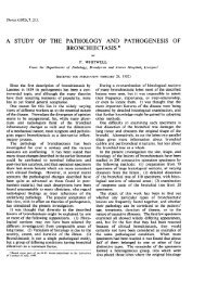
Figure 2 Right <strong>foot</strong> of the patient. (A) Ectrodactyly (<strong>split</strong> <strong>foot</strong> <strong>malformation</strong>) <strong>with</strong> apparent absence of the 2nd toe and<br />
syndactyly of toes 3 to 5. (B) Radiograph showing syndactyly of the first and second metatarsals and absence of the second<br />
phalanges and <strong>malformation</strong> of the third to fifth phalanges.(C) Ectrodactyly and pes planovalgus (severe talus verticalis<br />
deformity).<br />
families. 6 At least 15 patients have been<br />
reported to have cytogenetic abnormalities of<br />
chromosome 7q21.2-7q22.1, including nine<br />
patients <strong>with</strong> interstitial deletions. 7–9 In addition,<br />
mutations in the gene encoding the transactivation<br />
factor p63 on chromosome 3q27<br />
have been identified in familial and sporadic<br />
cases of EEC syndrome. 10 A third locus was<br />
mapped to chromosome 19q, 11 further delineating<br />
the genetic heterogeneity of this syndrome.<br />
The reason for the phenotypic<br />
heterogeneity in EEC syndrome patients <strong>with</strong><br />
7q abnormalities is unclear but may relate to<br />
the size of the deletion.<br />
Case report<br />
Our patient is the fifth child of healthy, consanguineous,<br />
fourth cousin, Austrian parents. The<br />
father and the mother were 41 and 36 years,<br />
respectively, at the time of his birth. His four<br />
sibs are healthy. He was born after an uneventful<br />
pregnancy in the 41st week of gestation and<br />
weighed 2840 g (10th centile), was 48 cm long<br />
(10th centile), and had a head circumference of<br />
31.5 cm (10th centile). Ectrodactyly of the<br />
right <strong>foot</strong> was noted and transient evoked<br />
otoacoustic emission screening indicated hearing<br />
impairment. Further examinations were at<br />
first declined by the mother. At 15 months of<br />
asc<br />
co<br />
va es<br />
co<br />
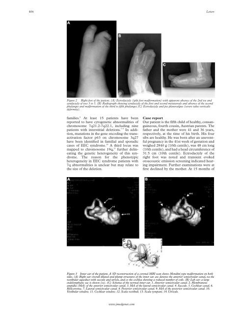
Figure 3 Inner ear of the patient. A 3D reconstruction of a coronal MRI scan shows Mondini type <strong>malformation</strong> on both<br />
sides. (A) Right ear: overall dilated and plump structures of the inner ear. asc denotes the anterior semicircular canal, va the<br />
vestibular aqueduct <strong>with</strong> saccule and utricle, and co the cochlea showing a reduced number of coils. (B) Left ear: a large<br />
endolymphatic sac is shown (es). (C) Schema of the normal inner ear. 1. Anterior semicircular canal. 2. Membranous<br />
ampulla (MA) of the anterior semicircular canal. 3. MA of the lateral semicircular canal. 4. Saccule. 5. Cochlear canal. 6.<br />
Helicotrema. 7. Lateral semicircular canal. 8. Posterior semicircular canal. 9. MA of the posterior semicircular canal. 10.<br />
Vestibular window. 11. Cochlear window. 12. Scala vestibuli. 13. Scala tympani. 14. Utricule.<br />
www.jmedgenet.com<br />
asc