Split hand/split foot malformation associated with sensorineural ...
Split hand/split foot malformation associated with sensorineural ...
Split hand/split foot malformation associated with sensorineural ...

You also want an ePaper? Increase the reach of your titles
YUMPU automatically turns print PDFs into web optimized ePapers that Google loves.
Letters 405<br />
J Med Genet<br />
2001;38:405–409<br />
Department of<br />
Paediatrics, University<br />
Hospital of Innsbruck,<br />
Austria<br />
E Haberlandt<br />
H Fischer<br />
P Heinz-Erian<br />
T Müller<br />
Institute of Medical<br />
Biology and Human<br />
Genetics, University of<br />
Innsbruck,<br />
Schöpfstrasse 41, 6020<br />
Innsbruck, Austria<br />
J Löffler<br />
G Utermann<br />
A R Janecke<br />
Departments of<br />
Hearing, Speech and<br />
Voice Disorders/ENT,<br />
University Hospital of<br />
Innsbruck, Austria<br />
A Hirst-Stadlmann<br />
Department of<br />
Orthopaedics,<br />
University Hospital of<br />
Innsbruck, Austria<br />
B Stöckl<br />
Institute of MR<br />
Imaging and<br />
Spectroscopy,<br />
University Hospital of<br />
Innsbruck, Austria<br />
W Judmaier<br />
Department of<br />
Otolaryngology,<br />
University of Iowa<br />
Hospitals, Iowa City,<br />
USA<br />
R J H Smith<br />
Correspondence to:<br />
Dr Janecke,<br />
Andreas.Janecke@uibk.ac.at<br />
26 Finnilä S, Hassinen IE, Majamaa K. Restriction fragment<br />
analysis as a source of error in detection of heteroplasmic<br />
mtDNA mutations. Mutat Res 1999;406:109-14.<br />
27 Richards MB, Macaulay VA, Bandelt HJ, Sykes BC. Phylogeography<br />
of mitochondrial DNA in Western Europe. Ann<br />
Hum Genet 1998;62:241-60.<br />
28 Tanno Y, Okuizumi K, Tsuji S. mtDNA polymorphisms in<br />
Japanese sporadic Alzheimer’s disease. Neurobiol Aging<br />
1998;19(suppl):S47-51.<br />
29 Macaulay V, Richards M, Hickey E, Vega E, Cruciani F,<br />
Guida V, Scozzari R, Bonné-Tamir B, Sykes B, Torroni A.<br />
The emerging tree of west Eurasian mtDNAs: a synthesis<br />
of control-region sequences and RFLPs. Am J Hum Genet<br />
1999;64:232-49.<br />
30 Johns DR. Seminars in medicine of the Beth Israel Hospital,<br />
Boston. Mitochondrial DNA and disease. N Engl J Med<br />
1995;333:638-44.<br />
<strong>Split</strong> <strong>hand</strong>/<strong>split</strong> <strong>foot</strong> <strong>malformation</strong> <strong>associated</strong> <strong>with</strong><br />
<strong>sensorineural</strong> deafness, inner and middle ear<br />
<strong>malformation</strong>, hypodontia, congenital vertical<br />
talus, and deletion of eight microsatellite markers<br />
in 7q21.1-q21.3<br />
Edda Haberlandt, Judith LöZer, Almut Hirst-Stadlmann, Bernd Stöckl, Werner Judmaier,<br />
Helmut Fischer, Peter Heinz-Erian, Thomas Müller, Gerd Utermann, Richard J H Smith,<br />
Andreas R Janecke<br />
EDITOR—The <strong>split</strong> <strong>hand</strong>/<strong>split</strong> <strong>foot</strong> <strong>malformation</strong><br />
(SHFM, MIM 183600) is a central reduction<br />
defect of the <strong>hand</strong>s and feet and occurs<br />
both as an isolated <strong>malformation</strong> and as part of<br />
several syndromes including the EEC syndrome<br />
(MIM 129900). We report on a2year<br />
old boy <strong>with</strong> SHFM <strong>associated</strong> <strong>with</strong> features of<br />
ectodermal hypoplasia, a submucous cleft palate,<br />
congenital vertical talus, <strong>malformation</strong>s of<br />
the middle ear, profound <strong>sensorineural</strong> hearing<br />
loss resulting from Mondini dysplasia, and a de<br />
novo deletion of the paternal chromosome<br />
7q21.1-q21.3. This patient <strong>with</strong> syndromic<br />
SHFM represents a case of atypical EEC<br />
syndrome, but also displays abnormalities<br />
previously not <strong>associated</strong> <strong>with</strong> SHFM or EEC<br />
syndrome.<br />
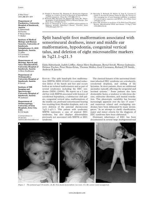
Figure 1 The proband aged 18 months. (A, B) Note facial dysmorphism (see text). (C) He cannot stand unsupported.<br />
www.jmedgenet.com<br />
The classical features of the autosomal dominant<br />
inherited EEC syndrome are ectrodactyly,<br />
ectodermal dysplasia, and clefting of the<br />
lip/palate. In most patients, there are additional<br />
anomalies typically aVecting the urogenital and<br />
lacrimal systems. 12 Some patients also have<br />
dysmorphic facies, a tendency to infectious disease,<br />
endocrine disorders, and mental retardation.<br />
This phenotypic variability has become<br />
increasingly apparent over the last 15 years 34<br />
and numerous related and overlapping syndromes<br />
have been delineated by many investigators.<br />
5 In an attempt to clarify classification,<br />
major and minor criteria for the diagnosis of<br />
EEC syndrome have been elaborated. 34<br />
Dominant inheritance of EEC has been<br />
documented in several large multigenerational
406 Letters<br />
Figure 2 Right <strong>foot</strong> of the patient. (A) Ectrodactyly (<strong>split</strong> <strong>foot</strong> <strong>malformation</strong>) <strong>with</strong> apparent absence of the 2nd toe and<br />
syndactyly of toes 3 to 5. (B) Radiograph showing syndactyly of the first and second metatarsals and absence of the second<br />
phalanges and <strong>malformation</strong> of the third to fifth phalanges.(C) Ectrodactyly and pes planovalgus (severe talus verticalis<br />
deformity).<br />
families. 6 At least 15 patients have been<br />
reported to have cytogenetic abnormalities of<br />
chromosome 7q21.2-7q22.1, including nine<br />
patients <strong>with</strong> interstitial deletions. 7–9 In addition,<br />
mutations in the gene encoding the transactivation<br />
factor p63 on chromosome 3q27<br />
have been identified in familial and sporadic<br />
cases of EEC syndrome. 10 A third locus was<br />
mapped to chromosome 19q, 11 further delineating<br />
the genetic heterogeneity of this syndrome.<br />
The reason for the phenotypic<br />
heterogeneity in EEC syndrome patients <strong>with</strong><br />
7q abnormalities is unclear but may relate to<br />
the size of the deletion.<br />
Case report<br />
Our patient is the fifth child of healthy, consanguineous,<br />
fourth cousin, Austrian parents. The<br />
father and the mother were 41 and 36 years,<br />
respectively, at the time of his birth. His four<br />
sibs are healthy. He was born after an uneventful<br />
pregnancy in the 41st week of gestation and<br />
weighed 2840 g (10th centile), was 48 cm long<br />
(10th centile), and had a head circumference of<br />
31.5 cm (10th centile). Ectrodactyly of the<br />
right <strong>foot</strong> was noted and transient evoked<br />
otoacoustic emission screening indicated hearing<br />
impairment. Further examinations were at<br />
first declined by the mother. At 15 months of<br />
asc<br />
co<br />
va es<br />
co<br />
Figure 3 Inner ear of the patient. A 3D reconstruction of a coronal MRI scan shows Mondini type <strong>malformation</strong> on both<br />
sides. (A) Right ear: overall dilated and plump structures of the inner ear. asc denotes the anterior semicircular canal, va the<br />
vestibular aqueduct <strong>with</strong> saccule and utricle, and co the cochlea showing a reduced number of coils. (B) Left ear: a large<br />
endolymphatic sac is shown (es). (C) Schema of the normal inner ear. 1. Anterior semicircular canal. 2. Membranous<br />
ampulla (MA) of the anterior semicircular canal. 3. MA of the lateral semicircular canal. 4. Saccule. 5. Cochlear canal. 6.<br />
Helicotrema. 7. Lateral semicircular canal. 8. Posterior semicircular canal. 9. MA of the posterior semicircular canal. 10.<br />
Vestibular window. 11. Cochlear window. 12. Scala vestibuli. 13. Scala tympani. 14. Utricule.<br />
www.jmedgenet.com<br />
asc
Letters 407<br />
A<br />
q21.1<br />
q21.3<br />
7 der(7)<br />
Figure 4 Cytogenetic and molecular findings. (A) High resolution cytogenetic analysis of both chromosomes 7 of the<br />
patient. The deletion is indicated by the arrow. (B) Preliminary analysis of microsatellite markers from chromosome 7q in<br />
the family of the patient. The deleted interval spans at least 8.9 cM on the paternal chromosome flanked by microsatellite<br />
markers D7S2443 and D7S2480. Data regarding microsatellite mapping are compiled from Dib et al 12 and Crackower et<br />
al. 13 14 The arrow indicates the position of the gene mutated in Pendred syndrome.<br />
age, he was referred to the hospital because of<br />
failure to thrive (weight 7200 g, below the 3rd<br />
centile; length 70 cm, below the 3rd centile;<br />
head circumference 43 cm, below the 3rd centile).<br />
Physical examination showed arched eyebrows,<br />
a small triangular nose <strong>with</strong> a depressed<br />
nasal bridge, and ears <strong>with</strong> overfolded helices<br />
and attached earlobes (fig 1). He also had<br />
hypertelorism, a large biparietal diameter,<br />
hypopigmented retina, micrognathia, a submucous<br />
cleft palate, carious primary teeth and<br />
hypodontia, sparse, light hair, pale skin,<br />
cryptorchidism, and bilateral severe congenital<br />
vertical talus, in addition to the previously<br />
noted ectrodactyly of the right <strong>foot</strong> (fig 2). CT<br />
and MRI scans showed Mondini dysplasia of<br />
the inner ear (fig 3) and cochlear implanting<br />
showed fixation of the ossicular chain. Audiometric<br />
examinations were consistent <strong>with</strong> these<br />
findings and showed conductive and profound<br />
<strong>sensorineural</strong> hearing loss. Laboratory investigations<br />
showed partial deficiency of growth<br />
hormone secretion. Mental and psychomotor<br />
developmental delay was noted.<br />
On GTG banding, we observed an interstitial<br />
deletion of chromosome 7 confined to the<br />
interval q21.1-q21.3 (fig 4A); parental karyotypes<br />
were normal. To delineate this deletion<br />
further, we used 21 chromosome 7q microsatellite<br />
markers to reconstruct parental and<br />
www.jmedgenet.com<br />
De novo<br />
deletion of<br />
about 8.9<br />
to 17.0 cM<br />
PDS gene<br />
B<br />
D7S2506<br />
D7S663<br />
D7S2455<br />
D7S634<br />
D7S2443<br />
D7S524<br />
D7S492<br />
D7S2410<br />
D7S657<br />
D7S2482<br />
D7S527<br />
D7S1812<br />
D7S821<br />
D7S2539<br />
D7S479<br />
D7S491<br />
D7S1796<br />
D7S2480<br />
D7S647<br />
D7S501<br />
D7S692<br />
3 2<br />
3 2<br />
3 1<br />
1 2<br />
2 2<br />
2 1<br />
2 2<br />
1 1<br />
1 1<br />
1 3<br />
2 1<br />
3 3<br />
2 2<br />
3 3<br />
1 2<br />
3 3<br />
2 3<br />
1 2<br />
2 2<br />
1 1<br />
3 2<br />
F<br />
D7S2506<br />
D7S663<br />
D7S2455<br />
D7S634<br />
D7S2443<br />
D7S524<br />
D7S492<br />
D7S2410<br />
D7S657<br />
D7S2482<br />
D7S527<br />
D7S1812<br />
D7S821<br />
D7S2539<br />
D7S479<br />
D7S491<br />
D7S1796<br />
D7S2480<br />
D7S647<br />
D7S501<br />
D7S692<br />
1 4<br />
4 1<br />
2 4<br />
2 3<br />
1 3<br />
2 3<br />
1 2<br />
1 1<br />
3 2<br />
2 3<br />
3 2<br />
2 1<br />
1 3<br />
2 1<br />
3 2<br />
2 1<br />
2 1<br />
2 2<br />
1 2<br />
2 3<br />
1 3<br />
SL<br />
3 1<br />
3 4<br />
3 2<br />
? 2<br />
2 1<br />
? 2<br />
- 1<br />
? 1<br />
- 3<br />
- 2<br />
? 2<br />
- 2<br />
- 1<br />
- 2<br />
- 3<br />
- 2<br />
? 2<br />
1 2<br />
2 1<br />
1 2<br />
3 1<br />
Distance<br />
Mb<br />
cM<br />
8.5<br />
10.3<br />
2.8<br />
0.7<br />
3.7<br />
3.1<br />
3.5<br />
1.5<br />
2.8<br />
1.3<br />
0.5<br />
7.8<br />
0.8<br />
patient haplotypes and found that for the eight<br />
markers flanked by D7S2443 and D7S2480,<br />
the patient had a deletion of the paternal allele<br />
(fig 4B). Two markers <strong>with</strong>in the interval<br />
(D7S2410 and D7S527) were uninformative,<br />
as were two flanking markers (D7S524 and<br />
D7S1796). These data define a deletion of 8.9<br />
to 17 cM , which includes the critical interval<br />
of
408 Letters<br />
association that has not previously been reported<br />
<strong>with</strong> either SHFM or a chromosome 7<br />
aberration. Mondini dysplasia is characterised<br />
by bony and membranous anomalies of the<br />
inner ear exhibiting a wide range of morphological<br />
and functional abnormalities. Typically,<br />
the cochlea is flat, the cochleal duct is short, the<br />
auditory and vestibular sense organs and nerves<br />
are immature, the vestibule is large, the semicircular<br />
canals are wide, small, or missing, and the<br />
endolymphatic sac is dilated. The anomaly can<br />
be unilateral or bilateral and occurs in isolation<br />
or in association <strong>with</strong> anomalies in other<br />
organs. 16 Familial examples of Mondini dysplasia<br />
generally represent examples of Pendred<br />
syndrome, an autosomal recessive disorder in<br />
which congenital <strong>sensorineural</strong> hearing impairment<br />
and goitre cosegregate.<br />
Because Pendred syndrome is caused by<br />
mutations in PDS, a gene that maps to<br />
chromosome 7q31, the simultaneous occurrence<br />
of atypical EEC syndrome and Pendred<br />
syndrome in our patient seemed an attractive<br />
possibility to explain the rare combination of<br />
physical findings. Although molecular analysis<br />
in our patient appears to place the distal<br />
breakpoint of the deletion about 9 cM centromeric<br />
to PDS, we cannot exclude a more complex<br />
chromosomal rearrangement. Assuming<br />
that the paternal copy of PDS could have been<br />
deleted, we completed a mutation screen for<br />
maternally inherited PDS allele variants. We<br />
were unable to identify any mutations by<br />
SSCP and direct sequencing as described previously<br />
17 and therefore could not establish a<br />
causal connection between the observed<br />
Mondini dysplasia and the chromosomal<br />
aberration. We also excluded an independent<br />
cause of the <strong>sensorineural</strong> hearing impairment,<br />
by sequencing the coding region and<br />
exon 1 of GJB2. 18 Mutations in this gene are<br />
the most common cause of autosomal recessive<br />
non-syndromic deafness.<br />
The simultaneous occurrence of an inner ear<br />
<strong>malformation</strong> and SHFM has rarely been<br />
reported. Berndorfer 19 noted absence of pinnae<br />
and lack of inner ears in one patient. Autosomal<br />
dominant ectrodactyly and deafness in a father<br />
and son were reported by Tolmie et al. 20 Both<br />
patients had CT verified cochlear abnormalities,<br />
which may have been consistent <strong>with</strong> Mondini<br />
dysplasia. There was no mention of any<br />
chromosomal anomaly. Moreover, <strong>sensorineural</strong><br />
hearing impairment has rarely been reported in<br />
syndromic SHFM. 21–26 In two of these cases,<br />
however, an apparently balanced translocation<br />
involving chromosome 7q was found to cosegre-<br />
gate <strong>with</strong> the disease.<br />
25 26<br />
Conductive hearing loss is observed in<br />
14-44% of cases of EEC syndrome, most commonly<br />
reflecting Eustachian tube dysfunction<br />
in association <strong>with</strong> the palatal clefting,<br />
although ossicular <strong>malformation</strong>s have been<br />
described. 27<br />
Of 10 patients <strong>with</strong> ectrodactyly in association<br />
<strong>with</strong> a deletion of 7q21-q22, microcephaly and<br />
general growth impairment have been reported<br />
in eight cases (80% 79 ) compared to only 2% and<br />
1%, respectively, in a survey of 230 patients <strong>with</strong><br />
EEC syndrome. 4 While adenohypophyseal<br />
www.jmedgenet.com<br />
3 4<br />
dysfunction in two sets of sibs has been reported<br />
in EEC syndrome, 28 29 partial growth hormone<br />
deficiency was identified in our patient as the<br />
aetiology of the growth retardation. However,<br />
growth retardation and microcephaly might<br />
also delineate a subtype of the EEC syndrome<br />
related to chromosomal aberrations involving<br />
chromosome 7q21-q22. We accordingly suggest<br />
initiating chromosomal and molecular<br />
investigations of this chromosomal region when<br />
growth retardation and microcephaly is present<br />
in patients <strong>with</strong> SHFM. Short stature, as well as<br />
low birth weight, abnormal skull shape, and ear<br />
<strong>malformation</strong>s were common findings among<br />
patients <strong>with</strong> proximal/intermediate deletions<br />
or rearrangements of chromosome 7q, <strong>with</strong> and<br />
<strong>with</strong>out SHFM. 8<br />
We believe that a specific pattern of facial<br />
anomalies characterises patients <strong>with</strong> aberrations<br />
of chromosome 7q21-q22. The facial<br />
phenotype consists of arched eyebrows, a<br />
small, triangular shaped nose <strong>with</strong> a depressed<br />
nasal bridge, abnormal ears <strong>with</strong> overfolded<br />
helices and attached earlobes, a large biparietal<br />
diameter, hypertelorism, and micrognathia. It<br />
was present in our patient and in at least six<br />
published case reports.<br />
9 30–34<br />
The <strong>split</strong> <strong>foot</strong> <strong>malformation</strong> in our patient<br />
was right sided, as has been mostly observed in<br />
cases of unilateral involvement of either the<br />
upper or lower limbs. 26 The presence of<br />
bilateral congenital vertical talus could not be<br />
explained by aplasia of the anterior calcaneus<br />
<strong>with</strong> loss of talar support or by a spinal defect,<br />
though the <strong>split</strong> <strong>foot</strong> <strong>malformation</strong> complicates<br />
the anomaly of the talus. Bilateral congenital<br />
vertical talus is an otherwise rare disorder, and<br />
to our knowledge has not been reported in the<br />
EEC syndrome or in related conditions.<br />
Our report suggests that patients <strong>with</strong><br />
syndromic SHFM should be examined for the<br />
findings we describe, and that the molecular<br />
analysis should include karyotyping and complementary<br />
studies to establish whether the<br />
critical interval of
Letters 409<br />
J Med Genet<br />
2001;38:409–411<br />
Division of<br />
Immunology/Allergy,<br />
The Infection,<br />
Immunity, Injury &<br />
Repair Program,<br />
Research Institute,<br />
Department of<br />
Pediatrics, The<br />
Hospital for Sick<br />
Children and the<br />
University of Toronto,<br />
Toronto, Canada<br />
E Grunebaum<br />
E Arpaia<br />
C M Roifman<br />
Department of<br />
Genetics, The Hospital<br />
for Sick Children and<br />
the University of<br />
Toronto, Toronto,<br />
Canada<br />
J J MacKenzie<br />
J Fitzpatrick<br />
PNRay<br />
Correspondence to:<br />
Dr Roifman, Infection,<br />
Immunity, Injury & Repair<br />
Program, Research Institute,<br />
The Hospital for Sick<br />
Children, 555 University<br />
Avenue, Toronto, Ontario<br />
M5G 1X8, Canada,<br />
chaim.roifman@sickkids.ca<br />
8 McElveen C, Carvajal MV, Moscatello D, Towner J, Lacassie<br />
Y. Ectrodactyly and proximal/intermediate interstitial<br />
deletion 7q. Am J Med Genet 1995;56:1-5.<br />
9 Marinoni JC, Stevenson RE, Evans JP, Geshuri D, Phelan<br />
MC, Schwartz CE. <strong>Split</strong> <strong>foot</strong> and developmental retardation<br />
<strong>associated</strong> <strong>with</strong> a deletion of three microsatellite markers<br />
in 7q21.2-q22.1. Clin Genet 1995;47:90-5.<br />
10 Celli J, Duijf P, Hamel BC, Bamshad M, Kramer B, Smits<br />
AP, Newbury-Ecob R, Hennekam RC, Van Buggenhout G,<br />
van Haeringen A, Woods CG, van Essen AJ, de Waal R,<br />
Vriend G, Haber DA, Yang A, McKeon F, Brunner HG,<br />
van Bokhoven H. Heterozygous germline mutations in the<br />
p53 homolog p63 are the cause of EEC syndrome. Cell<br />
1999;99:143-53.<br />
11 O’Quinn JR, Hennekam RCM, Jorde LB, Bamshad M.<br />
Syndromic ectrodactyly <strong>with</strong> severe limb, ectodermal, urogenital,<br />
and palatal defects maps to chromosome 19. Am J<br />
Hum Genet 1998;62:130-5.<br />
12 Dib C, Faure S, Fizames C, Samson D, Drouot N, Vignal A,<br />
Millasseau P, Marc S, Hazan J, Seboun E, Lathrop M,<br />
Gyapay G, Morissette J, Weissenbach J. A comprehensive<br />
genetic map of the human genome based on 5,264 microsatellites.<br />
Nature 1996;380:152-4.<br />
13 Crackower MA, Scherer SW, Rommens JM, Hui CC,<br />
Poorkaj P, Soder S, Cobben JM, Hudgins L, Evans JP, Tsui<br />
LC. Characterization of the <strong>split</strong> <strong>hand</strong>/<strong>split</strong> <strong>foot</strong> <strong>malformation</strong><br />
locus SHFM1 at 7q21.3-q22.1 and analysis of a candidate<br />
gene for its expression during limb development.<br />
Hum Mol Genet 1996;5:571-9.<br />
14 Crackower MA, Sinasac DS, Xia J, Motoyama J, Prochazka<br />
M, Rommens JM, Scherer SW, Tsui LC. Cloning and characterization<br />
of two cytoplasmic dynein intermediate chain<br />
genes in mouse and human. Genomics 1999;55:257-67.<br />
15 Akita S, Kuratomi H, Abe K, Harada N, Mukae N, Niikawa<br />
N. EC syndrome in a girl <strong>with</strong> paracentric inversion<br />
(7)(q22.1;q36.3). Clin Dysmorphol 1993;2:62-7.<br />
16 Ormerod FC. The pathology of congenital deafness. J<br />
Laryngol 1960;74:919-50.<br />
17 Van Hauwe P, Everett LA, Coucke P, Scott DA, Kraft ML,<br />
Ris-Stalpers C, Bolder C, Otten B, de Vijlder JJ, Dietrich<br />
NL, Ramesh A, Srisailapathy SC, Parving A, Cremers CW,<br />
Willems PJ, Smith RJ, Green ED, Van Camp G. Two<br />
frequent missense mutations in Pendred syndrome. Hum<br />
Mol Genet 1998;7:1099-104.<br />
18 Scott DA, Kraft ML, Carmi R, Ramesh A, Elbedour K, Yairi<br />
Y, Srisailapathy CR, Rosengren SS, Markham AF, Mueller<br />
RF, Lench NJ, Van Camp G, Smith RJ, SheYeld VC. Identification<br />
of mutations in the connexin 26 gene that cause<br />
autosomal recessive nonsyndromic hearing loss. Hum<br />
Mutat 1998;11:387-94.<br />
19 Berndorfer A. Gesichtsspalten gemeinsam mit Hand- und<br />
Fuspalten. Z Orthopad 1970;107:344-54.<br />
20 Tolmie J, Geddes NK, Knight S, Fredricks B. Autosomal<br />
dominant ectrodactyly and deafness. 5th Manchester Birth<br />
Defects Conference, 13-16 October 1992.<br />
21 Birch-Jensen A. Congenital deformities of the upper extremities.<br />
Copenhagen: Ejnar Munksgaards Forlag, 1949:19.<br />
22 Wildervanck LS. Perceptive deafness <strong>associated</strong> <strong>with</strong> <strong>split</strong><strong>hand</strong><br />
and <strong>foot</strong>, a new syndrome? Acta Genet 1963;13:161-9.<br />
23 Fraser GR. The causes of profound deafness in childhood.<br />
Baltimore: Johns Hopkins University Press, 1976.<br />
24 Anneren G, Andersson T, Lindgren PG, Kjartansson S.<br />
Ectrodactyly-ectodermal dysplasia-clefting syndrome<br />
(EEC): the clinical variation and prenatal diagnosis. Clin<br />
Genet 1991;40:257-62.<br />
25 Hasegawa T, Hasegawa Y, Asamura S, Nagai T, Tsuchiya Y,<br />
Ninomiya M, Fukushima Y. EEC syndrome (ectrodactyly,<br />
ectodermal dysplasia and cleft lip/palate) <strong>with</strong> a balanced<br />
reciprocal translocation between 7q11.21 and 9p12 (or<br />
7p11.2 and 9q12) in three generations. Clin Genet 1991;40:<br />
202-6.<br />
26 Genuardi M, Pomponi MG, Sammito V, Bellussi A, Zollino<br />
M, Neri G. <strong>Split</strong> <strong>hand</strong>/<strong>split</strong> <strong>foot</strong> anomaly in a family segregating<br />
a balanced translocation <strong>with</strong> breakpoint on 7q22.1.<br />
Am J Med Genet 1993;47:823-31.<br />
27 Robinson GC, Wildervanck LS, Chiang TP. Ectrodactyly,<br />
ectodermal dysplasia, and cleft lip-palate syndrome. Its<br />
association <strong>with</strong> conductive hearing loss. J Pediatr 1973;82:<br />
107-9.<br />
28 Knudtzon J, Aarskog D. Growth hormone deficiency <strong>associated</strong><br />
<strong>with</strong> the ectrodactyly-ectodermal dysplasia-clefting<br />
syndrome and isolated absent septum pellucidum. Pediatrics<br />
1987;79:410-12.<br />
29 Gershoni-Baruch R, Goldscher D, Hochberg Z.<br />
Ectrodactyly-ectodermal dysplasia-clefting syndrome and<br />
hypothalamo-pituitary insuYciency. Am J Med Genet<br />
1997;68:168-72.<br />
30 Young RS, Weaver DD, Kukolich MK, Heerema NA, Palmer<br />
CG, Kawira EL, Bender HA. Terminal and interstitial<br />
deletions of the long arm of chromosome 7: a review <strong>with</strong><br />
five new cases. Am J Med Genet 1984;17:437-50.<br />
31 Fryns JP, Kleczkowska A, Van den Berghe H. Moderate mental<br />
retardation and mild dysmorphic syndrome in proximal<br />
7q interstitial deletion. Ann Genet 1987;30:111-12.<br />
32 Tajara EH, Varella-Garcia M, Gusson AC. Interstitial longarm<br />
deletion of chromosome 7 and ectrodactyly. Am J Med<br />
Genet 1989;32:192-4.<br />
33 Rivera H, Sanchez-Corona J, Burgos-Fuentes VR,<br />
Melendez-Ruiz MJ. Deletion of 7q22 and ectrodactyly.<br />
Genet Couns 1991;2:27-31.<br />
34 Sharland M, Patton MA, Hill L. Ectrodactyly of <strong>hand</strong>s and<br />
feet in a child <strong>with</strong> a complex translocation including<br />
7q21.2. Am J Med Genet 1991;39:413-14.<br />
A missense mutation in the SEDL gene results in<br />
delayed onset of X linked spondyloepiphyseal<br />
dysplasia in a large pedigree<br />
E Grunebaum, E Arpaia, J J MacKenzie, J Fitzpatrick, P N Ray, C M Roifman<br />
EDITOR—Spondyloepiphyseal dysplasia (SED)<br />
is a rare osteochondroplasia, characterised by<br />
disproportionate short stature <strong>with</strong> a short<br />
neck and trunk and barrel chest. The pelvis<br />
tends to be narrow and deep, the femoral neck<br />
short, and the femoral head flattened. Mild to<br />
moderate epiphyseal dysplasia of the large<br />
joints may also be seen. The latter may lead to<br />
premature secondary osteoarthritis <strong>with</strong> significant<br />
morbidity. 1 SED may occur sporadically;<br />
however, in many cases the family<br />
history indicates an inherited condition. In<br />
some of these pedigrees, the inheritance<br />
pattern seems autosomal dominant, while in<br />
others it is consistent <strong>with</strong> autosomal recessive<br />
or X linked recessive. 2<br />
Recently, mutations in the gene designated<br />
SEDL, located on Xp22, were identified as the<br />
cause of X linked spondyloephiphyseal dysplasia<br />
tarda in three families. 3 We have previously<br />
described a large kindred of British descent<br />
www.jmedgenet.com<br />
spanning four generations aVected by SED. 1<br />
Briefly, 14 males between the ages of 10 and<br />
77 years were aVected, <strong>with</strong> early adolescence<br />
development of progressive decline in growth<br />
rate accompanied by short stature, short<br />
trunk, and barrel chest. Although some of<br />
them had to limit their activities because of hip<br />
or back limitation of movement or pain, many<br />
continued <strong>with</strong> normal activity and were able<br />
to perform in the work place <strong>with</strong>out impairment<br />
of function. There was no indication of<br />
other abnormalities previously reported in<br />
association <strong>with</strong> SED, such as mental retardation,<br />
2 immune abnormalities and retinopathy, 4<br />
cardiac dysfunction, 5 or hypogonadotrophic<br />
hypogonadism. 6 The female carriers in this<br />
pedigree had normal height. Although some of<br />
the females suVered from occasional mild back<br />
or hip pain, it did not aVect their daily activity,<br />
nor was there objective radiological evidence<br />
of spinal or joint involvement compatible <strong>with</strong>