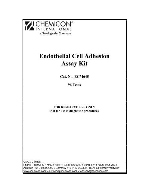
Endothelial Cell Adhesion Assay Kit - Millipore
Endothelial Cell Adhesion Assay Kit - Millipore
Endothelial Cell Adhesion Assay Kit - Millipore

You also want an ePaper? Increase the reach of your titles
YUMPU automatically turns print PDFs into web optimized ePapers that Google loves.
<strong>Endothelial</strong> <strong>Cell</strong> <strong>Adhesion</strong><br />
<strong>Assay</strong> <strong>Kit</strong><br />
Cat. No. ECM645<br />
96 Tests<br />
FOR RESEARCH USE ONLY<br />
Not for use in diagnostic procedures<br />
USA & Canada<br />
Phone: +1(800) 437-7500 • Fax: +1 (951) 676-9209 • Europe +44 (0) 23 8026 2233<br />
Australia +61 3 9839 2000 • Germany +49-6192-207300 • ISO Registered Worldwide<br />
www.chemicon.com • custserv@chemicon.com • techserv@chemicon.com
Introduction<br />
<strong>Endothelial</strong> cells form the inner lining of blood vessels and serve as the cellular<br />
interface between the circulating blood and the vessel wall. <strong>Endothelial</strong> cells are<br />
involved in many aspects of vascular biology, including inflammation and<br />
angiogenesis. The recruitment of leukocytes into inflammatory tissues is<br />
regulated by the interaction between blood cells and endothelial cells that is<br />
preceded by integrin-mediated cell adhesion. <strong>Endothelial</strong>-leukocyte cell adhesion<br />
plays a major role in cellular communication and regulation, and is of fundamental<br />
importance in the development and maintenance of tissues. Atherosclerosis, ulcer,<br />
myocardial infarction, stroke, and other inflammatory processes are all related<br />
to, and dependent on this endothelial-leukocyte adhesion process.<br />
Expression of surface molecules on the vascular endothelium is altered at sites of<br />
pathological inflammation. <strong>Endothelial</strong> <strong>Cell</strong> <strong>Adhesion</strong> Molecules (ECAMs) are<br />
important mediators of leukocyte recruitment and adherence to the endothelium.<br />
ECAMs such as E-selectin, VCAM-1, and ICAM-1 are upregulated during<br />
inflammation, which initiates leukocyte adhesion to the endothelium, and<br />
ultimately contributes to disease progression or tissue damage. Expression of<br />
these ECAMs is mediated by proinflammatory cytokines such as Interleukin-1<br />
beta and Tumor Necrosis Factor alpha, which propel the interaction of integrin<br />
multimers on the leukocyte cell surface and the ECAMs on the endothelial cells.<br />
The genes encoding ECAMs contain overlapping control mechanisms that allow<br />
a single signaling pathway to upregulate several genes with the potential to<br />
change the endothelial phenotype. Since ECAMs are regulated at the gene level<br />
by transcription factors, proteasome inhibitors can modulate cytokine-induced<br />
expression of ECAMs and subsequent leukocyte adhesion to the endothelium.<br />
Controlling and manipulating the endothelial-leukoctye adhesion process will<br />
create new avenues for drug therapies for vascular and inflammatory disease<br />
states.<br />
The CHEMICON ® <strong>Endothelial</strong> <strong>Cell</strong> <strong>Adhesion</strong> <strong>Assay</strong> kit (ECM645) allows the<br />
researcher to test a variety of cell types that interact with activated or inactivated<br />
endothelial cell layers. Human umbilical vein endothelial cells (HUVECs) from<br />
ATCC ® (CRL-1730) or Cambrex © (CC-2519) are ideal for measuring<br />
endothelial adhesion activity with the assay. The CHEMICON ® <strong>Endothelial</strong><br />
<strong>Cell</strong> <strong>Adhesion</strong> <strong>Assay</strong> kit includes a 96 well tissue culture treated fluorescence<br />
microtiter plate and reagents that allow for large-scale screening and quantitative<br />
comparison of multiple samples/cell line adhesion to an endothelial monolayer.<br />
Once endothelial cells are seeded in the tissue culture plate, they can be treated<br />
with cytokines, chemokines, transcription regulators, or selective adhesion<br />
inhibitors. The protein synthesis inhibitor Cycloheximide and the RNA<br />
synthesis inhibitor Actinomycin D are provided as controls for adhesion protein<br />
expression studies. Treatment of endothelial cells, such as human umbilical vein<br />
endothelial cells (HUVECs), with pro-inflammatory cytokines such as<br />
1
Interleukin-1β or Tumor Necrosis Factor-α can induce expression of ECAMs, as<br />
well as other adhesion molecules. The kit is supplied with both cytokines.<br />
The viable cell fluorescent compound Calcein-AM ® is provided for labeling test<br />
cells, such as leukocytes (granulocytes, monocytes, lymphocytes), fibroblasts,<br />
neural stem cells etc., and quantifying their adhesion by comparing bound versus<br />
unbound cells. The kit includes three Mouse anti-human monoclonal mAbs for<br />
adhesion blocking studies of ICAM-1, VCAM, and E-selectin endothelial cell<br />
surface markers. These may be used to verify changes in expression level of<br />
adhesion molecules, or effects of integrins upon endothelium activation. This<br />
simple and high-throughput kit format allows for the monitoring of the effects of<br />
a variety of conditions and compounds on endothelial adhesion. CHEMICON ®<br />
also offers a variety of antibodies to leukocyte adhesion molecules and surface<br />
antigens. See CHEMICON ® ‘s website at www.chemicon.com for additional<br />
information.<br />
CHEMICON ® continues to provide numerous migration, invasion, and adhesion<br />
products including:<br />
• <strong>Endothelial</strong> <strong>Cell</strong> Characterization <strong>Kit</strong> (SCR023)<br />
• In Vitro Angiogenesis <strong>Assay</strong> (ECM625)<br />
• Fibrin In Vitro Angiogenesis <strong>Assay</strong> (ECM630)<br />
• Blood Vessel Staining <strong>Kit</strong> (ECM590)<br />
• Alpha Integrin-Mediated <strong>Cell</strong> <strong>Adhesion</strong> Colorimetric Array <strong>Kit</strong> (ECM530)<br />
• Beta Integrin-Mediated <strong>Cell</strong> <strong>Adhesion</strong> Colorimetric Array <strong>Kit</strong> (ECM531)<br />
• Alpha/Beta Integrin-Mediated <strong>Cell</strong> <strong>Adhesion</strong> Colorimetric Array Combo <strong>Kit</strong><br />
(ECM532)<br />
• Alpha Integrin-Mediated <strong>Cell</strong> <strong>Adhesion</strong> Fluorimetric Array (ECM533)<br />
• Beta Integrin-Mediated <strong>Cell</strong> <strong>Adhesion</strong> Fluorimetric Array (ECM534)<br />
• Alpha/Beta Integrin-Mediated <strong>Cell</strong> <strong>Adhesion</strong> Array Combo Fluorimetric <strong>Kit</strong><br />
(ECM535)<br />
• ECM <strong>Cell</strong> <strong>Adhesion</strong> Colorimetric Array <strong>Kit</strong> (ECM540)<br />
• ECM <strong>Cell</strong> <strong>Adhesion</strong> Fluorimetric Array <strong>Kit</strong> (ECM545)<br />
• QCM 8µm 96-well Chemotaxis <strong>Cell</strong> Migration <strong>Assay</strong> (ECM510)<br />
• QCM 8µm 24-well Chemotaxis <strong>Cell</strong> Migration Colorimetric <strong>Assay</strong><br />
(ECM508)<br />
• QCM 8µm 24-well Chemotaxis <strong>Cell</strong> Migration Fluorimetric <strong>Assay</strong><br />
(ECM509)<br />
• QCM 5µm 96-well Chemotaxis <strong>Cell</strong> Migration <strong>Assay</strong> (ECM512)<br />
• QCM 3µm 96-well Chemotaxis <strong>Cell</strong> Migration <strong>Assay</strong> (ECM515)<br />
• QCM 96-well ECMatrix TM <strong>Cell</strong> Invasion <strong>Assay</strong> (ECM555)<br />
• QCM 96-well Collagen-based <strong>Cell</strong> Invasion <strong>Assay</strong> (ECM 556)<br />
2
Test Principle<br />
The CHEMICON ® <strong>Endothelial</strong> <strong>Cell</strong> <strong>Adhesion</strong> <strong>Assay</strong> utilizes a homogenous<br />
fluorescence detection format, allowing for large-scale screening and<br />
quantitative comparison of multiple samples with the flexibility for testing a<br />
variety of conditions. First, endothelial cells are seeded in the 96 well plate where<br />
they grow to confluency. Prior to activation, the endothelium can be treated with<br />
the protein synthesis inhibitor, (Cycloheximide) or the RNA synthesis inhibitor<br />
(Actinomycin D) to control endothelial adhesion protein expression.<br />
Subsequently, the endothelium is activated with one or two cytokines (TNF alpha<br />
and Interleukin-1 beta) provided. Upon activation, endothelial cells can be used<br />
for adhesion blocking studies with functional blocking mAbs to endothelial cell<br />
adhesion markers (e.g. E-selectin, ICAM-1, VCAM). Next, test cells are labeled<br />
with the Calcein AM ® green fluorescence viability cell marker and added to the<br />
endothelium on the plate. After a brief incubation, unbound cells are washed away<br />
while bound cells remain attached to the endothelium. Finally, the plate is read on<br />
a fluorescence plate reader. Relative test cell adhesion is directly related to the<br />
fluorescence of the wells. See the diagram on the next page.<br />
Application<br />
The CHEMICON ® <strong>Endothelial</strong> <strong>Cell</strong> <strong>Adhesion</strong> <strong>Assay</strong> <strong>Kit</strong> is a versatile tool for<br />
assessing and quantifying cell to cell interaction of human endothelial cells with<br />
leukocytes, or other cell types, under a variety of conditions. The kit is ideal for<br />
the assessment of specific cell surface integrins and surface receptors on human<br />
endothelial cells, monitoring in vitro cell differentiation, assessing endothelial<br />
adhesion blocking, or screening potential cell adhesion promoters/inhibitors. A<br />
variety of leukocytes or leukocyte-like cell lines can be tested in response to<br />
manipulation of the endothelial cell behavior. Each CHEMICON ® <strong>Endothelial</strong><br />
<strong>Cell</strong> <strong>Adhesion</strong> <strong>Assay</strong> <strong>Kit</strong> contains sufficient reagents for the evaluation of 96<br />
samples.<br />
The CHEMICON ® <strong>Endothelial</strong> <strong>Cell</strong> <strong>Adhesion</strong> <strong>Assay</strong> <strong>Kit</strong> is intended for research<br />
use only, not for diagnostic or therapeutic applications.<br />
3
Human <strong>Endothelial</strong><br />
cells are seeded in a<br />
black fluorescence tissue<br />
culture treated plate.<br />
<strong>Endothelial</strong> cell adhesion<br />
molecules express on<br />
the HUVEC surface.<br />
Blocking antibodies<br />
against ECAMs or<br />
integrins can be added.<br />
<strong>Endothelial</strong> <strong>Adhesion</strong> <strong>Assay</strong>(s) Procedure<br />
The cells are cultured 48<br />
to 72 hours or until they<br />
are confluent.<br />
4<br />
The cells are treated with<br />
a pro-inflammatory<br />
cytokine such as TNFα<br />
or IL1-β. The plate is<br />
incubated for 2-6 hours.<br />
TNFα<br />
TNFα<br />
TNFα<br />
TNFα<br />
TNFα TNFα<br />
Calcein-AM labeled cells, such as<br />
leukocytes, are added to the<br />
wells. The plate is incubated<br />
briefly to allow for cell binding.<br />
Non-specific cells are washed<br />
away. The plate is read at<br />
485nm/530nm in a<br />
fluorescent plate reader.<br />
Fluorescence is a direct<br />
correlation of cell adhesion.<br />
Blocking antibodies inhibit or<br />
reduce the leukocyte adhesion to<br />
activated endothelial cells<br />
For Research Use Only. Not for use in diagnostic procedures.<br />
TNFα<br />
TNFα<br />
TNFα<br />
TNFα<br />
TNFα<br />
Plate<br />
Reader<br />
Fluorescence
<strong>Kit</strong> Materials<br />
1. 96-Well <strong>Endothelial</strong> <strong>Cell</strong> Tissue Culture Fluorescence Plate: (Part No.:<br />
2004174) One 96-well tissue culture-treated black fluorescence plate with<br />
lid.<br />
2. Cycloheximide: (Part No. 2004165) One vial – 100 µL of solution.<br />
3. Actinomycin D: (Part No. 2004166) One vial – 50 µg of crystals.<br />
4. Tumor Necrosis Factor alpha: (Part No. 2004167) (Chemicon Cat. No.<br />
GF023) One vial – 20 µL of solution.<br />
5. Interleukin-1 beta: (Part No. 2004168) (Chemicon Cat. No. IL038) One<br />
vial – 12 µL of solution.<br />
6. 1X <strong>Assay</strong> Buffer: (Part No. 2004320) One bottle - 100 mL of solution.<br />
7. Calcein AM Dye ® : (Part No. 2004169) One vial – 50 µL of solution, ready<br />
to use.<br />
8. Mouse anti-Human ICAM-1 Monoclonal Antibody: (Part No. 2004181)<br />
(Chemicon Cat. No. MAB1379) One vial - 30 µL of solution.<br />
9. Mouse anti-Human VCAM Monoclonal Antibody: (Part No. 2004182)<br />
(Chemicon Cat. No. MAB2144) One vial - 30 µg in solution.<br />
10. Mouse anti-Human E-selectin Monoclonal Antibody: (Part No. 2004183)<br />
One vial – 37.5 µL of solution.<br />
11. Mouse Negative Control IgG: (Chemicon Cat. No. PP54-100UG) One vial<br />
- 100 µL of solution.<br />
Materials Not Supplied<br />
• <strong>Endothelial</strong> <strong>Cell</strong>s such as HUVEC.<br />
• <strong>Endothelial</strong> <strong>Cell</strong> Basal Medium (EBM) without FBS or FCS: Tissue culture<br />
basal medium appropriate for subject endothelial cells or test cells.<br />
• Tissue culture growth medium appropriate for subject endothelial cells or<br />
test cells, such as <strong>Endothelial</strong> <strong>Cell</strong> Growth Medium (EGM), DMEM,<br />
EMEM, or FBM (Fibroblast Basal Media) containing 10% FBS.<br />
• Harvesting Buffer: EDTA or trypsin cell detachment buffer. Suggested<br />
formulations include a) 2 mM EDTA/PBS, b) 0.05% trypsin in Hanks<br />
Balanced Salt Solution (HBSS) containing 25 mM HEPES, or other cell<br />
detachment formulations as optimized by individual investigators.<br />
5
Note: Trypsin cell detachment buffer may be required for difficult cell lines.<br />
Allow sufficient time for cell receptor recovery.<br />
• Quenching Medium: Serum-free medium, such as EGM, DMEM, EMEM,<br />
or FBM, containing 5% BSA<br />
Note: Quenching Medium must contain divalent cations (Mg 2+ , Ca 2+ )<br />
sufficient for quenching EDTA in the harvesting buffer<br />
• Sterile PBS or Hank’s Balanced Salt Solution (HBSS) for washing cells<br />
• Sterile pipettes and pipette tips sufficient for aliquoting cells<br />
• Sterile cell culture hood<br />
• Low speed centrifuge and tubes for cell harvesting<br />
• CO2 incubator appropriate for subject cells<br />
• Hemacytometer and microscope for counting cells<br />
• Trypan blue or equivalent viability stain<br />
• DMSO<br />
• Multi-channel and/or repeating pipettes<br />
• Deionized water<br />
• Conical and microfuge tubes<br />
• Fluorescence plate reader with 485 nm and 530 nm filters<br />
Precautions<br />
• No data is available on the biological toxicity of Calcein AM ® dye. This<br />
chemical is membrane permeable and, as such, should be treated as a<br />
potential mutagen, which may cause cancer and heritable genetic damage.<br />
Handle with caution. DMSO stock solution should be handled with special<br />
caution as DMSO can facilitate the entry of organic molecules into tissues.<br />
• Cycloheximide is highly toxic and should be handled with care. Use<br />
protective wear when handling. This chemical is membrane permeable and,<br />
as such, should be treated as a potential mutagen, which may cause cancer<br />
and heritable genetic damage.<br />
• Actinomycin D is highly toxic, carcinogenic, teratogenic and should be<br />
handled with care. Use protective wear when handling. This chemical is<br />
membrane permeable and, as such, should be treated as a potential mutagen,<br />
which may cause cancer and heritable genetic damage. DMSO stock<br />
6
solution should be handled with special caution as DMSO can facilitate the<br />
entry of organic molecules into tissues.<br />
Storage<br />
The <strong>Assay</strong> Buffer and the <strong>Endothelial</strong> <strong>Cell</strong> Culture Plate can be stored at 2° to 8°C<br />
up to their expiration dates. Store the plate in its foil pouch. If not using the entire<br />
plate, seal any used wells with a plate sealer to protect the unused wells. Ensure<br />
that the desiccant remains in the pouch, and that the pouch is securely closed.<br />
Store the remaining kit components at -20°C. Prepare undiluted aliquots for the<br />
Cycloheximide, TNF alpha, and IL-1 beta. Avoid repeated freeze-thaw cycles.<br />
Note: Actinomycin D adsorbs to plastic and glass upon standing in solution;<br />
however, frozen aliquots of the stock vial are expected to be stable for at least<br />
30 days at -20°C.<br />
Preparation of Reagents<br />
Important Note: During shipment, small volumes of product will occasionally<br />
become entrapped in the seal of the product vial. For products with volumes of<br />
200 µL or less, we recommend briefly centrifuging the vial in a tabletop<br />
centrifuge to dislodge any liquid in the container’s cap.<br />
Reagent Product Description Preparation<br />
1 Cycloheximide: Blocks translation of<br />
messenger RNA on cytosolic<br />
80S ribosomes, but does not<br />
inhibit organelle protein<br />
synthesis.<br />
2 Actinomycin D: Inhibits cell proliferation by<br />
forming a stable complex<br />
with dsDNA, thus inhibiting<br />
DNA-primed RNA synthesis.<br />
7<br />
Cycloheximide is<br />
provided at 10<br />
mg/mL. Prepare a<br />
solution of<br />
Cycloheximide to 1-<br />
20 µg/mL in <strong>Assay</strong><br />
Buffer or media as<br />
needed.<br />
Actinomycin D is<br />
provided as 50 µg<br />
crystals.<br />
Reconstitute the vial<br />
with 50 µL of<br />
DMSO. Vortex<br />
thoroughly. Prepare<br />
a solution of<br />
Actinomycin D to 1-<br />
10 µg/mL in <strong>Assay</strong><br />
Buffer or media as<br />
needed.
3 Tumor Necrosis<br />
Factor alpha<br />
(TNFα):<br />
4 Interleukin-1<br />
beta (IL-1β):<br />
5 Mouse anti-<br />
Human ICAM-1<br />
Monoclonal<br />
Antibody:<br />
TNFα is a pleiotropic<br />
inflammatory cytokine. The<br />
protein acts as a key mediary<br />
in the local inflammatory<br />
immune response. TNFα<br />
initiates a cascade of<br />
cytokines, ECAMs, and<br />
increases vascular<br />
permeability, thereby<br />
recruiting leukocytes to a site<br />
of infection.<br />
IL-1β is a pleotropic<br />
cytokine, secreted primarily<br />
by monocytes and<br />
macrophages, that mediates<br />
the pathophysiology of<br />
various acute and chronic<br />
inflammatory conditions. IL-<br />
1β’s pro-inflammatory effects<br />
are modulated by increased<br />
endothelial vascular<br />
permeability, accompanied<br />
by ECAM and cytokine<br />
expression.<br />
Mouse IgG 1 mAb recognizes<br />
the extracellular D1 domain<br />
of ICAM-1. Reacts with<br />
ICAM-1 antigen found on<br />
lyphocytes, monocytes,<br />
granulacytes, fibroblasts, and<br />
endothelial cells. Blocks<br />
ICAM-1 mediated adhesion<br />
to LFA-1.<br />
8<br />
TNFα is provided at<br />
0.1 mg/mL. Prepare<br />
a solution of TNFα<br />
to working<br />
concentration range<br />
of 10-1000 ng/mL in<br />
<strong>Assay</strong> Buffer or<br />
media as needed, just<br />
prior to use.<br />
IL-1 beta is provided<br />
at 0.1 mg/mL.<br />
Prepare a solution of<br />
Interleukin-1 beta to<br />
a working<br />
concentration range<br />
of 10-1000 ng/mL in<br />
<strong>Assay</strong> Buffer or<br />
media as needed, just<br />
prior to use.<br />
Ms x ICAM-1 is<br />
provided at 1 mg/mL.<br />
Dilute in <strong>Assay</strong><br />
Buffer or media as<br />
needed. Suggested<br />
working<br />
concentration is 10-<br />
50 µg/mL.<br />
Note: The end user<br />
must determine the<br />
optimal<br />
concentration and<br />
working dilutions for<br />
their particular<br />
application.
6 Mouse anti-<br />
Human VCAM<br />
Monoclonal<br />
Antibody:<br />
7 Mouse anti-<br />
Human<br />
E-selectin<br />
Monoclonal<br />
Antibody:<br />
Mouse IgG 1 mAb recognizes<br />
IL-1β activated endothelial<br />
cells and inhibits cell<br />
adhesion.<br />
Mouse IgG 1 mAb recognizes<br />
E-selectin expressed on<br />
cytokine activated HUVECs<br />
and inhibits adhesion of<br />
neutrophils, eosinophils, and<br />
skin-homing T-cells. This<br />
mAb has been shown to block<br />
U937, HL-60, THP-1, as well<br />
as HUVECs. This mAb<br />
binds specifically to Eselectin<br />
after being screened<br />
against COS cells transfected<br />
with cDNAs for E-selectin, Pselectin,<br />
L-selectin, PECAM-<br />
1, ICAM-1, and VCAM-1.<br />
9<br />
Ms x VCAM is<br />
provided at 1 mg/mL.<br />
Dilute in <strong>Assay</strong><br />
Buffer or media as<br />
needed. Suggested<br />
working<br />
concentration is 10-<br />
50 µg/mL.<br />
Note: The end user<br />
must determine the<br />
optimal<br />
concentration and<br />
working dilutions for<br />
their particular<br />
application.<br />
Ms x E-selectin is<br />
provided at 1 mg/mL.<br />
Dilute in <strong>Assay</strong><br />
Buffer or media as<br />
needed. Suggested<br />
working<br />
concentration is 20-<br />
50 µg/mL.<br />
Note: The end user<br />
must determine the<br />
optimal<br />
concentration and<br />
working dilutions for<br />
their particular<br />
application.
8 Mouse Negative<br />
Control IgG:<br />
Mouse IgG whole molecule<br />
can be used as a negative<br />
control on the endothelium to<br />
determine background<br />
affinity or non-specific<br />
binding of test cells.<br />
Preparation of <strong>Endothelial</strong> <strong>Cell</strong>s<br />
Perform the following steps in a sterile hood<br />
10<br />
Ms IgG is provided<br />
at 1 mg/mL. Dilute<br />
in <strong>Assay</strong> Buffer or<br />
media as needed.<br />
Note: The end user<br />
must determine the<br />
optimal<br />
concentration and<br />
working dilutions for<br />
their particular use.<br />
Prepare <strong>Endothelial</strong> cell line for investigation as desired. The following<br />
procedure is a suggestion for preparing endothelial cells used in cell layer<br />
formation. Human umbilical vein endothelial cells (HUVECs) from ATCC ®<br />
(CRL-1730) or Cambrex © (CC-2519) were used for generating endothelial<br />
adhesion activity data for this assay.<br />
1. Use cells that are at least 80-100% confluent.<br />
2. Visually inspect cells before harvest, taking note of relative cell numbers<br />
and morphology.<br />
3. Wash cells 2 times with sterile PBS or HBSS.<br />
4. Add 5 mL Harvesting Buffer (see Materials Not Supplied) per 100 mm dish<br />
and incubate at 37˚C for 5-15 minutes.<br />
5. Add 10-20 mL of Quenching Medium or <strong>Endothelial</strong> <strong>Cell</strong> Growth Medium<br />
(see Materials Not Supplied) to inactivate trypsin/EDTA from Harvesting<br />
Buffer and gently pipet the cells off the dish.<br />
6. Centrifuge cells gently to pellet (400 x g, 5-10 minutes).<br />
7. Carefully remove media from pellet.<br />
8. Gently resuspend the pellet in 1-5 mL of Quenching Medium or <strong>Endothelial</strong><br />
<strong>Cell</strong> Growth Medium, depending upon the size of the pellet. Optimum cell<br />
density may be determined by titration of the cells. Often it is best to harvest a<br />
greater number of cells than is needed.<br />
9. Count cells and bring to a volume that provides a concentration of<br />
1.0 –2.0 x 10 6 cells/mL with growth or basal media (EGM or EBM).<br />
10. OPTIONAL: If pretreatment of <strong>Endothelial</strong> <strong>Cell</strong>s is desired, add<br />
compound(s) (cytokines, pharmacological agents, etc.) at this time.
Preparation of Calcein AM ® Labeled Test <strong>Cell</strong>s<br />
Perform the following steps in a sterile hood<br />
Prepare cell line for investigation as desired. The following procedure is a<br />
suggestion for preparing leukocytic cell lines used in cell adhesion.<br />
Note: Use Calcein AM ® labeled cells within 4 –12 hours.<br />
1. Use cells that are at least 80-100% confluent.<br />
2. Visually inspect cells before harvesting, taking note of relative cell numbers<br />
and morphology.<br />
3. Dissociate adherent or non-adherent cell lines to be tested into separate,<br />
single cell suspensions.<br />
4. Dilute the leukocyte cell suspension(s) to 5 mL.<br />
5. Add 12.5 µL of Calcein AM ® to the cell suspension(s) for a 2.5 µM final<br />
concentration. Mix gently by inversion.<br />
6. Incubate the cells in a 37˚C CO2 incubator for 30 minutes.<br />
7. Centrifuge cells gently to pellet (400 xg, 5-10 minutes).<br />
8. Carefully remove the media from the cell suspension(s).<br />
Note: The cell pellet(s) should have a bright yellow appearance.<br />
9. Wash each labeled cell line with 5-10 mL of PBS or HBSS. Pipette to<br />
resuspend and wash the cells thoroughly.<br />
10. Centrifuge cells gently to pellet (400 xg, 5-10 minutes). Carefully remove<br />
and discard the wash.<br />
11. Repeat wash 2-3 times until all residual Calcein AM ® is removed.<br />
12. After the last wash, gently resuspend the cells in 1-5 mL of <strong>Assay</strong> Buffer,<br />
depending upon the size of the cell pellet.<br />
13. Count the cells and bring to a volume that provides a concentration of<br />
0.5 –2.0 x 10 6 cells/mL with <strong>Assay</strong> Buffer or media.<br />
Note: Resuspend the cells if they stand too long and settle prior to using<br />
them.<br />
<strong>Assay</strong> Instructions<br />
Perform the following steps in a sterile hood<br />
Note: Prepare cell lines and reagents for investigation as desired. The<br />
following procedure is a suggestion for performing leukocyte-endothelial cell<br />
adhesion. This procedure is intended as a guide. The user is encouraged to<br />
define their experimental parameters when evaluating endothelial adhesion.<br />
11
1. Rehydrate the desired number of plate wells with 100 µL of EGM per<br />
well and incubate the plate at room temperature until the cell<br />
suspensions are ready.<br />
2. Spin the final single endothelial cell suspension down one more time<br />
and gently resuspend in EGM. See <strong>Cell</strong> Harvesting.<br />
3. Prepare endothelial cells to desired concentration in EGM. A common<br />
starting range is 0.5 to 5.0 x 10 5 cells/mL.<br />
4. Add 100 µL of the endothelial cell suspension to each plate well to be<br />
tested. It is recommended that you seed 5,000 to 50,000 endothelial<br />
cells per well and prepare enough wells to assay each sample in<br />
duplicate or triplicate.<br />
5. Incubate the plate for 48-72 hrs at 37°C in a CO 2 incubator. During the<br />
incubation, monitor the cell growth microscopically to ensure cell<br />
viability, morphology and uniformity.<br />
6. Change the media every 24-48 hours.<br />
Optional: Perform cell starvation if desired. Incubate endothelial cells<br />
with basal media (EBM) with 1-2% serum for 4-24 hours prior to<br />
activation.<br />
7. After incubation, gently discard or aspirate the media from the wells.<br />
Note: Do not allow wells to dry.<br />
8. Add 100 µL of Cycloheximide or Actinomycin D solutions to control<br />
wells.<br />
9. Add 100 µL of media to the non-treated wells.<br />
10. Cover and incubate the plate 30 minutes at 37°C in a CO 2 incubator.<br />
11. Activate the endothelial cells by adding 10 µL of Tumor Necrosis<br />
Factor alpha or Interleukin-1 beta to the appropriate test wells. This<br />
dilutes the TNFalpha and IL-1beta to a final concentration range of ~1-<br />
100 ng/mL.<br />
12. Cover and incubate the plate at 37°C in a CO 2 incubator for 2-6 hours.<br />
13. Carefully remove 75-100% (~75 µL) of the solution from the wells and<br />
discard.<br />
Note: Do not allow wells to dry.<br />
14. Gently wash each well 1-2 times with 200 µL per well of <strong>Assay</strong> Buffer.<br />
Remove each 200 µL wash and discard. Try to leave about 20% (~50<br />
µL) of <strong>Assay</strong> Buffer per well when aspirating/removing the last wash<br />
solution so the cells remain hydrated at all times.<br />
Note: If performing endothelial adhesion blocking studies with a<br />
blocking mAb, go to step 15. If not performing endothelial adhesion<br />
blocking with a blocking mAb, go to step 17.<br />
15. Add adhesion-blocking mAb to the appropriate final concentration per<br />
well.<br />
16. Cover and incubate the plate at 37°C in a CO 2 incubator for an<br />
appropriate time (~30-60 minutes).<br />
12
Optional: The user may co-incubate the blocking mAb with the test<br />
cell line.<br />
17. Prepare test cell suspension as desired. Add 100 µL of test cell<br />
suspension (~50,000 to 200,000) to each well. See Preparation of<br />
Calcein AM ® Labeled <strong>Cell</strong>s.<br />
18. Cover and incubate the plate 30 minutes at 37°C in a CO 2 incubator.<br />
19. After the incubation period, verify microscopically that the test cells<br />
have settled onto the endothelium.<br />
20. Carefully aspirate 75% of the solution from the wells and discard.<br />
21. In order to remove test cells with non-specific binding, gently wash<br />
each well 2-3 times with 200 µL per well of <strong>Assay</strong> Buffer. Remove<br />
each 200 µL wash and discard. Try to leave about 50-100 µL of <strong>Assay</strong><br />
Buffer per well when aspirating/removing the wash solution so majority<br />
of the endothelial/test cell adhesion is maintained, and only nonspecifically<br />
bound test cells are removed.<br />
Note: Use caution when washing as forceful pipetting can dislodge<br />
cells and affect assay results.<br />
22. After washing, leave 100 µL of <strong>Assay</strong> Buffer in each well.<br />
23. Read the plate with a fluorescence plate reader using 485/530 nm<br />
excitation/emission filter sets.<br />
13
Row<br />
A<br />
B<br />
C<br />
D<br />
E<br />
F<br />
G<br />
H<br />
Note: No Txt = No treatment.<br />
Fig. 1 Suggested Experimental <strong>Adhesion</strong> Plate Layout: Suggested<br />
experimental design for testing four different cell lines utilizing all of the kit<br />
components. This plate layout is intended as a guide only. The end user must<br />
determine their optimal working conditions and protocols.<br />
Calculation of Results<br />
Suggested <strong>Adhesion</strong> Plate Layout<br />
Well Number<br />
Column 1 2 3 4 5 6 7 8 9 10 11 12<br />
Test <strong>Cell</strong> →<br />
-----------------<br />
Condition<br />
↓<br />
Cycloheximide<br />
→<br />
Actinomysin<br />
D →<br />
Ms x ICAM-1<br />
→<br />
Ms x VCAM<br />
→<br />
Ms x Eselectin<br />
→<br />
Ms IgG<br />
→<br />
Negative<br />
→<br />
Negative<br />
→<br />
Test<br />
<strong>Cell</strong><br />
#1<br />
↓<br />
TNF<br />
or<br />
IL1β<br />
TNF<br />
or<br />
IL1β<br />
TNF<br />
or<br />
IL1β<br />
TNF<br />
or<br />
IL1β<br />
TNF<br />
or<br />
IL1β<br />
TNF<br />
or<br />
IL1β<br />
TNF<br />
or<br />
IL1β<br />
TNF<br />
or<br />
IL1β<br />
Test<br />
<strong>Cell</strong><br />
#1<br />
↓<br />
TNF<br />
or<br />
IL1β<br />
TNF<br />
or<br />
IL1β<br />
TNF<br />
or<br />
IL1β<br />
TNF<br />
or<br />
IL1β<br />
TNF<br />
or<br />
IL1β<br />
TNF<br />
or<br />
IL1β<br />
TNF<br />
or<br />
IL1β<br />
TNF<br />
or<br />
IL1β<br />
Test<br />
<strong>Cell</strong><br />
#2<br />
↓<br />
TNF<br />
or<br />
IL1β<br />
TNF<br />
or<br />
IL1β<br />
TNF<br />
or<br />
IL1β<br />
TNF<br />
or<br />
IL1β<br />
TNF<br />
or<br />
IL1β<br />
TNF<br />
or<br />
IL1β<br />
TNF<br />
or<br />
IL1β<br />
TNF<br />
or<br />
IL1β<br />
Test<br />
<strong>Cell</strong><br />
#2<br />
↓<br />
TNF<br />
or<br />
IL1β<br />
TNF<br />
or<br />
IL1β<br />
TNF<br />
or<br />
IL1β<br />
TNF<br />
or<br />
IL1β<br />
TNF<br />
or<br />
IL1β<br />
TNF<br />
or<br />
IL1β<br />
TNF<br />
or<br />
IL1β<br />
TNF<br />
or<br />
IL1β<br />
Test<br />
<strong>Cell</strong><br />
#3<br />
↓<br />
TNF<br />
or<br />
IL1β<br />
TNF<br />
or<br />
IL1β<br />
TNF<br />
or<br />
IL1β<br />
TNF<br />
or<br />
IL1β<br />
TNF<br />
or<br />
IL1β<br />
TNF<br />
or<br />
IL1β<br />
TNF<br />
or<br />
IL1β<br />
TNF<br />
or<br />
IL1β<br />
Optimal assay timing and performance may vary for different cell lines but<br />
generally can be obtained using subconfluent cell cultures. Subconfluent cultures<br />
can be achieved by splitting cells 1 to 2 days prior to performing the assay.<br />
Results of the <strong>Endothelial</strong> <strong>Cell</strong> <strong>Adhesion</strong> <strong>Assay</strong> may be illustrated graphically by<br />
the use of a "bar" chart. A typical endothelial cell adhesion experiment will<br />
compare the activated endothelium against inactivated cells. Results from<br />
negative control wells are typically used as fluorescence background for<br />
interpretation of data. A small amount of background fluorescence, or "noise,"<br />
is expected from the negative wells.<br />
14<br />
Test<br />
<strong>Cell</strong><br />
#3<br />
↓<br />
TNF<br />
or<br />
IL1β<br />
TNF<br />
or<br />
IL1β<br />
TNF<br />
or<br />
IL1β<br />
TNF<br />
or<br />
IL1β<br />
TNF<br />
or<br />
IL1β<br />
TNF<br />
or<br />
IL1β<br />
TNF<br />
or<br />
IL1β<br />
TNF<br />
or<br />
IL1β<br />
Test<br />
<strong>Cell</strong><br />
#4<br />
↓<br />
TNF<br />
or<br />
IL1β<br />
TNF<br />
or<br />
IL1β<br />
TNF<br />
or<br />
IL1β<br />
TNF<br />
or<br />
IL1β<br />
TNF<br />
or<br />
IL1β<br />
TNF<br />
or<br />
IL1β<br />
TNF<br />
or<br />
IL1β<br />
TNF<br />
or<br />
IL1β<br />
Test<br />
<strong>Cell</strong><br />
#4<br />
↓<br />
TNF<br />
or<br />
IL1β<br />
TNF<br />
or<br />
IL1β<br />
TNF<br />
or<br />
IL1β<br />
TNF<br />
or<br />
IL1β<br />
TNF<br />
or<br />
IL1β<br />
TNF<br />
or<br />
IL1β<br />
TNF<br />
or<br />
IL1β<br />
TNF<br />
or<br />
IL1β<br />
Test<br />
<strong>Cell</strong><br />
#1<br />
↓<br />
No<br />
Txt<br />
No<br />
Txt<br />
No<br />
Txt<br />
No<br />
Txt<br />
No<br />
Txt<br />
No<br />
Txt<br />
No<br />
Txt<br />
No<br />
Txt<br />
Test<br />
<strong>Cell</strong><br />
#2<br />
↓<br />
No<br />
Txt<br />
No<br />
Txt<br />
No<br />
Txt<br />
No<br />
Txt<br />
No<br />
Txt<br />
No<br />
Txt<br />
No<br />
Txt<br />
No<br />
Txt<br />
Test<br />
<strong>Cell</strong><br />
#3<br />
↓<br />
No<br />
Txt<br />
No<br />
Txt<br />
No<br />
Txt<br />
No<br />
Txt<br />
No<br />
Txt<br />
No<br />
Txt<br />
No<br />
Txt<br />
No<br />
Txt<br />
Test<br />
<strong>Cell</strong><br />
#4<br />
↓<br />
No<br />
Txt<br />
No<br />
Txt<br />
No<br />
Txt<br />
No<br />
Txt<br />
No<br />
Txt<br />
No<br />
Txt<br />
No<br />
Txt<br />
No<br />
Txt
The following charts illustrate typical results for the various cell lines<br />
tested. This data should be used for reference only and not be used to<br />
interpret actual assay results. The end user must determine optimization<br />
and experimental conditions.<br />
Relative Fluorescence Units (RFUs)<br />
140000<br />
120000<br />
100000<br />
80000<br />
60000<br />
40000<br />
20000<br />
0<br />
<strong>Adhesion</strong> of HL60 and THP-1 <strong>Cell</strong>s to HUVECs<br />
(+)<br />
Cycloheximide<br />
(+) Cytokine<br />
(+)<br />
Cycloheximide<br />
(-) Cytokine<br />
(-)<br />
Cycloheximide<br />
(+) Cytokine<br />
<strong>Adhesion</strong> <strong>Assay</strong> Conditions<br />
HL60 IL-1beta<br />
Treatment<br />
HL60 TNFalpha<br />
Treatment<br />
THP-1 IL-1beta<br />
Treatment<br />
THP-1 TNFalpha<br />
Treatment<br />
(-)<br />
Cycloheximide<br />
(-) Cytokine<br />
Fig. 2 Leukocyte-<strong>Endothelial</strong> <strong>Adhesion</strong> Inhibition. Human Vascular <strong>Endothelial</strong><br />
cells (HUVECs) were cultured until confluency and then treated with or without<br />
Cycloheximide. The cells were then activated with TNF alpha or IL-1 beta for 4<br />
hours at 37°C. Next, Calcein AM ® labeled HL60 and THP-1 cells were incubated for<br />
30 minutes at 37°C. Cycloheximide blocked >90% HL60 and THP-1 cell adhesion<br />
to the endothelium compared to treated and non-treated HUVECs. Relative<br />
Fluorescence Units were determined using a Perkin Elmer Victor 2 1420 Multilabel<br />
Counter.<br />
References<br />
1. Béchard, D. et al. (2001). J. Immunology. 167: 3099-3106.<br />
2. Bevilacqua, M. et al. (1985). J. Clin. Invest. 76: 2003-2011.<br />
3. Bevilacqua, M. et al. (1986). Proc. Natl. Acad. Sci. 83: 4533-4537.<br />
4. Carlos, T. and Harlan J. (1994). Blood. 84(7): 2068-2101.<br />
15
5. Dagia, N. and Goetz, D. (2003). Am. J. Physiol. <strong>Cell</strong> Physiol. 285: C813-<br />
C822.<br />
6. Etzioni A., Doerschuk C., and Harlan J. (1999). Blood. 94(10): 3281-3288.<br />
7. Harlan, J. and Winn, R. (2002) Crit. Care Med. 30: S214-S219.<br />
8. Katagiri, K. et al. (1996). Blood. 87(10): 4276-4285.<br />
9. Luscinskas F. and Gimbrone M. (1996) Annu. Rev. Med. 47: 413-421.<br />
10. Nozawa, F. et al. (2000). Pancreas. 21(4): 392-398.<br />
11. Panés J., Perry M., and Granger DN. (1999) British J. Pharm. 126: 537-<br />
550.<br />
12. Pober J. et al. (1986). J. Immunol. 136: 1680-1687.<br />
13. Read, M. et al. (1995). Immunity. 2: 493-506.<br />
14. Shen, J. et al. (1997). J. Virology. 71(12): 9323-9332.<br />
Warranty<br />
These products are warranted to perform as described in their labeling and in<br />
CHEMICON ® literature when used in accordance with their instructions.<br />
THERE ARE NO WARRANTIES, WHICH EXTEND BEYOND THIS<br />
EXPRESSED WARRANTY AND CHEMICON ® DISCLAIMS ANY IMPLIED<br />
WARRANTY OF MERCHANTABILITY OR WARRANTY OF FITNESS<br />
FOR PARTICULAR PURPOSE. CHEMICON ® ’s sole obligation and<br />
purchaser’s exclusive remedy for breach of this warranty shall be, at the option<br />
of CHEMICON ® , to repair or replace the products. In no event shall<br />
CHEMICON ® be liable for any proximate, incidental or consequential damages<br />
in connection with the products.<br />
©2006: CHEMICON ® International, Inc. - By CHEMICON ® International, Inc.<br />
All rights reserved. No part of these works may be reproduced in any form<br />
without permissions in writing.<br />
16
Cat No. ECM645<br />
March 2006<br />
Revision A: 4001980